. 2020 Jan 6;52(1):69-87.e8.
doi: 10.1016/j.devcel.2019.10.025. Epub 2019 Dec 5.

Galectin-3 Coordinates a Cellular System for Lysosomal Repair and Removal

Affiliations

Galectin-3 Coordinates a Cellular System for Lysosomal Repair and Removal

Jingyue Jia et al. Dev Cell. .

Abstract

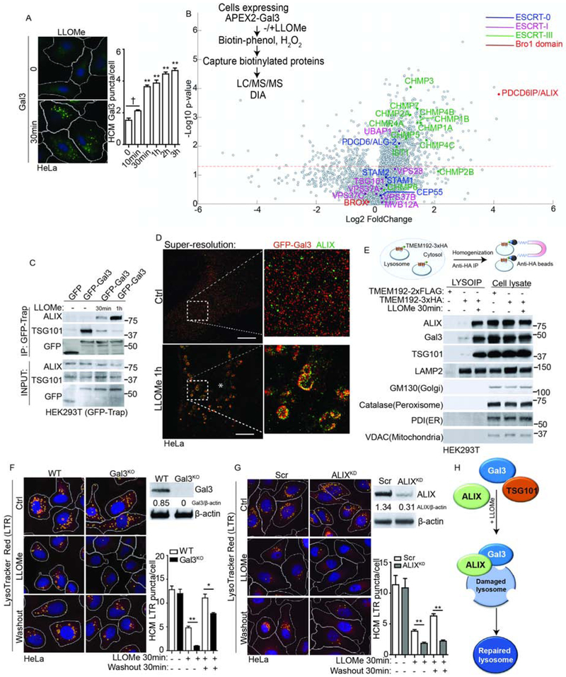
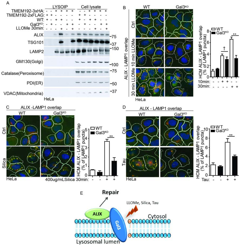
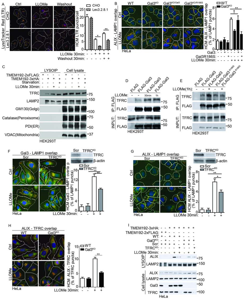
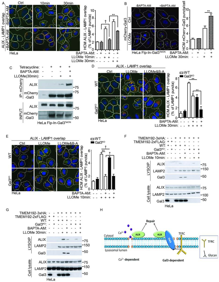
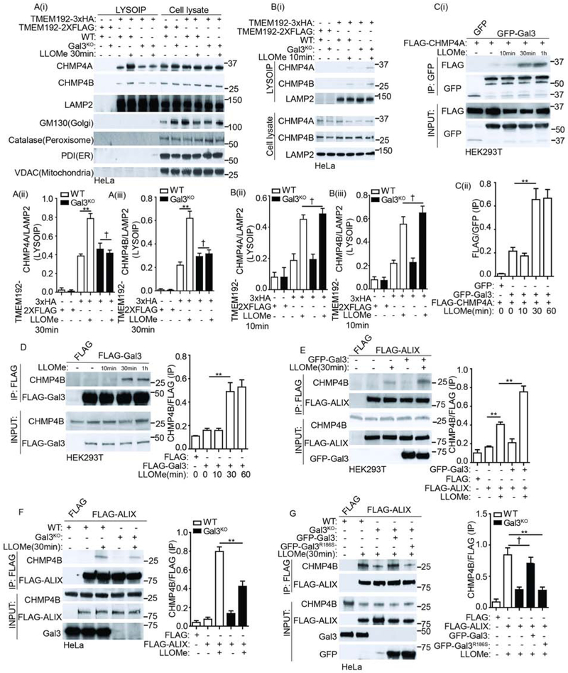
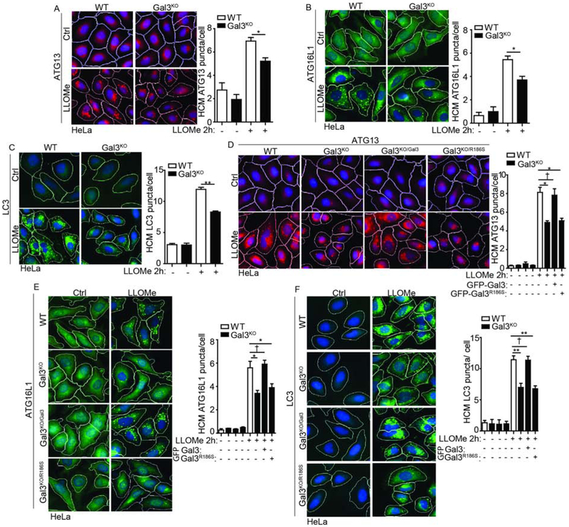
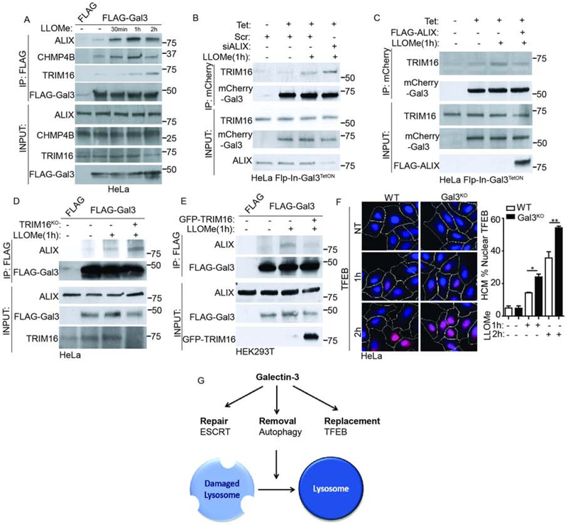
Endomembrane damage elicits homeostatic responses including ESCRT-dependent membrane repair and autophagic removal of damaged organelles. Previous studies have suggested that these systems may act separately. Here, we show that galectin-3 (Gal3), a β-galactoside-binding cytosolic lectin, unifies and coordinates ESCRT and autophagy responses to lysosomal damage. Gal3 and its capacity to recognize damage-exposed glycans were required for efficient recruitment of the ESCRT component ALIX during lysosomal damage. Both Gal3 and ALIX were required for restoration of lysosomal function. Gal3 promoted interactions between ALIX and the downstream ESCRT-III effector CHMP4 during lysosomal repair. At later time points following lysosomal injury, Gal3 controlled autophagic responses. When this failed, as in Gal3 knockout cells, lysosomal replacement program took over through TFEB. Manifestations of this staged response, which includes membrane repair, removal, and replacement, were detected in model systems of lysosomal damage inflicted by proteopathic tau and during phagosome parasitism by Mycobacterium tuberculosis.

Keywords: ESCRT; TFEB; autophagy; endosome; galectins; lysosome; membrane damage homeostasis.


Conflict of interest statement

DECLARATION OF INTERESTS

The authors declare no competing interests.

Figures

Figure 1.
Figure 1.. Galectin-3 and ESCRT component ALIX interact and protect lysosomes from damage.
(A) Time-response of Gal3 during lysosomal damage. HeLa cells were treated with 1mM LLOMe and endogenous Gal3 puncta quantified by high content microscopy (HCM). White masks, algorithm-defined cell boundaries (primary objects); Yellow masks, computer-identified Gal3 puncta (target objects). (B) EncyclopeDIA/scaffoldDIA analysis of Gal3 binding partners proximity-biotinylated by APEX2-Gal3 during lysosomal damage with 1mM LLOMe for 1h. Scatter (volcano) plot shows log2 fold change and −log10 P value for the proteins identified and quantified (LC/MS/MS) in 3 independent experiments (see Table S1, Tabs1 and 2). (C) Co-IP analysis of interactions between Gal3 and ALIX/TSG101 during lysosomal damage. (D) Superresolution microscopy analysis of ALIX and Gal3. HeLa cells transiently expressing GFP-Gal3 were treated with 1mM LLOMe for 1h. Scale bar, 5 μm. (E) Analysis of lysosomes purified by anti-HA immunoprecipitation (LysoIP; TMEM192-3xHA) from HEK293T cells treated with 1mM LLOMe for 30min. TMEM192-2xFLAG, control. (F) Status of acidified organelles in parental HeLa (WT) and Gal3-KO HeLa cells (Gal3KO) assessed by LysoTracker HCM during lysosomal damage (1mM LLOMe for 30min followed by 30min washout). Ctrl, control (untreated cells). Yellow masks, computer-identified LTR puncta. (G) As in F, ALIX knockdown (ALIXKD). Scr, scrambled siRNA. (H) Schematic summary of the findings in Figure 1. Data, means ± SEM; HCM: n ≥ 3 (each experiment: 500 valid primary objects/cells per well, ≥ 5 wells/sample). † p ≥ 0.05 (not significant), *p < 0.05, **p < 0.01, ANOVA. See also Figure S1 and S2.
Figure 2.
Figure 2.. Galectin-3 is required for efficient recruitment of ALIX to damaged lysosomes.
(A) LysoIP analysis for indicated proteins in cell lysates or lysosomes purified from parental HeLa (WT) and Gal3-knockout HeLa cells (Gal3KO) subjected to the 1mM LLOMe treatment for 30min. (B) Quantification by HCM of overlaps between ALIX and LAMP1 in parental HeLa (WT) and Gal3-knockout HeLa cells (Gal3KO) during lysosomal damage. (C) Quantification by HCM of overlaps between ALIX and LAMP1 in parental HeLa (WT) and Gal3-knockout HeLa cells (Gal3KO) during 400 μg/mL silica treatment. (D) Quantification by HCM of overlaps between ALIX and LAMP1 in parental HeLa (WT) and Gal3-knockout HeLa cells (Gal3KO) during Tau oligomer treatment. Cells were treated with 1μg/mL Tau oligomer overnight and subjected to HCM analysis. (E) Schematic summary of the findings in Figure 2. None-treated cells were used as control (Ctrl). White masks, algorithm defined cell boundaries; Yellow masks, computer-identified overlap of ALIX and LAMP1. See also Figure S2.
Figure 3.
Figure 3.. Glycosylation and specific glycosylated proteins play a role in Gal3 recognition of lysosomal damage.
(A) HCM Analysis of the status of acidified organelles in wild-type CHO cells and mutant Lec3.2.8.1 by LysoTracker during lysosomal damage. As in Fig. 1F. (B) Quantification by HCM of overlaps between ALIX and LAMP1 in parental HeLa (WT) and Gal3-knockout HeLa cells (Gal3KO). As in Fig. 2B. (C) LysoIP analysis for indicated proteins in cell lysates or lysosomes purified from parental HeLa cells subjected to 1mM LLOMe or starvation (EBSS medium) treatment for 30min. (D) Co-immunoprecipitation analysis of changes in interactions between Gal3 and TFRC during the process of lysosomal damage. (E) Co-immunoprecipitation analysis of changes in interactions between Gal3/Gal3R186S and TFRC during the process of lysosomal damage. (F) Quantification by HCM of overlaps between Gal3 and LAMP1 in parental HeLa (WT) and TFRC-knockdown HeLa cells (TFRCKD) during LLOMe treatment. (G) Quantification by HCM of overlaps between ALIX and LAMP1 in parental HeLa (WT) and TFRC-knockdown HeLa cells (TFRCKD) during LLOMe treatment. (H) Quantification by HCM of overlaps between ALIX and TFRC in parental HeLa (WT) and Gal3-knockout HeLa cells (Gal3KO) during LLOMe treatment. (I) LysoIP analysis for indicated proteins in cell lysates or lysosomes purified from parental HeLa (WT) and Gal3-knockout HeLa cells (Gal3KO) with or without TFRC-knockdown (TFRCKD). None-treated cells were used as control (Ctrl). White masks, algorithm defined cell boundaries; Yellow masks, computer-identified overlap of two proteins. See also Figure S3.
Figure 4.
Figure 4.. Optimal ALIX recruitment to damaged lysosomes requires two signals.
(A) Cells were treated with 15μM BAPTA-AM for 1h, subjected to 1mM LLOMe treatment for the indicated time, followed by HCM analysis of overlaps between ALIX and LAMP1. As in Fig. 2B. (B) The constructed HeLa Flp-In-Gal3TetON cells stably expressing mCherry-Gal3 induced by tetracycline were subject to 15μM BAPTA-AM treatment for 1h and then treated with LLOMe. (C) Co-immunoprecipitation analysis of changes in interactions between Gal3 and ALIX under BAPTA-AM treatment during lysosomal damage. (D) Quantification by HCM of overlaps between ALIX and LAMP1 in parental HeLa (WT) and Gal3-knockout HeLa cells (Gal3KO) treated with BAPTA-AM during lysosomal damage. (E) Quantification by HCM of overlaps between ALIX and LAMP1 in parental HeLa (WT) and Gal3-knockout HeLa cells (Gal3KO) treated with BAPTA-AM during lysosomal damage. (F) LysoIP analysis for indicated proteins in cell lysates or lysosomes purified from parental HeLa (WT) and Gal3-knockout HeLa cells (Gal3KO) treated with 15μM BAPTA-AM for 1h subjected to the 1mM LLOMe treatment for 10min. (G) LysoIP analysis for indicated proteins in cell lysates or lysosomes purified from parental HeLa (WT) and Gal3-knockout HeLa cells (Gal3KO) treated with 15μM BAPTA-AM for 1h subjected to LLOMe treatment. (H) Schematic summary of the findings in Figure 4.
Figure 5.
Figure 5.. Galectin-3 promotes response of core ESCRT-III effectors during lysosomal damage.
(A)(i) LysoIP analysis for indicated proteins in cell lysates or lysosomes purified from parental HeLa (WT) and Gal3-knockout HeLa cells (Gal3KO) after 1mM LLOMe treatment for 30min. (ii-iii) Quantification of LysoIP analysis for CHMP4A and CHMP4B. (B)(i) LysoIP analysis for indicated proteins in cell lysates or lysosomes purified from parental HeLa (WT) and Gal3-knockout HeLa cells (Gal3KO) after 1mM LLOMe treatment for 10min. (ii-iii) Quantification of LysoIP analysis for CHMP4A and CHMP4B. (C) Co-immunoprecipitation analysis of changes in interactions between Gal3 and CHMP4A during the process of lysosomal damage. (D) Co-immunoprecipitation analysis of changes in interactions between Gal3 and CHMP4B during the process of lysosomal damage. (E) Co-immunoprecipitation analysis of the effect of Gal3 on the interaction between ALIX and CHMP4B during lysosomal damage. (F) Co-immunoprecipitation analysis of the interaction between ALIX and CHMP4B in parental HeLa (WT) and Gal3-knockout HeLa cells (Gal3KO) during lysosomal damage. (G) Co-immunoprecipitation analysis of the effect of Gal3 and its mutant Gal3R186S on the interaction between ALIX and CHMP4B. See also Figure S5.
Figure 6.
Figure 6.. Galectin-3 is important for autophagy response to lysosomal damage.
(A) HeLa cells were treated with 1mM LLOMe for 2h and subjected to HCM analysis of ATG13 puncta. (B) HeLa cells were treated with 1mM LLOMe for 2h and subjected to HCM analysis of ATG16L1 puncta. (C) HeLa cells were treated with 1mM LLOMe for 2h and subjected to HCM analysis of LC3. (D) Gal3-knockout HeLa cells (Gal3KO) transfected with GFP-tagged WT Gal3 or Gal3R186S were treated with 1mM LLOMe for 2h and subjected to HCM analysis of ATG13 puncta. (E) Gal3-knockout HeLa cells (Gal3KO) transfected with GFP-tagged WT Gal3 or Gal3R186S were treated with 1mM LLOMe for 2h and subjected to HCM analysis of ATG16L1 puncta. (F) Gal3-knockout HeLa cells (Gal3KO) transfected with GFP-tagged WT Gal3 or -Gal3R186S were treated with 1mM LLOMe for 2h and the puncta of LC3 was quantified using HCM. None-treated cells were used as control (Ctrl). White masks, algorithm defined cell boundaries; Red masks, computer-identified target protein puncta. See also Figure S5.
Figure 7.
Figure 7.. Galectin-3 serves as a switch between ESCRT and autophagy responses to lysosomal damage.
(A) Co-immunoprecipitation analysis of changes in interactions between Gal3 and ESCRT components during the process of lysosomal damage. (B) The constructed HeLa Flp-In-Gal3TetON cells stably expressing Gal3 induced by tetracycline (Tet) were subject to ALIX knockdown and then treated with 1mM LLOMe for 1h. (C) Co-immunoprecipitation analysis of the effect of ALIX on interaction between Gal3 and TRIM16 during lysosomal damage. (D) Co-immunoprecipitation analysis of the effect of TRIM16 on the interaction between ALIX and Gal3 during lysosomal damage. (E) Co-immunoprecipitation analysis of the effect of TRIM16 on interaction between Gal3 and ALIX during lysosomal damage. (F) TFEB nuclear translocation in parental HeLa (WT) and Gal3-knockout HeLa cells (Gal3KO) during lysosomal damage. Blue: nuclei, Hoechst 33342; Red: anti-TFEB antibody, Alexa-568. White masks, computer algorithm-defined cell boundaries; Pink masks, computer-identified nuclear TFEB based on the average intensity of Alexa-568 fluorescence. (G) Schematic summary of the findings in Figure 7. See also Figure S6.

References

    1. Abu-Remaileh M, Wyant GA, Kim C, Laqtom NN, Abbasi M, Chan SH, Freinkman E, and Sabatini DM (2017). Lysosomal metabolomics reveals V-ATPase- and mTOR-dependent regulation of amino acid efflux from lysosomes. Science 358, 807–813. - PMC - PubMed
    1. Aits S, Kricker J, Liu B, Ellegaard AM, Hamalisto S, Tvingsholm S, Corcelle-Termeau E, Hogh S, Farkas T, Holm Jonassen A, et al. (2015). Sensitive detection of lysosomal membrane permeabilization by lysosomal galectin puncta assay. Autophagy 11, 1408–1424. - PMC - PubMed
    1. Baietti MF, Zhang Z, Mortier E, Melchior A, Degeest G, Geeraerts A, Ivarsson Y, Depoortere F, Coomans C, Vermeiren E, et al. (2012). Syndecan-syntenin-ALIX regulates the biogenesis of exosomes. Nat Cell Biol 14, 677–685. - PubMed
    1. Bakula D, Muller AJ, Zuleger T, Takacs Z, Franz-Wachtel M, Thost AK, Brigger D, Tschan MP, Frickey T, Robenek H, et al. (2017). WIPI3 and WIPI4 beta-propellers are scaffolds for LKB1-AMPK-TSC signalling circuits in the control of autophagy. Nature communications 8, 15637. - PMC - PubMed
    1. Baskaran S, Carlson LA, Stjepanovic G, Young LN, Kim do J, Grob P, Stanley RE, Nogales E, and Hurley JH (2014). Architecture and dynamics of the autophagic phosphatidylinositol 3-kinase complex. Elife 3. - PMC - PubMed

Publication types