. 2020 Mar 13;29(4):580-590.
doi: 10.1093/hmg/ddz271.

G2019S-LRRK2 mutation enhances MPTP-linked Parkinsonism in mice

Affiliations

G2019S-LRRK2 mutation enhances MPTP-linked Parkinsonism in mice

Nicolas Arbez et al. Hum Mol Genet. .

Abstract

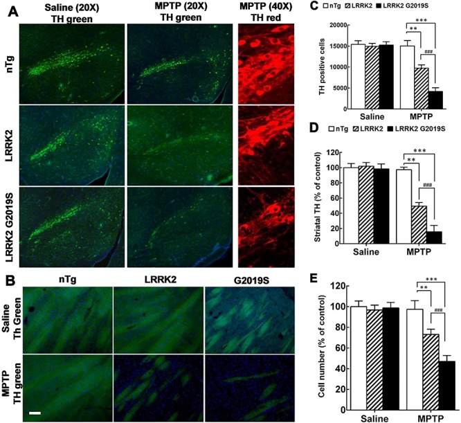
Parkinson's disease (PD) is a common neurodegenerative disease with a heterogeneous etiology that involves genetic and environmental factors or exogenous. Current LRRK2 PD animal models only partly reproduce the characteristics of the disease with very subtle dopaminergic neuron degeneration. We developed a new model of PD that combines a sub-toxic MPTP insult to the G2019S-LRRK2 mutation. Our newly generated mice, overexpressing mutant G2019S-LRRK2 protein in the brain, displayed a mild, age-dependent progressive motor impairment, but no reduction of lifespan. Cortical neurons from G2019S-LRRK2 mice showed an increased vulnerability to stress insults, compared with neurons overexpressing wild-type WT-LRRK2, or non-transgenic (nTg) neurons. The exposure of LRRK2 transgenic mice to a sub-toxic dose of MPTP resulted in severe motor impairment, selective loss of dopamine neurons and increased astrocyte activation, whereas nTg mice with MPTP exposure showed no deficits. Interestingly, mice overexpressing WT-LRRK2 showed a significant impairment that was milder than for the mutant G2019S-LRRK2 mice. L-DOPA treatments could partially improve the movement impairments but did not protect the dopamine neuron loss. In contrast, treatments with an LRRK2 kinase inhibitor significantly reduced the dopaminergic neuron degeneration in this interaction model. Our studies provide a novel LRRK2 gene-MPTP interaction PD mouse model, and a useful tool for future studies of PD pathogenesis and therapeutic intervention.


Figures

Figure 1
Figure 1
Generation of human wild-type WT-LRRK2 and G2019S-LRRK2 transgenic mice. (A) Flag-WT-LRRK2 or G2019S-LRRK2 gene was cloned into a mouse prion promotor (MoPrP) vector to generate transgenic mice expressing LRRK2 variants predominantly in neurons. (B) Lysates derived from whole brain of nTg, WT-, and G2019S-LRRK2 (Flag-tagged) heterozygous (Hetero) and homozygous (Homo) transgenic mice were analyzed by western blot using anti-LRRK2 antibodies. (C) Quantitation of LRRK2 protein levels in transgenic mice normalized to nTg mice based on three independent western blot experiments using anti-LRRK2. The LRRK2 protein level in the brain of G2019S homozygous transgenic mice is about 2–3-fold of heterozygous mice and about 5-fold of nTg mice. (D) Average weight of the mice at 4, 9 and 12 months. No difference has been observed by ANOVA.
Figure 2
Figure 2
G2019S-LRRK2 mice display motor impairment with aging. (A) Locomotor activity was monitored by open field testing for transgenic mice at ages of 6 and 12 months. Twenty-four mice in each group, half male and half female. G2019S mice showed significant reductions in both locomotor activity and rearing activities. P < 0.05 by ANOVA versus nTg mice. (B) Rotarod testing was done for mice at ages of 4, 9 and 12 months. n=24, half male and half female. G2019S mice showed an age-dependent impairment. T-test, ***P < 0.001 by ANOVA versus nTg mice.
Figure 3
Figure 3
Primary cortical neurons from LRRK2 transgenic mice were more vulnerable to stress insults. Primary cortical neurons isolated from nTg, LRRK2 and G2019S-LRRK2 at 7 DIV were treated with different stressors. (A) Autophagy inhibitor, 3MA, (B) excitotoxic stressor, NMDA, (C) depolarization agent, KCl and (D) ER stressor, DTT were applied at the indicated concentrations for 48 h. (E) Primary neurons were deprived of trophic factor supplement, B27 in the culture medium for 24 h. (F) Primary neurons were exposed to increasing concentration of MPP+ for 48 h. Quantification of cell death was done by nuclear condensation assay. Two-way ANOVA analysis, *P < 0.05 versus nTg, **P < 0.01 versus nTg and ***P < 0.001 versus nTg; #P < 0.05 versus G2019S, ##P < 0.01 versus G2019S and ###P < 0.001 versus G2019S.
Figure 4
Figure 4
Sub-toxic dose of MPTP exposure induced motor impairment in LRRK2 transgenic mice. (A) Experimental flow chart. Mice at age of 4 months (nTg, LRRK2 and G2019S-LRRK2 mice, n = 10 per group) were injected with MPTP (2.5 mg/kg, sc, 2 doses 24 h apart). Rotarod testing was performed from 5 to 7 days post-injection (3 trials per day, 3 days in a row). (B) Shown are the average latency to fall ± sem. Two-way ANOVA *P < 0.05 versus nTg, ***P < 0.001 versus nTg and ###P < 0.001 versus G2019S.
Figure 5
Figure 5
Sub-toxic dose of MPTP exposure induced the loss of TH-positive immunoreactivity in LRRK2 transgenic mouse brains. (A) Representative images of TH neurons in the substantia nigra after MPTP exposure. Left and middle: TH green 20X; right: TH Red 40X. (B) Quantification of TH-positive cells in the substantia nigra. Results are normalized to control and expressed as mean ± sem. (C) and (D) Quantification of TH-positive immunoreactivity in the striatum. (E) DAPI and anti-TH staining of substantia nigra. The number of cells was quantified by counting DAPI. Results are normalized to control and expressed as mean ± sem. Two-way ANOVA analysis, *P < 0.05 versus nTg, **P < 0.01 versus nTg and ***P < 0.001 versus nTg; #P < 0.05 versus G2019S, ##P < 0.01 versus G2019S and ###P < 0.001 versus G2019S.
Figure 6
Figure 6
Sub-toxic dose of MPTP exposure increased astrocytic activation in the brains of LRRK2 transgenic mice. (A) Representative images of GFAP (Red) staining in brains of transgenic mice. (B) Quantification of (A). Results are normalized to control and expressed as mean ± sem. Two-way ANOVA analysis, ***P < 0.001 versus nTg; ###P < 0.001 versus G2019S.
Figure 7
Figure 7
Treatment of LRRK-IN-1 or L-DOPA attenuated MPTP and G2019S-LRRK2-induced PD-like phenotypes in mice. (A) Experimental flow chart. A cohort of mice from each group (n = 10) at age of 4 months were injected with MPTP (2.5 mg/kg, sc, 2 doses 24 h apart), and treated with a LRRK2 Kinase inhibitor (LRRK2-IN-1) or L-DOPA. (B) Rotarod testing was performed from 5 to 7 days post-injection (3 trials per day, 3 days in a row). (C) Quantification of TH-positive cells in the substantia nigra. (D) Quantification of GFAP. Two-way ANOVA analysis, *P < 0.05 versus nTg, **P < 0.01 versus nTg and ***P < 0.001 versus nTg; #P < 0.05 versus G2019S, ##P < 0.01 versus G2019S and ###P < 0.001 versus G2019S.

References

    1. Tysnes O.B. and Storstein A. (2017) Epidemiology of Parkinson's disease. J. Neural Transm., 124, 901–905. - PubMed
    1. Brug M.P., Singleton A., Gasser T. and Lewis P.A. (2015) Parkinson's disease: From human genetics to clinical trials. Sci. Transl. Med., 7, 205–220. - PMC - PubMed
    1. Poewe W. (2017) Parkinson's disease and the quest for preclinical diagnosis: an interview with Professor Werner Poewe. Neurodegener Dis. Manag., 7, 273–277. - PubMed
    1. Deng H., Wang P. and Jankovic J. (2018) The genetics of Parkinson disease. Ageing Res. Rev., 42, 72–85. - PubMed
    1. Smith W.W., Pei Z., Jiang H., Dawson V.L., Dawson T.M. and Ross C.A. (2006) Kinase activity of mutant LRRK2 mediates neuronal toxicity. Nat. Neurosci., 9, 1231–1233. - PubMed

Publication types

Substances