Review
. 2020 Mar;48(3):253-264.
doi: 10.1002/dc.24354. Epub 2019 Dec 9.

Role of TFF3 as an adjunct in the diagnosis of Barrett's esophagus using a minimally invasive esophageal sampling device-The CytospongeTM

Affiliations
Review

Role of TFF3 as an adjunct in the diagnosis of Barrett's esophagus using a minimally invasive esophageal sampling device-The CytospongeTM

Anna L Paterson et al. Diagn Cytopathol. 2020 Mar.

Abstract

The incidence of esophageal carcinoma continues to increase whilst its prognosis remains poor. The most dramatic reduction in mortality is likely to follow early diagnosis of the preinvasive precursor lesion, Barrett's esophagus (BE), coupled with treatment of dysplastic lesions. The major risk factor for BE is gastroesophageal reflux disease, however this is highly prevalent and only a small proportion of individuals have BE, therefore an endoscopy-based screening strategy to detect BE is unfeasible. Minimally invasive esophageal sampling devices offer an alternative, cost-effective strategy which can be deployed within an at-risk population in a primary care setting to identify individuals with probable BE who can then be referred for endoscopic confirmation. The device that has currently progressed furthest in clinical trials is the CytospongeTM which collects cells from the gastric cardia, gastroesophageal junction and along the whole esophageal length. The cell sample is processed into a formalin-fixed paraffin-embedded block and sections assessed for the presence of intestinal metaplasia. TFF3 immunohistochemistry has consistently been shown to be a valuable adjunct that increases the accuracy of the CytospongeTM test by highlighting early goblet cells which may be missed on morphological assessment and by allowing pseudogoblet cells to be differentiated from true goblet cells.

Keywords: Barrett's esophagus; biomarkers; esophageal adenocarcinoma; screening.


Conflict of interest statement

Conflict of interest statement: Patents and a trademark were filed on Cytosponge™ in 2010 by the Medical Research Council (MRC). RCF and MOD are named inventors on patents pertaining to the Cytosponge™ and related assays. In 2013 the MRC licensed the technology to Covidien GI Solutions (now Medtronic).

Figures

Figure 1
Figure 1
Schematic outlining the steps of the Cytosponge™-TFF3 assay. Step 1 - the sponge is swallowed, the coating dissolves in the stomach and the sponge deploys. The Cytosponge™ is then withdrawn using the attached string collecting cells from the gastroesophageal junction and the length of the oesophagus. Step 2 – the sampled cells are retrieved from the sponge by a series of washing steps then a cell pellet is made by centrifugation. Plasma-thrombin is added to the cell pellet making a cell clot. Step 3 – the cell clot is fixed in formalin and processed into a paraffin block using standard laboratory protocols. Step 4 – sections are cut from the paraffin block and stained for assessment by a pathologist.
Figure 2
Figure 2
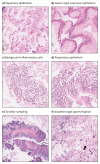
Normal components that may be seen in a Cytosponge™ sample, all images at 10x magnification. (a) Squamous epithelial cells from the esophagus and oropharynx. (b) Gastric-type columnar epithelium from the stomach and/or a hiatus hernia. (c) Mixed inflammatory cells which provided that they are separate from epithelial cell groups are not considered to indicate clinically significant esophagitis. (d) Strips of respiratory epithelium recognised by their terminal bars and cilia. (e) Tonsillar sampling characterised by keratinous material and actinomyces organisms. (f) Fungal spores and occasional hyphae (arrow) with appearances consistent with Candida. As there is no associated significant acute inflammation they are considered to represent commensal organisms rather than being suggestive of Candida esophagitis.
Figure 3
Figure 3
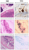
Challenges in identifying true goblet cells using hematoxylin and eosin stained preparations alone and the benefits of TFF3 immunohistochemistry, all images at 20x magnification. (a,b) Classic, mature goblet cell. (c,d) Subtle goblet cells which are highlighted on TFF3 staining. (e,f) Columnar cells with features suggestive of an associated goblet cell however no staining is seen with TFF3 showing that this is not a true goblet cell but a “pseudogoblet” cell.
Figure 4
Figure 4
Interpreting TFF3 staining patterns. (a) TFF3 staining in true goblet cells should be dense dark brown with a crisp edge. (b) Weak non-specific cytoplasmic staining can occasionally be seen in strips of columnar cells however it lacks the dense, well-defined pattern of staining in individual cells seen in true goblet cell staining. (c) Strips of respiratory epithelium can contain goblet cells which will also be TFF3 positive. The respiratory nature of these groups can be recognised on the parallel hematoxylin and eosin stain by the presence of terminal bars and cilia.

References

    1. Edgren G, Adami HO, Weiderpass E, Weiderpass Vainio E, Nyrén O. A global assessment of the oesophageal adenocarcinoma epidemic. Gut. 2013;62(10):1406–1414. - PubMed
    1. Wong MCS, Hamilton W, Whiteman DC, et al. Global Incidence and mortality of oesophageal cancer and their correlation with socioeconomic indicators temporal patterns and trends in 41 countries. Sci Rep. 2018;8(1) 4522. - PMC - PubMed
    1. Cancerresearchuk.org. Cancer Research UK Statistics. [accessed March 2019];
    1. Qiao Y, Hyder A, Bae SJ, et al. Surveillance in Patients With Barrett's Esophagus for Early Detection of Esophageal Adenocarcinoma: A Systematic Review and Meta-Analysis. Clin Transl Gastroenterol. 2015;6:e131. - PMC - PubMed
    1. Hamouda A, Forshaw M, Rohatgi A, Mirnezami R, Botha A, Mason R. Presentation and survival of operable esophageal cancer in patients 55 years of age and below. World J Surg. 2010;34(4):744–749. - PubMed

MeSH terms