Review
. 2019 Nov 13:2019:1389289.
doi: 10.1155/2019/1389289. eCollection 2019.

Precursors to Cholangiocarcinoma

Affiliations
Review

Precursors to Cholangiocarcinoma

Tomas Hucl. Gastroenterol Res Pract. .

Abstract

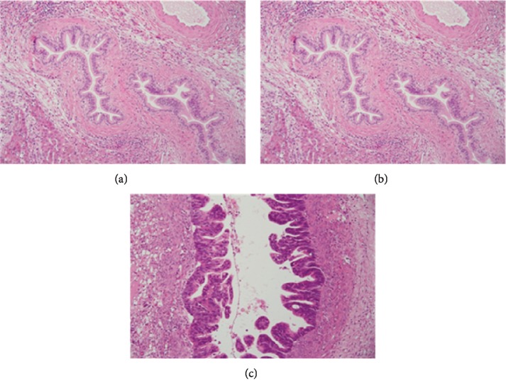
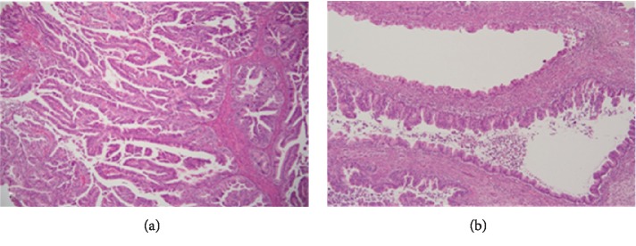
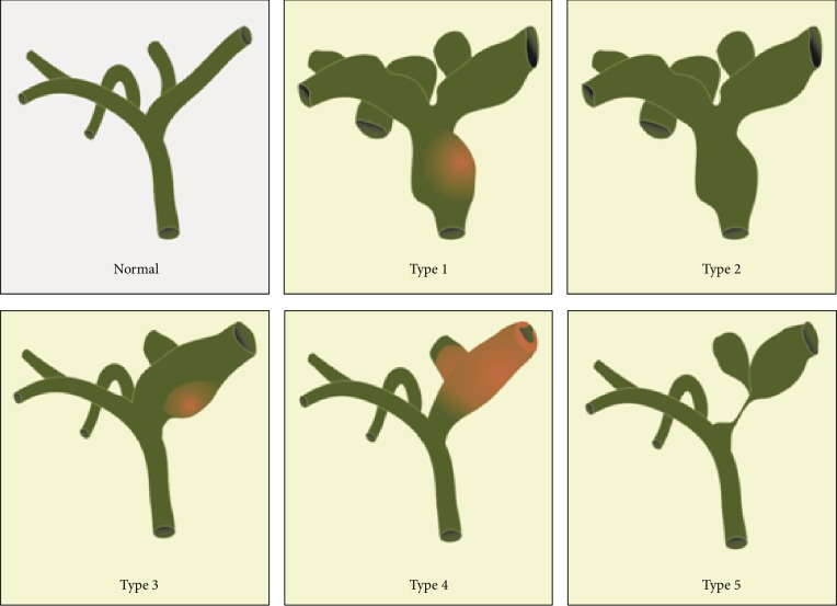
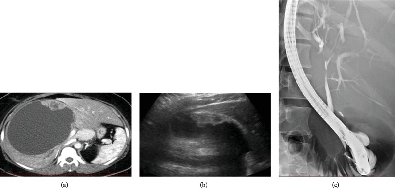
Cancers of the biliary tract include intra- and extrahepatic cholangiocarcinomas and gallbladder cancer. Biliary tract cancers are diseases with unfavorable prognoses. In recent years, several lesions have been described as precursors that precede biliary cancers. They include flat and microscopic lesions known as biliary intraepithelial neoplasia, macroscopic and tumor-forming intraductal papillary biliary neoplasia, intraductal tubular neoplasia, and mucinous cystic neoplasm of the bile duct. These conditions are rarely diagnosed, while their natural history and progression to cancer have yet to be adequately characterized. This review examines the epidemiology, pathology, molecular biology, diagnosis, and therapy of these various precursors. Further research is required if we are to better understand this evolving field and improve the prevention and early detection of bile duct cancer.


Conflict of interest statement

There is no conflict of interest regarding the publication of this paper.

Figures

Figure 1
Figure 1
Histology image of biliary cancer precursors. (a) Biliary intraepithelial neoplasia grade 1 (BilIN-1)—bile duct with tall columnar cells with mild nuclear abnormalities. (b) Biliary intraepithelial neoplasia grade 2 (BilIN-2)—bile duct with columnar cells with moderate nuclear abnormalities and focal pseudopapillary or micropapillary architecture of the epithelium. (c) Biliary intraepithelial neoplasia grade 3 (BilIN-3)—bile duct with columnar cells with severe nuclear abnormalities and pseudopapillary and cribriform architecture of the epithelial layer. Haematoxylin-eosin staining (courtesy of Dr. Jana Maluskova).
Figure 2
Figure 2
Histology image of biliary cancer precursors. (a) Intraductal papillary neoplasia of the bile duct (IPNB)—bile duct with papillary architecture of the epithelium and cytological nuclear abnormalities. (b) Biliary mucinous cystic neoplasia (BMCN)—cyst with the epithelium of the mucinous type and variable grade of nuclear abnormalities. Haematoxylin-eosin staining (courtesy of Dr. Jana Maluskova).
Figure 3
Figure 3
Anatomical classification of intraductal papillary neoplasms of the bile duct (figure based on Wan XS, WJG 2013).
Figure 4
Figure 4
Imaging findings in a patient with biliary mucinous cystic neoplasm. (a) CT image of a large BMCN in the right liver lobe with a hypodense solid component. (b) Ultrasound image of the same BMCN with a hypoechoic solid component by the wall. (c) ERCP image of the same patient showing s smooth stricture of the hepatic duct caused by extrinsic compression of the cyst.

References

    1. Kloppel G., Adsay V., Konukiewitz B., Kleeff J., Schlitter A. M., Esposito I. Precancerous lesions of the biliary tree. Best Practice & Research. Clinical Gastroenterology. 2013;27(2):285–297. doi: 10.1016/j.bpg.2013.04.002. - DOI - PubMed
    1. Razumilava N., Gores G. J. Classification, diagnosis, and management of cholangiocarcinoma. Clinical Gastroenterology and Hepatology. 2013;11(1):13–21.e1. doi: 10.1016/j.cgh.2012.09.009. - DOI - PMC - PubMed
    1. Wan X. S., Xu Y. Y., Qian J. Y., et al. Intraductal papillary neoplasm of the bile duct. World Journal of Gastroenterology. 2013;19(46):8595–8604. doi: 10.3748/wjg.v19.i46.8595. - DOI - PMC - PubMed
    1. Burak K., Angulo P., Pasha T. M., Egan K., Petz J., Lindor K. D. Incidence and risk factors for cholangiocarcinoma in primary sclerosing cholangitis. The American Journal of Gastroenterology. 2004;99(3):523–526. doi: 10.1111/j.1572-0241.2004.04067.x. - DOI - PubMed
    1. Singham J., Yoshida E. M., Scudamore C. H. Choledochal cysts: part 1 of 3: classification and pathogenesis. Canadian Journal of Surgery. 2009;52:434–440. - PMC - PubMed

LinkOut - more resources