Review
. 2019 Jul 26;10(10):1678-1691.
doi: 10.1039/c9md00292h. eCollection 2019 Oct 1.

Glycans in drug discovery

Affiliations
Review

Glycans in drug discovery

Pablo Valverde et al. Medchemcomm. .

Abstract

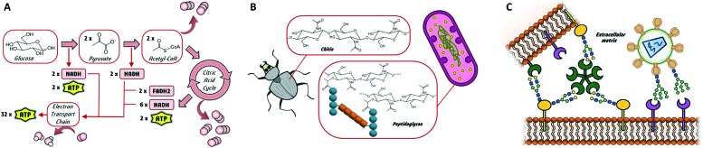
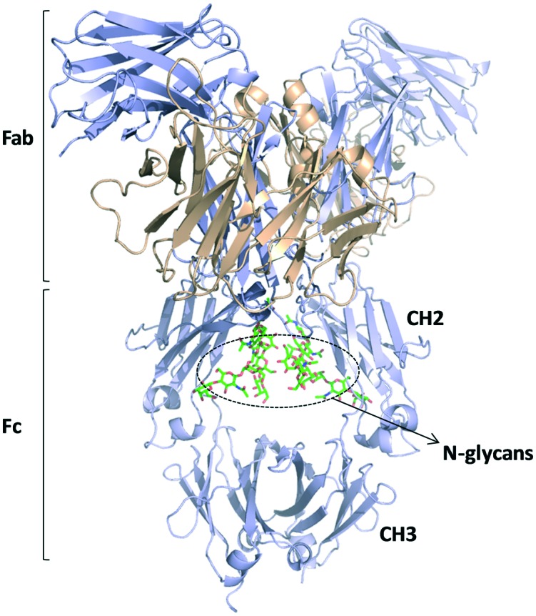
Glycans are key players in many biological processes. They are essential for protein folding and stability and act as recognition elements in cell-cell and cell-matrix interactions. Thus, being at the heart of medically relevant biological processes, glycans have come onto the scene and are considered hot spots for biomedical intervention. The progress in biophysical techniques allowing access to an increasing molecular and structural understanding of these processes has led to the development of effective therapeutics. Indeed, strategies aimed at designing glycomimetics able to block specific lectin-carbohydrate interactions, carbohydrate-based vaccines mimicking self- and non-self-antigens as well as the exploitation of the therapeutic potential of glycosylated antibodies are being pursued. In this mini-review the most prominent contributions concerning recurrent diseases are highlighted, including bacterial and viral infections, cancer or immune-related pathologies, which certainly show the great promise of carbohydrates in drug discovery.


Figures

Fig. 1
Fig. 1. Three main biological roles of glycans as (A) energy source, (B) structural elements and (C) molecular recognition elements in cell–cell and cell-pathogen interactions.
Scheme 1
Scheme 1. Chemical structure of fondaparinux sodium salt, commercially known as Arixtra.
Fig. 2
Fig. 2. C-glycoside inhibitors of LecB bearing sulfonamide moieties. X-ray crystal structures of the interaction with LecB (pdb 5MB1 and ; 5MAY).
Fig. 3
Fig. 3. Covalent inhibitor described for LecA. The crystal structure for the non-covalent mode interaction is depicted (pdb 5MIM).
Fig. 4
Fig. 4. Mannoside inhibitors of FimH displaying aromatic aglycones.
Fig. 5
Fig. 5. Chemical structure of selectin inhibitors rivipansel and uproleselan. The chemical structure of sLex antigen and the crystal structure of the interaction with selectin are depicted at the bottom (pdb ; 1G1T).
Fig. 6
Fig. 6. Trivalent modified N-glycans described as pM inhibitors of Siglec-2.
Fig. 7
Fig. 7. (A) First- and second-generation DC-SIGN glycomimetics. The X-ray crystal structure of DC-SIGN bound to a second-generation glycomimetic is also represented (pdb 2XR5). (B) DC-SIGN multivalent ligand.
Scheme 2
Scheme 2. High-affinity and selective thiodigalactoside glycomimetics of galectin-3.
Fig. 8
Fig. 8. Schematic representation of the employed approach to design anti-HIV immunogenic glycopeptides.
Fig. 9
Fig. 9. (A) Model of normal densely glycosylated MUC1 and tumor-associated (TA) MUC1 with truncated glycosylation. (B) MUC1 synthetic glycopeptide bearing modifications at the peptide backbone and at the glycosidic linkage.
Fig. 10
Fig. 10. Schematic representation of an IgG1 antibody structure (pdb 1HZH). Fab and Fc regions are indicated and the glycans present in the CH2 domains are highlighted.
Fig. 11
Fig. 11. Biosynthetic approaches for the preparation of homogeneous glycoproteins. (A) Previous and post enzyme-based remodeling of the overexpressed glycoforms. (B) Cleavage and subsequent incorporation of the desired oligosaccharide chain.

References

    1. Cummings R. D. and Etzler M. E., Essentials of Glycobiology, ed. A. Varki, R. D. Cummings, J. D. Esko, H. H. Freeze, P. Stanley, C. R. Bertozzi, G. W. Hart and M. E. Etzler, Cold Spring Harbor (NY): Cold Spring Harbor Laboratory Press, New York, 2nd edn, 2009, ch. 6. - PubMed
    1. Varki A. Glycobiology. 2017;27:3–49. - PMC - PubMed
    1. Reily C., Stewart T. J., Renfrow M. B., Novak J. Nat. Rev. Nephrol. 2019;15:346–366. - PMC - PubMed
    1. Joshi H. J., Narimatsu Y., Schjoldager K. T., Tytgat H. L. P., Aebi M., Clausen H., Halim A. Cell. 2018;172:632–632.e2. - PubMed
    1. Schwarz F., Aebi M. Curr. Opin. Struct. Biol. 2011;21:576–582. - PubMed