. 2019 Nov 1;9(11):2442-2455.
eCollection 2019.

Disulfiram combined with copper induces immunosuppression via PD-L1 stabilization in hepatocellular carcinoma

Affiliations

Disulfiram combined with copper induces immunosuppression via PD-L1 stabilization in hepatocellular carcinoma

Binghai Zhou et al. Am J Cancer Res. .

Abstract

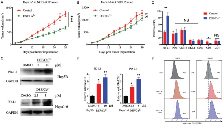
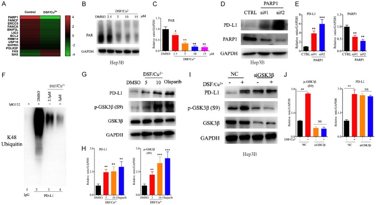
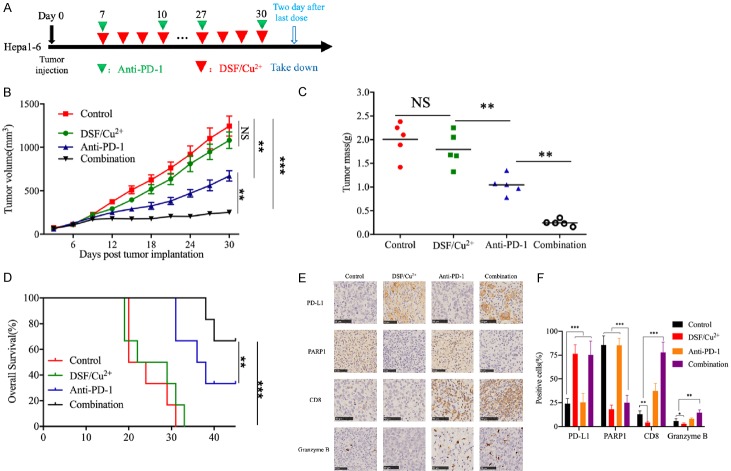
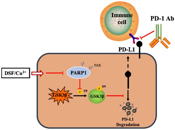
As a potential antitumor drug and chemotherapeutic sensitizer, disulfiram combined with Copper (DSF/Cu2+) does not exert considerable antitumor effects on an immunocompetent hepatocellular carcinoma (HCC) model. In this article, we will explore the mechanism underlying the resistance to DSF in HCC. We analyzed the antitumor effect of DSF/Cu2+ in vivo studies. Tumor and immune cells collected from mice were analyzed by flow cytometry. Then, we analyzed the transcriptional changes in liver cancer cells after DSF/Cu2+ treatment by transcriptional expression profiling. The expression of PD-L1 was detected by real-time PCR, Western blotting and flow cytometry. The expression of PARP1 and GSK3β was knocked down by small interfering RNAs (siRNAs). A subcutaneous Hepa1-6 tumor model was used for single-drug or combined-drug studies. Tissue chips (268 samples of liver cancer tissue) were used to analyze the relationship among PARP1, p-GSK3β and PD-L1. We found that DSF/Cu2+ failed to inhibit HCC tumor growth in C57BL/6 mice. DSF/Cu2+ upregulated PD-L1 expression by inhibiting PARP1 activity and enhancing GSK3β phosphorylation at Ser9 and ultimately inhibited T cell infiltration. The combination of DSF/Cu2+ and an anti-PD-1 antibody produced an additive effect that slowed HCC growth in mice. In addition, we observed negative associations between PARP1 and p-GSK3β (Ser9) or PD-L1 expression in tumor tissue samples from HCC patients. Through in vitro and in vivo studies, we found that DSF/Cu2+ could restrain GSK3β activity by inhibiting PARP1, leading to the upregulation of PD-L1 expression. Combination therapy with DSF/Cu2+ and an anti-PD-1 antibody showed much better antitumor efficacy than monotherapy.

Keywords: GSK3; PARP1; PD-L1; disulfiram; hepatocellular carcinoma.


Conflict of interest statement

None.

Figures

Figure 1
Figure 1
DSF/Cu2+ inhibited proliferation and upregulated PD-L1 expression in HCC tumor cells. A. The growth of tumors generated by Hepa1-6 cells in NOD-SCID/CrlSlac immunodeficient mice following drug intervention with DSF/Cu2+. Tumors were measured at the indicated time points. B. Hepa1-6 tumor growth in C57BL/6 mice following drug intervention with DSF/Cu2+. C. DSF/Cu2+-treated mice. Tumor-infiltrating immune cells and inflammatory cells, including T cells (CD8+), natural killer (NK) cells (NK1.1+), and B lymphocytes (CD19+), as well as PD-L1+ tumor cells were detected by flow cytometry. D. PD-L1 protein expression after DSF/Cu2+ treatment. Hep3B and Hep1-6 cells were treated with DSF/Cu2+ (0, 5, or 10 μM DSF in Hep3B cells and 0, 2.5, or 5 μM DSF in Hep1-6 cells; 1 µM Cu2+), and PD-L1 protein levels were analyzed by Western blotting. E. Quantitative analysis of PD-L1 expression after DSF/Cu2+ treatment through ImageJ intensity measurements. F. Cell-surface PD-L1 expression with DSF/Cu2+ treatment in both Hep3B cells and Hep1-6 cells. Cell-surface PD-L1 levels were measured by flow cytometry. Error bars: mean ± SD. *P < 0.05, **P < 0.01, ***P < 0.001.
Figure 2
Figure 2
DSF/Cu2+ promoted PD-L1 expression via the PARP1/GSK3β pathway. A. Heatmap of a selected list of DNA damage repair-related genes showing fold changes in expression between DSF/Cu2+-treated and control Hep3B cells. B. Activity of PARP1 after DSF/Cu2+ treatment. PAR expression in Hep3B cells was detected by Western blot analysis after DSF/Cu2+ treatment for 16 h (2.5, 5, 10, or 15 μM DSF and 1 µM Cu2+). C. Quantitative analysis of PAR expression after DSF/Cu2+ treatment through ImageJ intensity measurements. D. PARP1 and PD-L1 expression after treatment with PARP1-specific siRNA. PARP1 and PD-L1 protein expression in Hep3B cells was evaluated by Western blotting after treatment with PARP1-specific siRNA. E. Quantitative analysis of PARP1 and PD-L1 expression after treatment with PARP1-specific siRNA through ImageJ intensity measurements. F. Ubiquitination assay evaluating PD-L1 in Hep3B cells. Cell lysates were immunoprecipitated with an anti-PD-L1 antibody and subjected to Western blot analysis with an antibody against K48-linked ubiquitin. The cells were treated with DSF/Cu2+ or MG132 prior to the ubiquitination analysis. G, I. PD-L1 and p-GSK3β (Ser9) expression after treatment with DSF/Cu2+, olaparib or GSK3β-specific siRNA. PD-L1 and p-GSK3β (Ser9) protein expression in Hep3B cells was evaluated by Western blotting after treatment with DSF/Cu2+ (5 or 10 μM DSF and 1 µM Cu2+), olaparib (10 μM for 24 h) or GSK3β-specific siRNA. H, J. Quantitative analysis of PD-L1 and p-GSK3β (Ser9) expression after treatment with DSF/Cu2+, olaparib or GSK3β-specific siRNA through ImageJ intensity measurements. Error bars: mean ± SD. *P < 0.05, **P < 0.01, ***P < 0.001.
Figure 3
Figure 3
Anti-PD-1 therapy in combination with DSF/Cu2+ improved antitumor activity. A. Schematic diagram of the drug intervention protocol for disulfiram plus copper gluconate and/or anti-PD-1 antibody treatment of C57BL/6 mice. B. The growth of subcutaneous Hepa1-6 tumors in disulfiram plus copper gluconate and/or anti-PD-1 antibody-treated C57BL/6 mice. Tumors were measured at the indicated time points. C. Tumor weights after the drug intervention endpoints. D. Survival of mice bearing Hepa1-6 tumors following treatment with disulfiram plus copper gluconate and/or anti-PD-1 antibody. Significance was evaluated using the log-rank test. E. Immunohistochemical staining for PD-L1, PARP1, CD8, and granzyme B protein expression patterns in Hepa1-6 tumors. Scale bar, 50 μm. F. The positive cells of PD-L1, PARP1, CD8, and granzyme B in Hepa1-6 tumors following treatment with disulfiram plus copper gluconate and/or anti-PD-1 antibody. Error bars: mean ± SD. *P < 0.05, **P < 0.01, ***P < 0.001.
Figure 4
Figure 4
Correlation among the expression of PARP1, p-GSK3β (Ser9) and PD-L1 in human tumor tissue samples. A. Representative pictures of IHC staining of HCC tumors for PARP1, PD-L1 and p-GSK3β (Ser9) in HCC tumors. Patient tissue samples were stained for PARP1, PD-L1 and p-GSK3β (Ser9). B. The correlations between PARP1 and PD-L1 or p-GSK3β (Ser9) expression levels in liver cancer patients. P, Pearson chi-square test; -/+, negative or low expression; ++/+++, medium or high expression. Scale bar, 100 μm.
Figure 5
Figure 5
An illustration of the proposed working model. The phosphorylation of GSK3β at Ser9 induced by PARP inhibition is a key step in stabilizing PD-L1.

References

    1. Torre LA, Bray F, Siegel RL, Ferlay J, Lortet-Tieulent J, Jemal A. Global cancer statistics, 2012. CA Cancer J Clin. 2015;65:87–108. - PubMed
    1. El-Serag HB. Hepatocellular carcinoma. N Engl J Med. 2011;365:1118–1127. - PubMed
    1. Llovet JM, Ricci S, Mazzaferro V, Hilgard P, Gane E, Blanc JF, de Oliveira AC, Santoro A, Raoul JL, Forner A, Schwartz M, Porta C, Zeuzem S, Bolondi L, Greten TF, Galle PR, Seitz JF, Borbath I, Haussinger D, Giannaris T, Shan M, Moscovici M, Voliotis D, Bruix J. Sorafenib in advanced hepatocellular carcinoma. N Engl J Med. 2008;359:378–390. - PubMed
    1. Bruix J, Qin S, Merle P, Granito A, Huang YH, Bodoky G, Pracht M, Yokosuka O, Rosmorduc O, Breder V, Gerolami R, Masi G, Ross PJ, Song T, Bronowicki JP, Ollivier-Hourmand I, Kudo M, Cheng AL, Llovet JM, Finn RS, LeBerre MA, Baumhauer A, Meinhardt G, Han G. Regorafenib for patients with hepatocellular carcinoma who progressed on sorafenib treatment (RESORCE): a randomised, double-blind, placebo-controlled, phase 3 trial. Lancet. 2017;389:56–66. - PubMed
    1. Kudo M, Finn RS, Qin S, Han KH, Ikeda K, Piscaglia F, Baron A, Park JW, Han G, Jassem J, Blanc JF, Vogel A, Komov D, Evans TRJ, Lopez C, Dutcus C, Guo M, Saito K, Kraljevic S, Tamai T, Ren M, Cheng AL. Lenvatinib versus sorafenib in first-line treatment of patients with unresectable hepatocellular carcinoma: a randomised phase 3 non-inferiority trial. Lancet. 2018;391:1163–1173. - PubMed

LinkOut - more resources