. 2019 Dec 3:2:449.
doi: 10.1038/s42003-019-0693-y. eCollection 2019.

Preserved wake-dependent cortical excitability dynamics predict cognitive fitness beyond age-related brain alterations

Affiliations

Preserved wake-dependent cortical excitability dynamics predict cognitive fitness beyond age-related brain alterations

Maxime Van Egroo et al. Commun Biol. .

Erratum in

Abstract

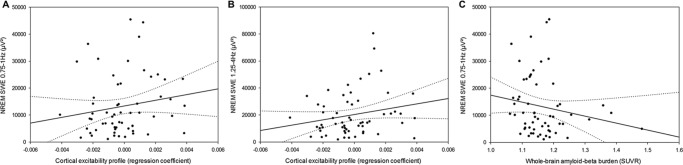
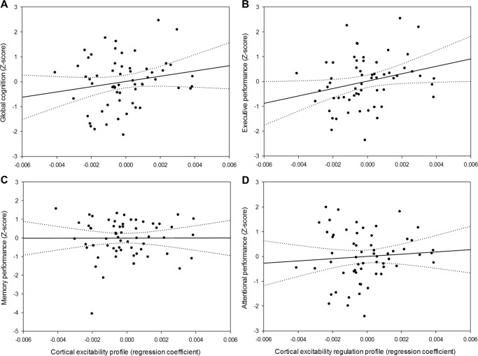
Age-related cognitive decline arises from alterations in brain structure as well as in sleep-wake regulation. Here, we investigated whether preserved wake-dependent regulation of cortical function could represent a positive factor for cognitive fitness in aging. We quantified cortical excitability dynamics during prolonged wakefulness as a sensitive marker of age-related alteration in sleep-wake regulation in 60 healthy older individuals (50-69 y; 42 women). Brain structural integrity was assessed with amyloid-beta- and tau-PET, and with MRI. Participants' cognition was investigated using an extensive neuropsychological task battery. We show that individuals with preserved wake-dependent cortical excitability dynamics exhibit better cognitive performance, particularly in the executive domain which is essential to successful cognitive aging. Critically, this association remained significant after accounting for brain structural integrity measures. Preserved dynamics of basic brain function during wakefulness could therefore be essential to cognitive fitness in aging, independently from age-related brain structural modifications that can ultimately lead to dementia.

Keywords: Cognitive ageing; Wakefulness.


Conflict of interest statement

Competing interestsThe authors declare no competing interests.

Figures

Fig. 1
Fig. 1
Study design and cortical excitability assessment. a Overview of the whole experimental protocol and the timing of the different steps for a representative subject with bedtime at 11:00PM and wake time at 07:00AM. AN: adaptation night with polysomnography to screen for sleep apnea; BN: baseline night under EEG recording. b Cortical excitability over the frontal cortex was assessed using neuronavigation-based TMS coupled to EEG. Left: reconstructed head with electrodes position; Right: representative location of TMS coil and stimulation hotspot with electric field orientation. c Butterfly plot of TMS-evoked EEG response over the 60 electrodes (−100 ms pre-TMS to 300 ms post-TMS; average of ~250 trials). d Representative TMS-evoked EEG potential (0–32 ms post-TMS) in the five TMS-EEG sessions with indicative clock time and circadian phase (15° = 1 h). Cortical excitability was computed as the slope (µV/ms) of the first component of the TMS-evoked EEG response at the electrode closest to the hotspot (dotted line: example for 10:00AM session).
Fig. 2
Fig. 2
Cortical excitability dynamics as a marker of sleep–wake regulation processes. a Average cortical excitability dynamics (mean ± SEM) during 20 h of prolonged wakefulness over the entire sample (n = 60). Gray background represents the average melatonin secretion profile (0° indicating dim-light melatonin onset, i.e. the beginning of the biological night; 15° = 1 h). *padj < 0.01. b Detrended cortical excitability values of all individuals and their respective linear regression lines across the five TMS-EEG measurements.
Fig. 3
Fig. 3
Cortical excitability, slow wave energy, and brain structural integrity. a Positive association between CEP and cumulated frontal NREM SWE in the lower range (0.75–1 Hz) during habitual sleep (n = 60; F1,55 = 5.35, p = 0.02, β* = 0.09). b Positive association between CEP and cumulated frontal NREM SWE in the higher range (1.25–4 Hz) during habitual sleep (n = 60; F1,55 = 5.47, p = 0.02, β* = 0.09). c Negative association between NREM SWE (0.75–1 Hz range) and whole-brain amyloid-beta burden (n = 60; F1,53 = 5.15, p = 0.03, β* = 0.09). Simple regressions were used only for a visual display and do not substitute the GLMM outputs. Dotted lines represent 95% confidence interval of these simple regressions.
Fig. 4
Fig. 4
Relationships between CEP and cognition. a Positive association between CEP and global cognition (n = 60; F1,55 = 6.76, p = 0.01, β* = 0.11). b Domain-specific positive association between CEP and performance to tasks probing executive functions (n = 60; F1,55 = 8.47, p = 0.005, β* = 0.13). c No significant association between CEP and memory performance (n = 60; F1,55 = 0.39, p = 0.54). d No significant association between CEP and attentional performance (n = 60; F1,55 = 2.44, p = 0.12). Simple regressions were used only for a visual display and do not substitute the GLMM outputs. Dotted lines represent 95% confidence interval of these simple regressions.

References

    1. Giorgio A, et al. Age-related changes in grey and white matter structure throughout adulthood. Neuroimage. 2010;51:943–951. doi: 10.1016/j.neuroimage.2010.03.004. - DOI - PMC - PubMed
    1. Jack CR, et al. Tracking pathophysiological processes in Alzheimer’s disease: an updated hypothetical model of dynamic biomarkers. Lancet Neurol. 2013;12:207–216. doi: 10.1016/S1474-4422(12)70291-0. - DOI - PMC - PubMed
    1. Reuter-Lorenz PA, Park DC. How does it STAC up? revisiting the scaffolding theory of aging and cognition. Neuropsychol. Rev. 2014;24:355–370. doi: 10.1007/s11065-014-9270-9. - DOI - PMC - PubMed
    1. Jack CR, et al. NIA-AA research framework: toward a biological definition of Alzheimer’s disease. Alzheimer’s Dement. 2018;14:535–562. doi: 10.1016/j.jalz.2018.02.018. - DOI - PMC - PubMed
    1. Braak H, Thal DR, Ghebremedhin E, Tredici KDel. Stages of the pathologic process in Alzheimer disease: age categories from 1 to 100 years. J. Neuropathol. Exp. Neurol. 2011;70:960–969. doi: 10.1097/NEN.0b013e318232a379. - DOI - PubMed

Publication types