

Wilms Tumor of the Ovary: Review of the Literature and Report of 2 Cases
- PMID: 31815892
- PMCID: PMC6546561
- DOI: 10.1097/PGP.0000000000000565
Wilms Tumor of the Ovary: Review of the Literature and Report of 2 Cases
Abstract
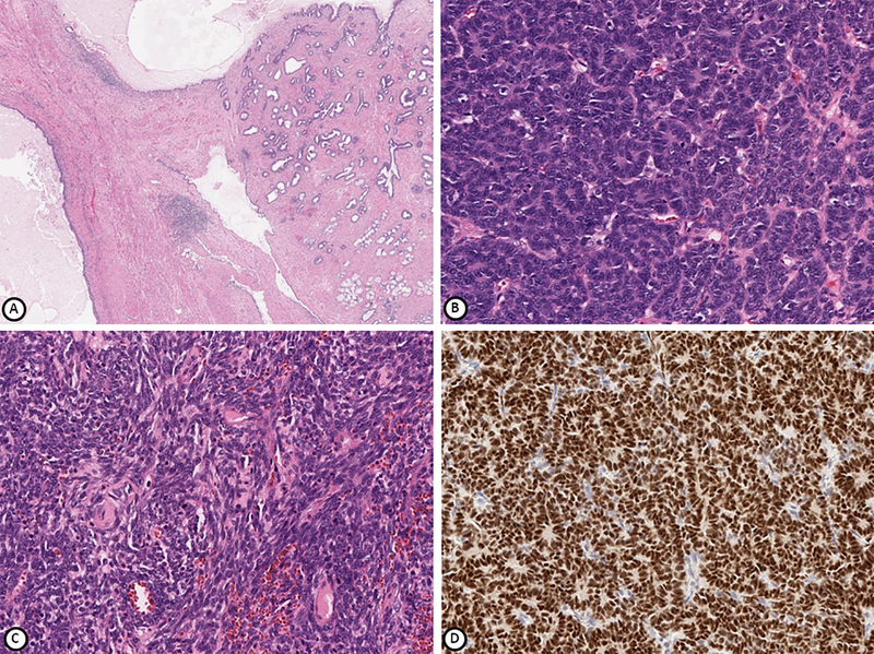
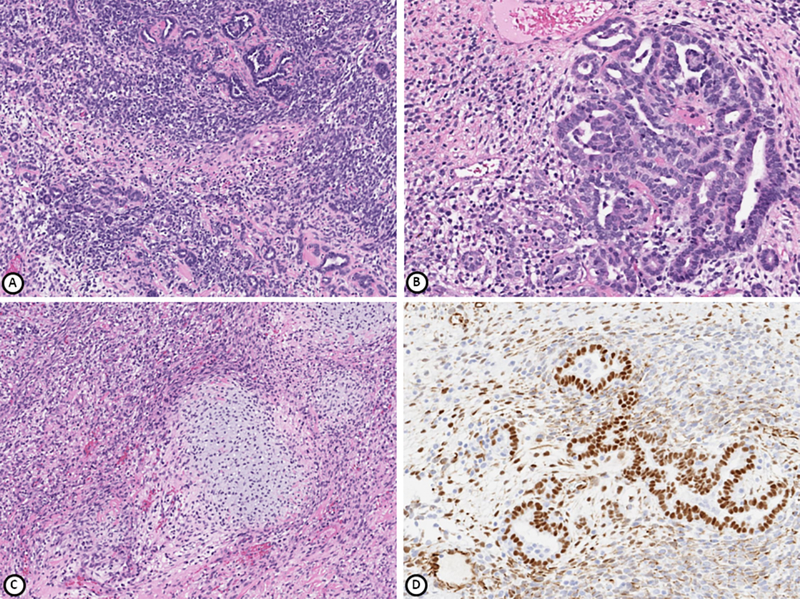
Primary extrarenal Wilms tumor of the gynecologic tract is extremely rare with scattered case reports occurring in the ovary, uterine corpus and cervix. Only 9 cases of primary ovarian Wilms tumor have been reported to date. Here, we provide an extensive literature review and describe 2 patients with ovarian Wilms tumor: a 36-yr-old female (patient 1) and a 16-yr-old female (patient 2), both presenting with abdominal pain and suspected ovarian torsion. They were each found to have unilateral ovarian masses measuring >15 cm in size which were removed by unilateral salpingo-oophorectomy. Microscopically, the tumors exhibited the typical triphasic histology of Wilms tumor. In addition, the tumor from patient 1 contained elements of mature cystic teratoma, while an extensive rhabdomyosarcomatous component was identified in patient 2. Both tumors were diffusely and strongly positive for WT1 with variable staining for other biomarkers. The cases were diagnostically challenging and referred to our center for an expert opinion. Teratoid Wilms tumor in patient 1 is the second reported case of ovarian Wilms tumor arising in association with teratoma. Recognition of primary ovarian Wilms tumor requires a high index of suspicion and exclusion of other entities based on tumor morphology and immunohistochemical studies.
Conflict of interest statement
CONFLICT OF INTEREST
The authors have no conflicts of interest to declare.
Figures
References
-
- Popov SD, Sebire NJ, Vujanic GM. Wilms’ Tumour - Histology and Differential Diagnosis. In: van den Heuvel-Eibrink MM, editor. Wilms Tumor. Brisbane (AU)2016. - PubMed
-
- Faria P, Beckwith JB, Mishra K, et al. Focal versus diffuse anaplasia in Wilms tumor--new definitions with prognostic significance: a report from the National Wilms Tumor Study Group. Am J Surg Pathol. 1996;20(8):909–20. Epub 1996/08/01. - PubMed
-
- Andrews PE, Kelalis PP, Haase GM. Extrarenal Wilms’ tumor: results of the National Wilms’ Tumor Study. J Pediatr Surg. 1992;27(9):1181–4. Epub 1992/09/01. - PubMed
Publication types
MeSH terms
Grants and funding
LinkOut - more resources
Full Text Sources
Medical
Molecular Biology Databases
