Review
. 2019 Dec 3;9(12):1725.
doi: 10.3390/nano9121725.

Graphene Oxide-Based Biosensors for Liquid Biopsies in Cancer Diagnosis

Affiliations
Review

Graphene Oxide-Based Biosensors for Liquid Biopsies in Cancer Diagnosis

Shiue-Luen Chen et al. Nanomaterials (Basel). .

Abstract

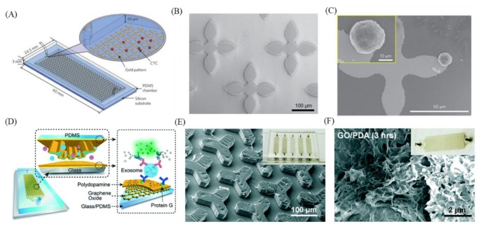
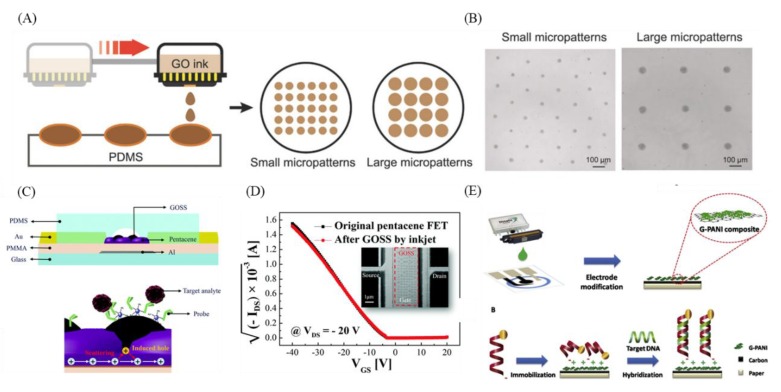
Liquid biopsies use blood or urine as test samples, which are able to be continuously collected in a non-invasive manner. The analysis of cancer-related biomarkers such as circulating tumor cells (CTCs), circulating tumor DNA (ctDNA), microRNA, and exosomes provides important information in early cancer diagnosis, tumor metastasis detection, and postoperative recurrence monitoring assist with clinical diagnosis. However, low concentrations of some tumor markers, such as CTCs, ctDNA, and microRNA, in the blood limit its applications in clinical detection and analysis. Nanomaterials based on graphene oxide have good physicochemical properties and are now widely used in biomedical detection technologies. These materials have properties including good hydrophilicity, mechanical flexibility, electrical conductivity, biocompatibility, and optical performance. Moreover, utilizing graphene oxide as a biosensor interface has effectively improved the sensitivity and specificity of biosensors for cancer detection. In this review, we discuss various cancer detection technologies regarding graphene oxide and discuss the prospects and challenges of this technology.

Keywords: circulating tumor DNA; circulating tumor cells; exosome; graphene oxide; liquid biopsy.


Conflict of interest statement

The authors declare no conflicts of interest.

Figures

Figure 1
Figure 1
Example of antibody-modified graphene oxide for capturing CTCs. (A) A reduce graphene oxide film efficiently captures circulating tumor cells (CTCs) from clinical blood samples. (B) Environmental Scanning Electron Microscope (ESEM) image of the reduced graphene oxide (rGO) layer-by-layer structure, and (C) an anti- Epithelial cell adhesion molecule (EpCAM) -rGO film after capture CTCs. (D) The modification steps of anti-EpCAM-rGO film. (E) Schematic of CTC capture system using functionalized graphene oxide (GO) nanosheets on a patterned gold surface. (F) Schematic of a polymer–GO microfluidic device. Figures (AD) reproduced with permission of [77], Wiley©, 2015; (E) [76], Springer Nature©, 2013; (F) [81] Wiley©, 2016.
Figure 2
Figure 2
GO-based DNA-based optical sensors. (A) Schematic of fluorescent sensors using DNA-functionalized graphene oxide. (B) Molecular dynamics simulation of FAM-tagged single—stranded DNA (ssDNA) absorbed on the surface of GO (left) and double—stranded DNA (dsDNA) detached from the surface of GO (right). (C) Photographs showing GO and rGO had strong fluorescence quenching ability. (D) Schematic of using a DNA-functionalized graphene oxide sensor for deletion mutation in the EFGR gene in lung cancer. (E) Fluorescence spectra for fDNA after the detection of various concentrations of cDNA. Figures (A,B) reproduced with permission of [84], Wiley©, 2010; (C) [100], ACS©, 2010; (D,E) [91], Elsevier©, 2016.
Figure 3
Figure 3
GO-based DNA-based electrochemical sensors. (A) Schematic of MoS2/graphene nanosheets electrode for ctDNA detection. (B) Scanning Electron Microscope (SEM) image of MoS2/graphene composites. (C) The Differential Pulse Voltammetry (DPV) plots change after hybridization of various concentrations of ctDNA. (D) Schematic of sensing steps of graphene-DNA electrochemical sensor with AuNPs functionalized report DNA. (E) SEM image of sensor without adding DNA-r AuNPs (left) and adding DNA-r AnNPs (right). Figures (AC) reproduced with permission of [93], RSC©, 2016; (D,E) [95], Elsevier©, 2014.
Figure 4
Figure 4
Application of GO-based biosensors for exosome detection. (A) Schematic of antibody immobilization on rGO surface. (B) Atomic Force Microscope (AFM) image (5 × 5 µm2) of antibody-immobilized surface. (Scale bar is 1 µm). (C) Efficiency of antibody immobilization on different rGO surfaces. (D) Resistance change (Rab-R)/R before and after immobilization and counting the number of immobilized antibodies with a particular size on the AFM image (7–9 nm). (E) New nanoplasmonic sensor (NPS) platform and heatmap analysis. Figures (AD) reproduced with permission of [102], Elsevier©, 2017; (E), [104], Elsevier©, 2018.
Figure 5
Figure 5
Examples of microfluidic devices with microposts for capturing tumor cells. (A) Schematic of circulating tumor cell capturing system with microposts. (B) SEM image of flower shaped microposts. (C) SEM image of flower shaped micropost with captured tumor cell. (D) Schematic of nano-interfaced microfluidic exosome platform and Graphene oxide/polydopamine (GO/PDA) coated interface. (E) SEM image of Y shaped microposts with GO/PDA coating. (F) SEM image of GO/PDA-coated channel. Figures (AC) reproduced with permission of [76], Springer Nature©, 2013; and (DF) [101], RSC©, 2016. This article is licensed under a Creative Commons Attribution-NonCommercial 3.0 Unported Licence.
Figure 6
Figure 6
Examples of inkjet printing utilized in tumor-related molecule sensing. (A) Schematic of inkjet printing graphene oxide. (B) Uniformly inkjet-printed different sizes of graphene oxide micropattern. (C) Schematic of graphene oxide support system (GOSS) and its detection mechanism. (D) Electrical performance of original pentacene field-effect transistor (FET) and inkjet-printed pentacene FET. (E) Schematic of paper-based electrochemical biosensor and its sensing mechanism. Figures (A,B) reproduced with permission of [127], Wiley©, 2018; (C,D) [120], RSC©, 2017; and (E) [122], Elsevier©, 2017.

References

    1. Bouck N. Tumor angiogenesis: The role of oncogenes and tumor suppressor genes. Cancer Cells (Cold Spring Harbor) 1990;2:179–185. - PubMed
    1. Feitelson M.A., Arzumanyan A., Kulathinal R.J., Blain S.W., Holcombe R.F., Mahajna J., Marino M., Martinez-Chantar M.L., Nawroth R., Sanchez-Garcia I., et al. Sustained proliferation in cancer: Mechanisms and novel therapeutic targets. Semin. Cancer Biol. 2015;35:S25–S54. doi: 10.1016/j.semcancer.2015.02.006. - DOI - PMC - PubMed
    1. Inagaki J., Rodriguez V., Bodey G.P. Causes of death in cancer patients. Cancer. 1974;33:568–573. doi: 10.1002/1097-0142(197402)33:2<568::AID-CNCR2820330236>3.0.CO;2-2. - DOI - PubMed
    1. Edge S.B., Compton C.C. The American Joint Committee on Cancer: The 7th edition of the AJCC cancer staging manual and the future of TNM. Ann. Surg. Oncol. 2010;17:1471–1474. doi: 10.1245/s10434-010-0985-4. - DOI - PubMed
    1. Gerlinger M., Rowan A.J., Horswell S., Larkin J., Endesfelder D., Gronroos E., Martinez P., Matthews N., Stewart A., Tarpey P. Intratumor heterogeneity and branched evolution revealed by multiregion sequencing. N. Engl. J. Med. 2012;366:883–892. doi: 10.1056/NEJMoa1113205. - DOI - PMC - PubMed

LinkOut - more resources