. 2019 Dec 3;8(12):2134.
doi: 10.3390/jcm8122134.

TMPRSS4: A Novel Tumor Prognostic Indicator for the Stratification of Stage IA Tumors and a Liquid Biopsy Biomarker for NSCLC Patients

Affiliations

TMPRSS4: A Novel Tumor Prognostic Indicator for the Stratification of Stage IA Tumors and a Liquid Biopsy Biomarker for NSCLC Patients

Maria Villalba et al. J Clin Med. .

Abstract

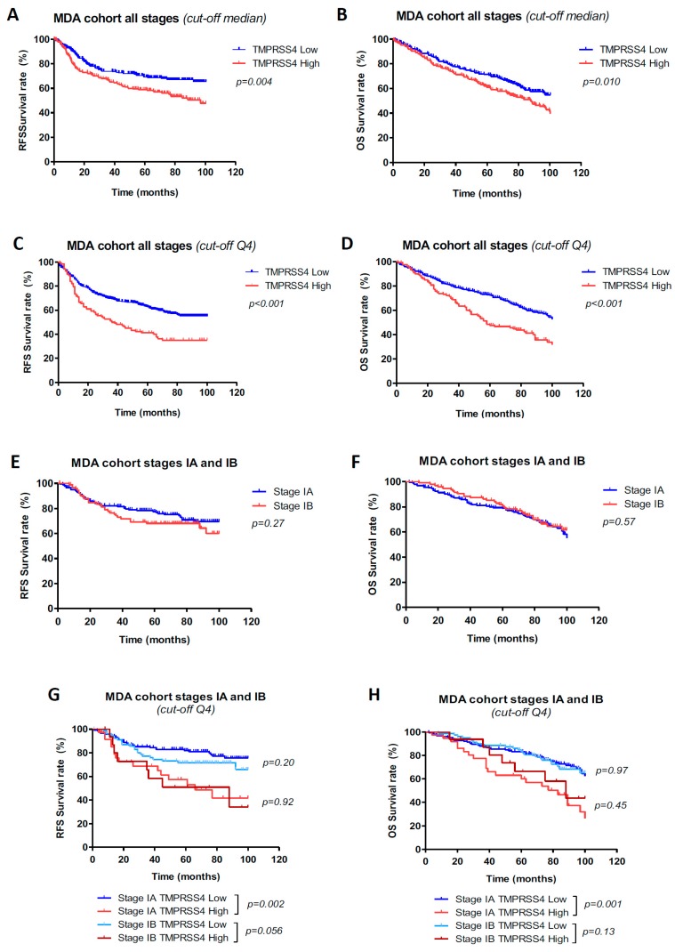
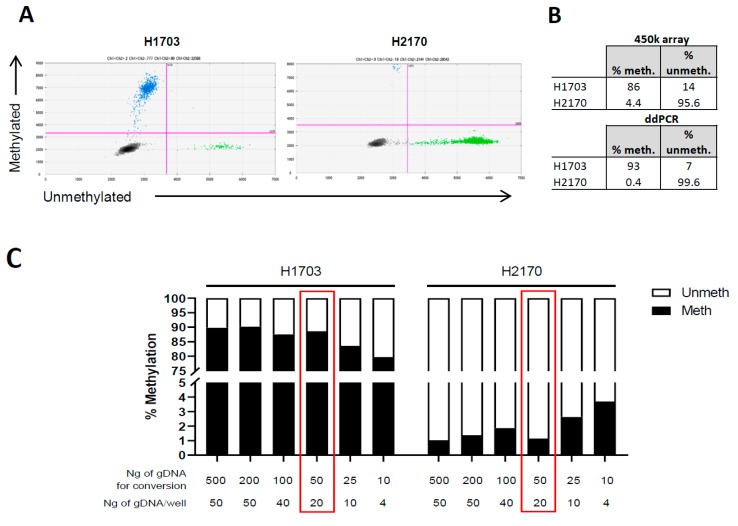
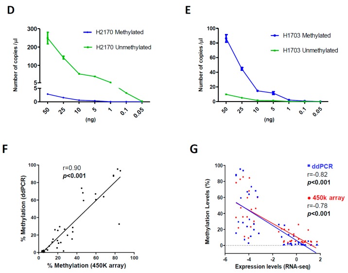
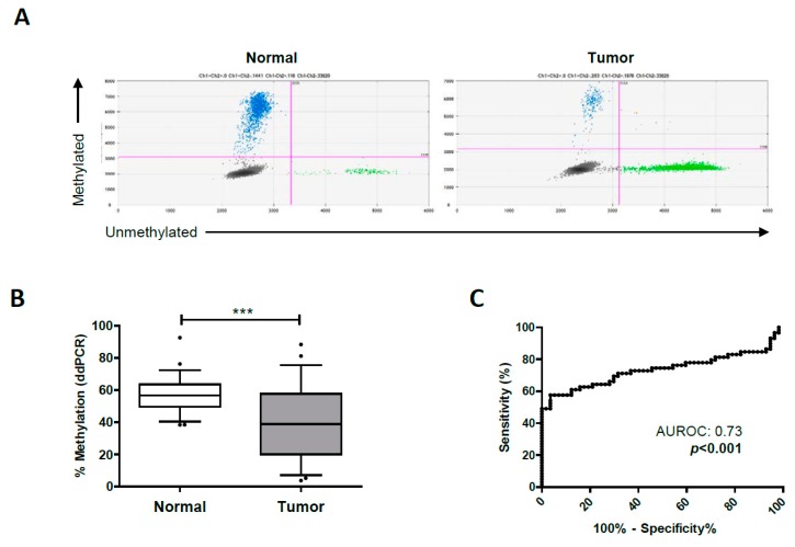
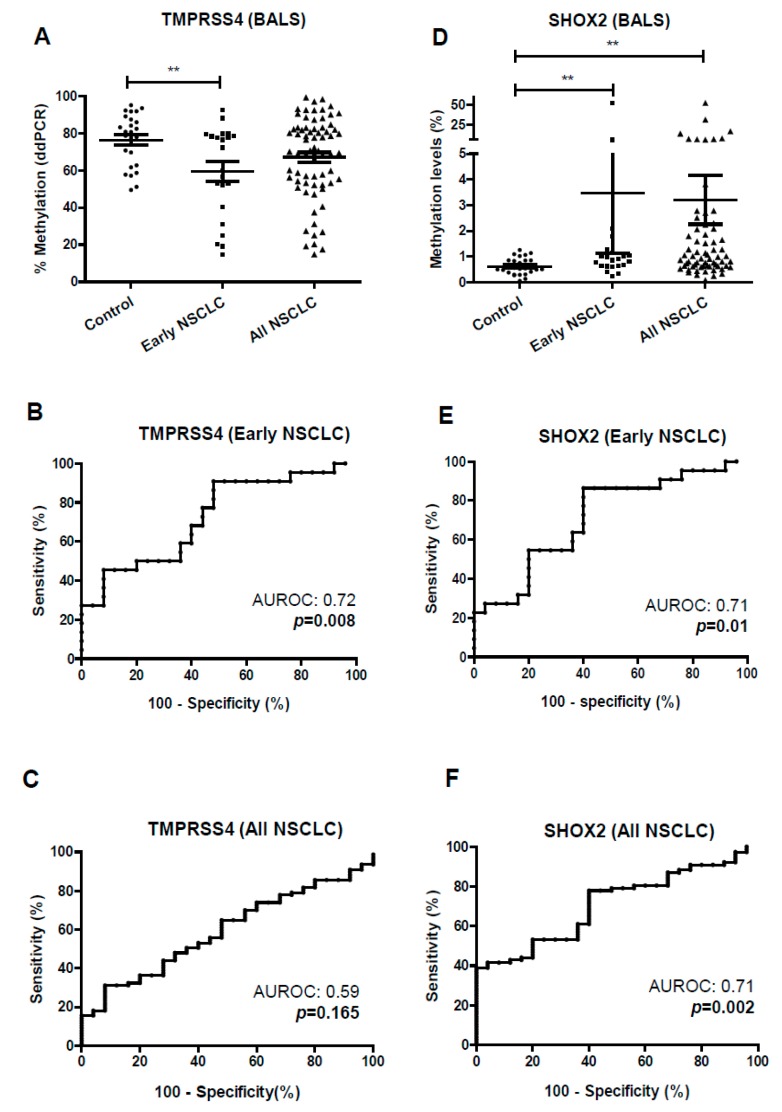
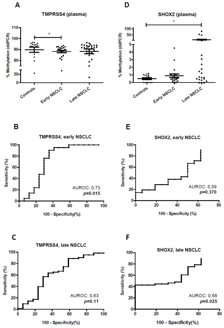
Relapse rates in surgically resected non-small-cell lung cancer (NSCLC) patients are between 30% and 45% within five years of diagnosis, which shows the clinical need to identify those patients at high risk of recurrence. The eighth TNM staging system recently refined the classification of NSCLC patients and their associated prognosis, but molecular biomarkers could improve the heterogeneous outcomes found within each stage. Here, using two independent cohorts (MDA and CIMA-CUN) and the eighth TNM classification, we show that TMPRSS4 protein expression is an independent prognostic factor in NSCLC, particularly for patients at stage I: relapse-free survival (RFS) HR, 2.42 (95% CI, 1.47-3.99), p < 0.001; overall survival (OS) HR, 1.99 (95% CI, 1.25-3.16), p = 0.004). In stage IA, high levels of this protein remained associated with worse prognosis (p = 0.002 for RFS and p = 0.001 for OS). As TMPRSS4 expression is epigenetically regulated, methylation status could be used in circulating tumor DNA from liquid biopsies to monitor patients. We developed a digital droplet PCR (ddPCR) method to quantify absolute copy numbers of methylated and unmethylated CpGs within the TMPRSS4 and SHOX2 (as control) promoters in plasma and bronchoalveolar lavage (BAL) samples. In case-control studies, we demonstrated that TMPRSS4 hypomethylation can be used as a diagnostic tool in early stages, with an AUROC of 0.72 (p = 0.008; 91% specificity and 52% sensitivity) for BAL and 0.73 (p = 0.015; 65% specificity and 90% sensitivity) for plasma, in early stages. In conclusion, TMPRSS4 protein expression can be used to stratify patients at high risk of relapse/death in very early stages NSCLC patients. Moreover, analysis of TMPRSS4 methylation status by ddPCR in blood and BAL is feasible and could serve as a non-invasive biomarker to monitor surgically resected patients.

Keywords: DNA methylation; NSCLC; TMPRSS4; liquid biopsy; prognosis.


Conflict of interest statement

The authors declare no conflict of interest.

Figures

Figure 1
Figure 1
Prognostic value of the Transmembrane protease serine 4 (TMPRSS4) including patients from all stages (I–IV) in the MDA cohort. Relapse-free survival (RFS) (A) and overall survival (OS) (B) after stratifying the patients based on median protein expression of TMPRSS4. Patients with levels above the median showed worse prognosis. When patients were stratified by quartiles, prognostic differences between both groups were larger: patients with levels above the top 25% (Q4) were associated with lower RFS (p < 0.001) (C) and OS (p < 0.001) (D). RFS (E) and OS (F) comparing stage IA and IB patients. (G) RFS analysis in stage IA and IB patients upon stratification by TMPRSS4 protein expression. The top 25% protein expression (Q4) was considered as high level. In the case of stage IA, TMPRSS4 levels in Q4 were significantly associated with lower RFS (p = 0.002) as compared to patients within the same stage where TMPRSS4 levels were low. In the case of stage IB, the same tendency was observed for RFS, but results were not statistically different. (H) Evaluation of OS rendered similar results to those found for RFS; n = 187 stage IA, n = 95 stage IB.
Figure 2
Figure 2
Optimization of conditions for TMPRSS4 methylation analysis by ddPCR and evaluation of methylation status in lung cancer cell lines. (A) Representative two-dimensional (2D) ddPCR plots of number of methylated (blue) and unmethylated (green) events in the cell lines H1703 and H2170. (B) Percentages of methylated/unmethylated copies in the cell lines selected as controls (H1703 and H2170). (C) Assessment of the amount of DNA for bisulfite conversion and for loading that is necessary for an accurate quantification. The expected percentage of methylated and unmethylated DNA was maintained when at least 50 ng of DNA was converted and 20 ng was loaded. (D,E) Dilution assays in H2170 and H1703. Upon bisulfite conversion of 50 ng of DNA, serially diluted DNA (indicated in the X-axis) was loaded. (F) Methylation status of the TMPRSS4 promoter in a panel of 46 lung cancer cell lines, using 50 ng of DNA as the starting amount and 20 ng for loading. A very significant correlation between ddPCRs and data from the 450k methylation array was found. (G) Inverse correlation between methylation status (450k in red and ddPCR in blue) and expression of TMPRSS4 in the lung cancer cell panel.
Figure 2
Figure 2
Optimization of conditions for TMPRSS4 methylation analysis by ddPCR and evaluation of methylation status in lung cancer cell lines. (A) Representative two-dimensional (2D) ddPCR plots of number of methylated (blue) and unmethylated (green) events in the cell lines H1703 and H2170. (B) Percentages of methylated/unmethylated copies in the cell lines selected as controls (H1703 and H2170). (C) Assessment of the amount of DNA for bisulfite conversion and for loading that is necessary for an accurate quantification. The expected percentage of methylated and unmethylated DNA was maintained when at least 50 ng of DNA was converted and 20 ng was loaded. (D,E) Dilution assays in H2170 and H1703. Upon bisulfite conversion of 50 ng of DNA, serially diluted DNA (indicated in the X-axis) was loaded. (F) Methylation status of the TMPRSS4 promoter in a panel of 46 lung cancer cell lines, using 50 ng of DNA as the starting amount and 20 ng for loading. A very significant correlation between ddPCRs and data from the 450k methylation array was found. (G) Inverse correlation between methylation status (450k in red and ddPCR in blue) and expression of TMPRSS4 in the lung cancer cell panel.
Figure 3
Figure 3
Quantification of the TMPRSS4 promoter methylation status in normal and tumor samples by ddPCR. (A) Representative 2D ddPCR plots of the number of methylated (blue) and unmethylated (green) events in a normal and a malignant sample. (B) Tumors from NSCLC patients show a significant hypomethylation in the TMPRSS4 promoter. (C) ROC curve and area under the ROC curve (AUROC) analyzing methylation status of the TMPRSS4 promoter in normal vs. tumors tissue samples. *** p < 0.001.
Figure 4
Figure 4
Methylation status of the TMPRSS4 and SHOX2 promoters in BAL samples from control individuals and NSCLC patients. Early stages: I–II; all stages: I–IV. (A) Significant hypomethylation of TMPRSS4 (p < 0.01) was found in BAL from early-stage NSCLC patients in comparison with controls. (B) ROC curve and area under the ROC (AUROC) resulting from the analysis of TMPRSS4 methylation status in early stages. (C) ROC curve and AUROC for TMPRSS4 methylation status in all stages. (D) Significant hypermethylation of SHOX2 (p < 0.01) was observed in BAL for early and late NSCLC stages. (E) ROC curve and AUROC for the methylation status of SHOX2 in early stages. (F) The same analysis but considering all stages. ** p < 0.01.
Figure 5
Figure 5
Methylation status of the TMPRSS4 and SHOX2 promoter in plasma samples from control individuals and NSCLC patients. Early stages: I–II; late stages: III–IV. (A) Significant hypomethylation of the TMPRSS4 promoter was found in plasma from early stages in comparison with control individuals. (B) ROC curve and area under the ROC (AUROC) for TMPRSS4 methylation status in early stages. (C) The same analysis but for late stages. (D). Significant hypermethylation of the SHOX2 promoter was found in plasma only in the case of late stages. (E,F) ROC curves and AUROC for the SHOX2 methylation status in the case of early (E) or late (F) stages. * p < 0.05.

References

    1. Siegel R.L., Miller K.D., Jemal A. Cancer statistics, 2019. CA Cancer J. Clin. 2019;69:7–34. doi: 10.3322/caac.21551. - DOI - PubMed
    1. Uramoto H., Tanaka F. Recurrence after surgery in patients with NSCLC. Transl. Lung Cancer Res. 2014;3:242–249. - PMC - PubMed
    1. Goldstraw P., Chansky K., Crowley J., Rami-Porta R., Asamura H., Eberhardt W.E.E., Nicholson A.G., Groome P., Mitchell A., Bolejack V., et al. The IASLC Lung Cancer Staging Project: Proposals for Revision of the TNM Stage Groupings in the Forthcoming (Eighth) Edition of the TNM Classification for Lung Cancer. J. Thorac. Oncol. 2016;11:39–51. doi: 10.1016/j.jtho.2015.09.009. - DOI - PubMed
    1. Burotto M., Thomas A., Subramaniam D., Giaccone G., Rajan A. Biomarkers in Early-Stage Non–Small-Cell Lung Cancer: Current Concepts and Future Directions. J. Thorac. Oncol. 2014;9:1609–1617. doi: 10.1097/JTO.0000000000000302. - DOI - PMC - PubMed
    1. Exposito F., Villalba M., Redrado M., de Aberasturi A.L., Cirauqui C., Redin E., Guruceaga E., de Andrea C., Vicent S., Ajona D., et al. Targeting of TMPRSS4 sensitizes lung cancer cells to chemotherapy by impairing the proliferation machinery. Cancer Lett. 2019;453:21–33. doi: 10.1016/j.canlet.2019.03.013. - DOI - PubMed