Meta-Analysis
. 2019 Dec 4;10(12):1005.
doi: 10.3390/genes10121005.

Transcriptome Meta-Analysis Deciphers a Dysregulation in Immune Response-Associated Gene Signatures during Sepsis

Affiliations
Meta-Analysis

Transcriptome Meta-Analysis Deciphers a Dysregulation in Immune Response-Associated Gene Signatures during Sepsis

Shaniya Ahmad et al. Genes (Basel). .

Abstract

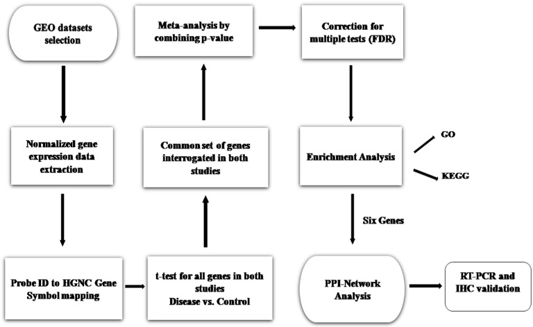
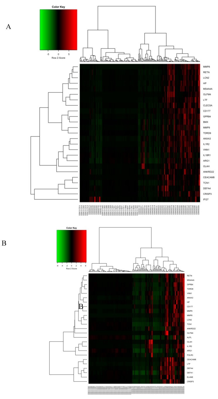
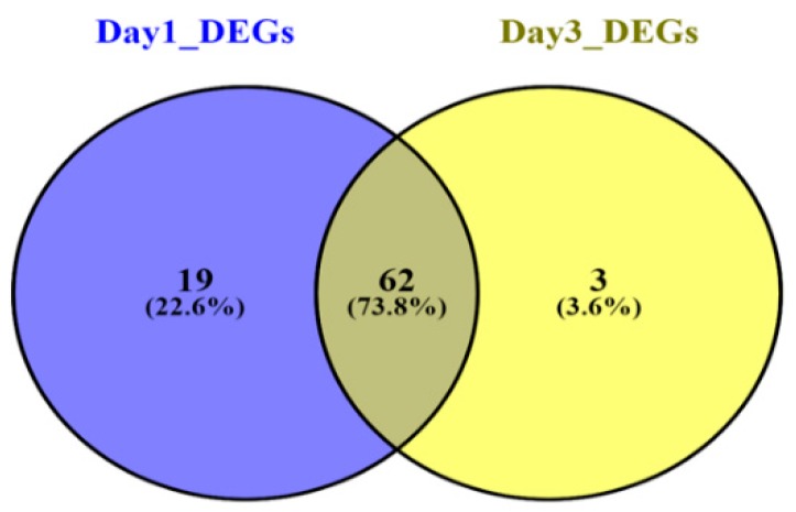
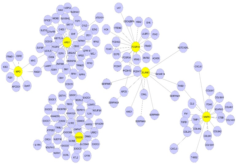
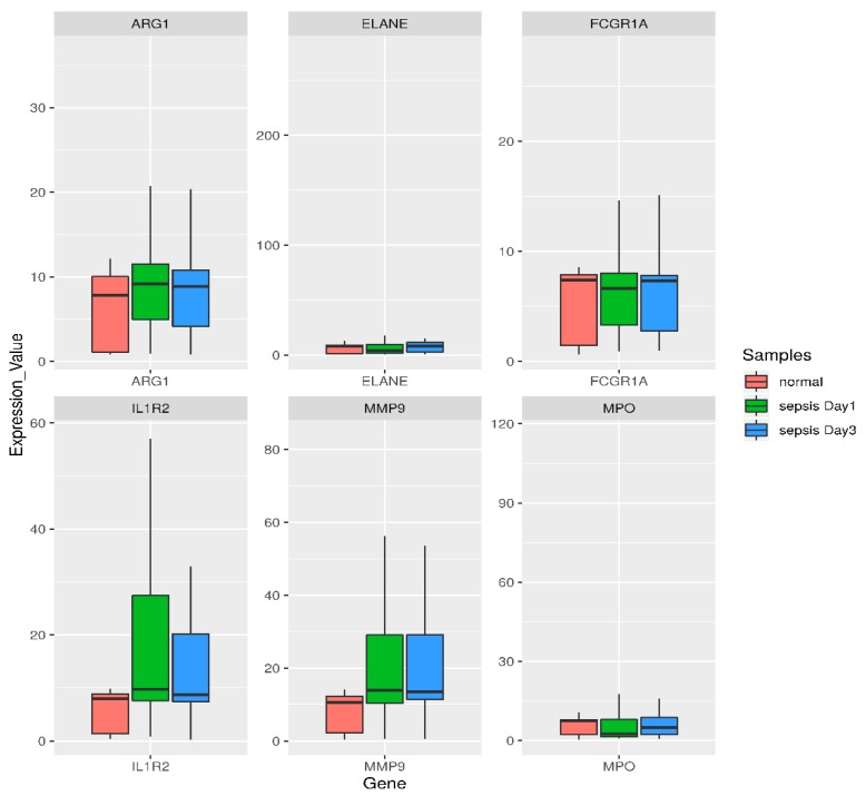
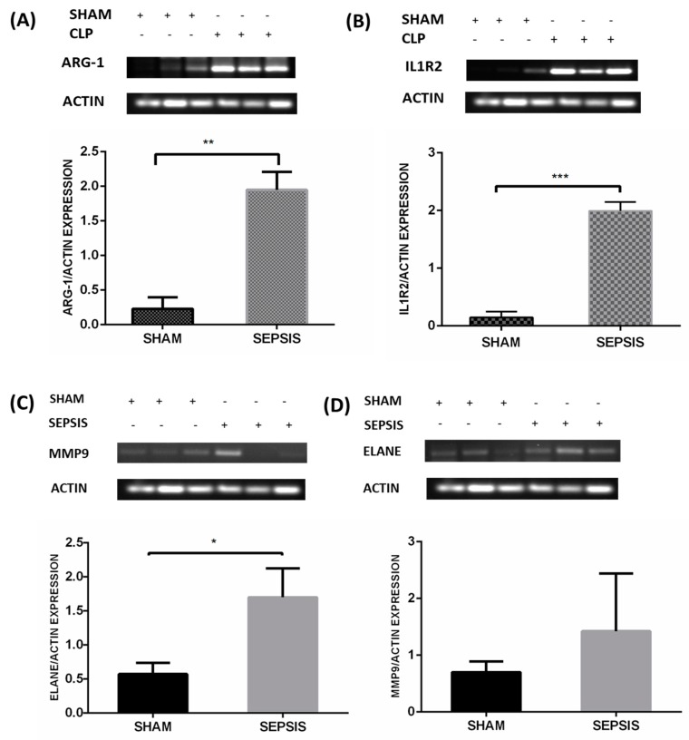
Sepsis is a life-threatening disease induced by a systemic inflammatory response, which leads to organ dysfunction and mortality. In sepsis, the host immune response is depressed and unable to cope with infection; no drug is currently available to treat this. The lungs are frequently the starting point for sepsis. This study aimed to identify potential genes for diagnostics and therapeutic purposes in sepsis by a comprehensive bioinformatics analysis. Our criteria are to unravel sepsis-associated signature genes from gene expression datasets. Differentially expressed genes (DEGs) were identified from samples of sepsis patients using a meta-analysis and then further subjected to functional enrichment and protein‒protein interaction (PPI) network analysis for examining their potential functions. Finally, the expression of the topmost upregulated genes (ARG1, IL1R2, ELANE, MMP9) was quantified by reverse transcriptase-PCR (RT-PCR), and myeloperoxidase (MPO) expression was confirmed by immunohistochemistry (IHC) staining in the lungs of a well-established sepsis mouse model. We found that all the four genes were upregulated in semiquantitative RT-PCR studies; however, MMP9 showed a nonsignificant increase in expression. MPO staining showed strong immunoreactivity in sepsis as compared to the control. This study demonstrates the role of significant and widespread immune activation (IL1R2, MMP9), along with oxidative stress (ARG1) and the recruitment of neutrophils, in sepsis (ELANE, MPO).

Keywords: DEG; PPI; meta-analysis; sepsis.


Conflict of interest statement

The authors declare no conflict of interest.

Figures

Figure 1
Figure 1
Proposed methodology workflow. Analysis of genes was performed by comparing sepsis with control. From these two datasets, the p-vaules were calculated and then combined to compute a single p-vaule per gene, adjusted for multiple testing (FDR). One hundred and forty-six genes with significant p-vaule < 0.05 and FC > 2 were considered as differentially expressed (upregulated) in sepsis. The 146 upregulated genes were subjected to enrichment analysis. A protein‒protein interaction (PPI) network of the identified differentially expressed genes (DEGs) based on the pathway and Gene Ontology (GO) term enrichment analysis was constructed. Thereafter, RT-PCR and immunohistochemistry (IHC) validation studies were performed.
Figure 2
Figure 2
Heatmap for the top 25 upregulated DEGs. Clustering of the top 25 significant DEGs was performed and shown as a heatmap plot in (A) the Sepsis day1 group and (B) the Sepsis day3 group. A hierarchical clustering algorithm uses an average linkage method and Pearson’s correlation coefficient. Green and red in the plot represent lower and higher expression vaules, respectively.
Figure 3
Figure 3
Overlapping DEGs in Sepsis day1 group and Sepsis day3 group. The Venn diagram shows the intersections between the Sepsis day1 group (purple circle) and the Sepsis day3 group (yellow circle). Nineteen genes were included exclusively in “Sepsis day1”, three genes were included exclusively in “Sepsis day3”, and 62 genes were in both groups.
Figure 4
Figure 4
Protein-protein interaction (PPI) network analysis of significantly enriched sepsis-associated DEGs. The network of PPI interaction was constructed from the six identified DEGs. In total, 143 proteins were involved in this network. The six DEGs are in yellow and their interacting partners are purple.
Figure 5
Figure 5
Box- and −whisker plot of six highly upregulated DEGs based on the functional enrichment and PPI analysis for comparing their expression levels. Green corresponds to day1 sepsis, blue to day3 sepsis, and red to control subjects. Genes are shown at the bottom. For individual genes, the vaules of gene expression in log intensities have been normalized to the median of the control group expression. A box plot displays the five-number summary of a set of data: the minimum, first quartile, median, third quartile, and maximum. Endpoints of the axis are labeled by the minimum and maximum vaules. The first and third quartile marks one end and the other end of the box, respectively. The median can be between the first and third quartiles.
Figure 6
Figure 6
Validation of mRNA expression of selected genes in the lung tissue of an animal sepsis model. Mice were CLP operated for 24 h and then sacrificed; lung tissues were collected for analysis. The figure shows the semi-quantitative mRNA expression and densitometry of (A) ARG1, (B) IL1R2, (C) ELANE, and (D) MMP9 in the lung tissue of the sham and CLP groups. A minimum of three animals were used for each group of animals. Data are presented as mean±SEM, p < 0.05. Note: + means treated and − means non-treated.
Figure 7
Figure 7
The expression of Myeloperoxidase in neutrophil granulocyte within lung alveoli was assessed by the immunohistochemical technique in the sham and CLP groups. (A)This represents strong immunoreactivity in the well-established CLP sepsis animal model as compared to the sham group; (B) the significant increase in neutrophil numbers in sepsis as compared to the control. Data are represented as mean ± SEM; experiments were performed in triplicate, with statistical significance at p-vaule < 0.0001.

References

    1. Singer M., Deutschman C.S., Seymour C.W., Shankar-Hari M., Annane D., Bauer M., Bellomo R., Bernard G.R., Chiche J.D., Coopersmith C.M., et al. The Third International Consensus Definitions for Sepsis and Septic Shock (Sepsis-3) JAMA. 2016;315:801–810. doi: 10.1001/jama.2016.0287. - DOI - PMC - PubMed
    1. Remick D.G. Pathophysiology of Sepsis. Am. J. Pathol. 2007;170:1435–1444. doi: 10.2353/ajpath.2007.060872. - DOI - PMC - PubMed
    1. Fleischmann C., Scherag A., Adhikari N.K., Hartog C.S., Tsaganos T., Schlattmann P., Angus D.C., Reinhart K. Assessment of Global Incidence and Mortality of Hospital-treated Sepsis. Current Estimates and Limitations. Am. J. Respir. Crit. Care Med. 2016;193:259–272. doi: 10.1164/rccm.201504-0781OC. - DOI - PubMed
    1. Gotts J.E., Matthay M.A. Sepsis: Pathophysiology and clinical management. BMJ. 2016;353:i1585. doi: 10.1136/bmj.i1585. - DOI - PubMed
    1. Sørensen T.I.A., Nielsen G.G., Andersen P.K., Teasdale T.W. Genetic and Environmental Influences on Premature Death in Adult Adoptees. N. Engl. J. Med. 1988;318:727–732. doi: 10.1056/NEJM198803243181202. - DOI - PubMed

Publication types

MeSH terms