. 2019 Dec 6;24(24):4477.
doi: 10.3390/molecules24244477.

PEGylated Purpurin 18 with Improved Solubility: Potent Compounds for Photodynamic Therapy of Cancer

Affiliations

PEGylated Purpurin 18 with Improved Solubility: Potent Compounds for Photodynamic Therapy of Cancer

Vladimíra Pavlíčková et al. Molecules. .

Abstract

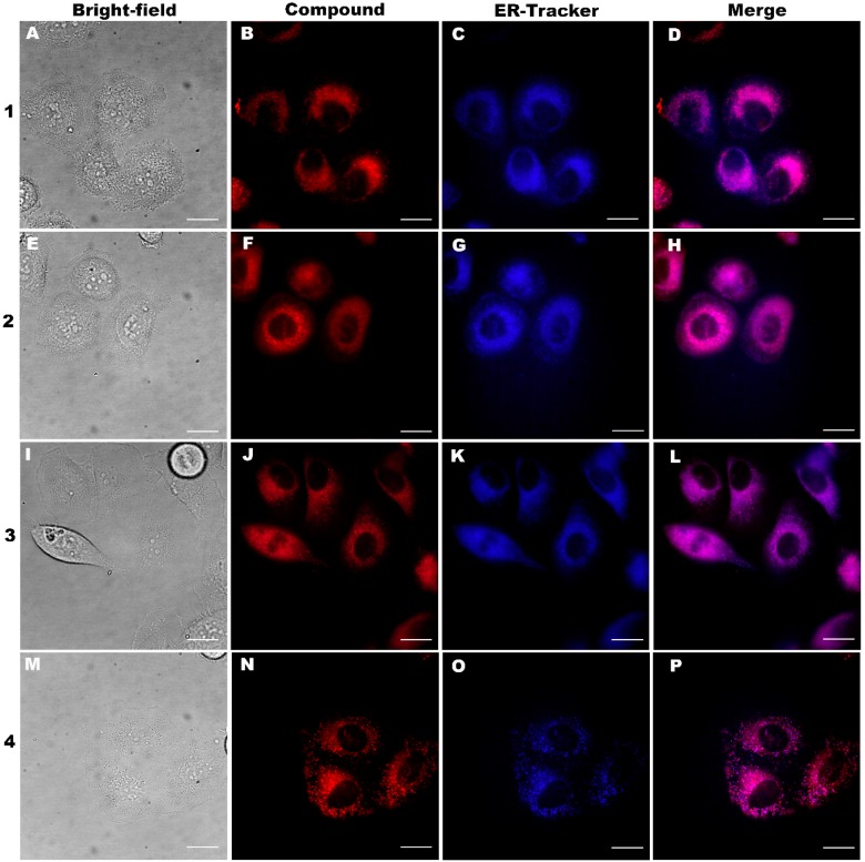
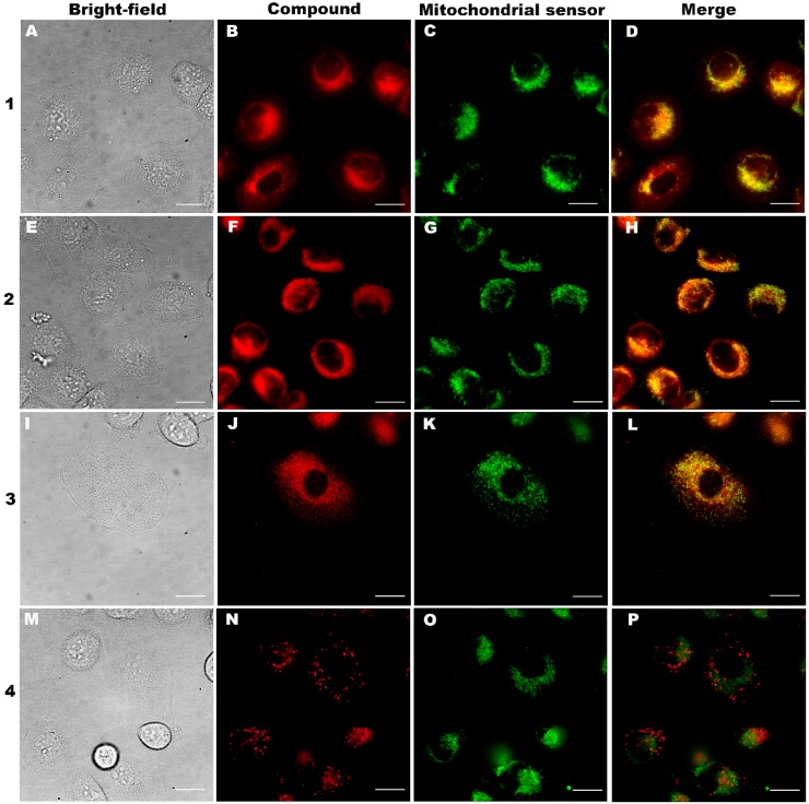
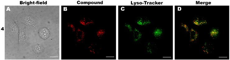
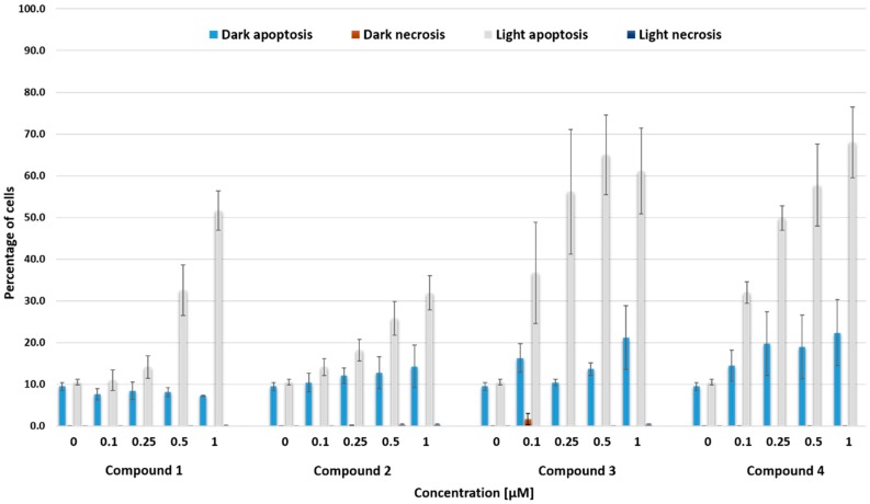
Purpurin 18 derivatives with a polyethylene glycol (PEG) linker were synthesized as novel photosensitizers (PSs) with the goal of using them in photodynamic therapy (PDT) for cancer. These compounds, derived from a second-generation PS, exhibit absorption at long wavelengths; considerable singlet oxygen generation and, in contrast to purpurin 18, have higher hydrophilicity due to decreased logP. Together, these properties make them potentially ideal PSs. To verify this, we screened the developed compounds for cell uptake, intracellular localization, antitumor activity and induced cell death type. All of the tested compounds were taken up into cancer cells of various origin and localized in organelles known to be important PDT targets, specifically, mitochondria and the endoplasmic reticulum. The incorporation of a zinc ion and PEGylation significantly enhanced the photosensitizing efficacy, decreasing IC50 (half maximal inhibitory compound concentration) in HeLa cells by up to 170 times compared with the parental purpurin 18. At effective PDT concentrations, the predominant type of induced cell death was apoptosis. Overall, our results show that the PEGylated derivatives presented have significant potential as novel PSs with substantially augmented phototoxicity for application in the PDT of cervical, prostate, pancreatic and breast cancer.

Keywords: PEGylated purpurin 18; apoptosis; cancer cells; cytotoxicity; flow cytometry; live-cell fluorescence microscopy; photodynamic therapy; photosensitizer; phototoxicity; singlet oxygen.


Conflict of interest statement

The authors declare no conflict of interest. The funders had no role in the design of the study; in the collection, analyses, or interpretation of data; in the writing of the manuscript, or in the decision to publish the results.

Figures

Scheme 1
Scheme 1
Synthesis of derivatives of PEGylated purpurin 18 (compound 1). Reagents and conditions: (a) Zn(OAc)2·2H2O, MeOH, CHCl3, 50 °C, 13 h; yield of compound 2 was 61%; (b) DIC, EDIPA, THF, HOBt, 24 h, RT (22 °C); yield of compound 3 was 41% over two steps; (c) TFA, wet DCM, 1 h, RT (22 °C); yield of compound 4 was 56%.
Figure 1
Figure 1
Depletion of 9,10-anthracenediyl-bis(methylene)dimalonic acid (AB, 7 × 10⁻⁵ M) with photosensitizer-generated singlet oxygen in Dulbecco’s Modified Eagle Medium with 10% fetal bovine serum. Photosensitizers: (A) compound 1 (7.7 × 10⁻⁶ M, 1.5 × 10⁻⁵ M), (B) compound 2 (8.0 × 10⁻⁶ M, 1.6 × 10⁻⁵ M), (C) compound 3 (7.2 × 10⁻⁶ M, 1.4 × 10⁻⁵ M), (D) compound 4 (7.5 × 10⁻⁶ M, 1.5 × 10⁻⁵ M). The experiments were duplicated. formula image formula image—Solution exposed to light, formula image formula image—Solution kept in dark. crel,AB—Relative concentration of AB (actual concentration with respect to concentration at experiment start).
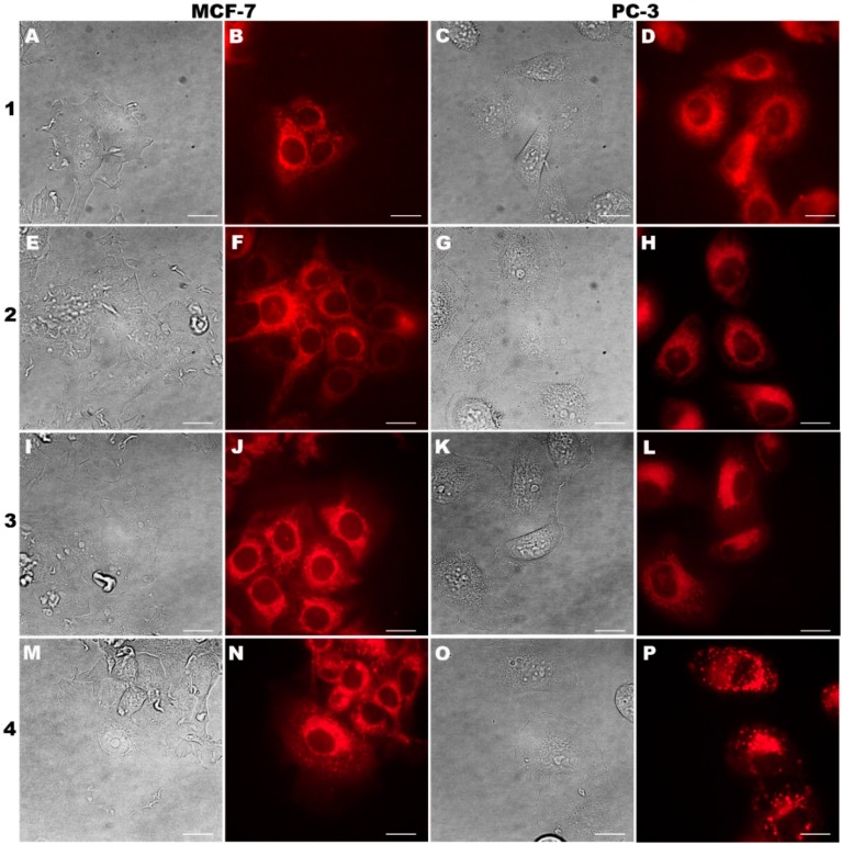
Figure 2
Figure 2
Fluorescence microscopy images of intracellular localization of purpurin 18 (compound 1) and its derivatives (compounds 24) at 0.5 µM concentration in human cancer cell lines of MCF-7 (breast carcinoma) and PC-3 (prostate carcinoma) after 24 h incubation. In the first and third columns, there are bright field images; the second and fourth columns show compound localization. The scale bars represent 20 µm.
Figure 3
Figure 3
Fluorescence microscopy images of localization of purpurin 18 (compound 1) and its derivatives (compounds 24) in the endoplasmic reticulum of human PC-3 cells derived from prostate carcinoma. Colocalization of compounds 12 (0.5 µM, 24 h) or compounds 34 (0.5 µM, 24 h) with ER-Tracker™ Blue-White DPX (70 nM, 30 min). (A,E,I,M) Bright-field images; (B,F,J,N) localization of the tested compounds; (C,G,K,O) ER-Tracker™ Blue-White DPX; (D,H,L,P) merged fluorescent images. The scale bars represent 20 µm.
Figure 4
Figure 4
Fluorescence microscopy images of localization of purpurin 18 (compound 1) and its derivatives (compounds 24) in the mitochondria of human PC-3 cells derived from prostate carcinoma. Colocalization of compounds 12 (0.5 µM, 3 h) or compounds 34 (0.5 µM, 3 h) with a mitosensor (70 nM, 10 min) based on our patented dimethinium salt [47]. (A,E,I,M) Bright-field images; (B,F,J,N) localization of the tested compounds; (C,G,K,O) mitosensor; (D,H,L,P) merge of the fluorescent images. The scale bars represent 20 µm.
Figure 5
Figure 5
Fluorescence microscopy images of compound 4 localization in lysosomes of human PC-3 cells derived from prostate carcinoma. Colocalization of compound 4 (0.5 µM, 24 h) with LysoTracker Green DND-26 (70 nM, 20 min). (A) Bright-field images; (B) localization of compound 4; (C) LysoTracker Green DND-26; (D) merge of the fluorescent images. The scale bars represent 20 µm.
Figure 6
Figure 6
Dose-dependent mechanisms of cell death in MCF-7 cells induced by compounds 14 after 24-h treatment and light induction (Light) measured by flow cytometry. Control represents untreated cells and cells incubated with the same compounds without illumination (Dark). Total light dose was 4 J·cm−2. The data values and the errors are stated in Table S1 in Supplementary information.
Figure 7
Figure 7
Best positions of docked ligands 14 shown in licorice representation. Carbon atoms are shown in light blue, hydrogen atoms in white, nitrogen atoms in dark blue, oxygen atoms in red and zinc atoms in grey. Human serum albumin is depicted as a ribbon. The images were captured by VMD software (Theoretical and Computational Biophysics Group, NIH Center for Macromolecular Modeling and Bioinformatics at the Beckman Institute, University of Illinois at Urbana-Champaign, USA, version 1.9.2).
Figure 8
Figure 8
Structure of compound 2.
Figure 9
Figure 9
Structure of compound 3.
Figure 10
Figure 10
Structure of compound 4.

References

    1. Lim S.H., Yam M.L., Lam M.L., Kamarulzaman F.A., Samat N., Kiew L.V., Chung L.Y., Lee H.B. Photodynamic characterization of amino acid conjugated 15(1)-hydroxypurpurin-7-lactone for cancer treatment. Mol. Pharm. 2014;11:3164–3173. doi: 10.1021/mp500351s. - DOI - PubMed
    1. Bible K.C., Buytendorp M., Zierath P.D., Rinehart K.L. Tunichlorin: A nickel chlorin isolated from the caribbean tunicate Trididemnum solidum. Proc. Natl. Acad. Sci. USA. 1988;85:4582–4586. doi: 10.1073/pnas.85.13.4582. - DOI - PMC - PubMed
    1. Tang P.M., Chan J.Y., Au S.W., Kong S.K., Tsui S.K., Waye M.M., Mak T.C., Fong W.P., Fung K.P. Pheophorbide a, an active compound isolated from Scutellaria barbata, possesses photodynamic activities by inducing apoptosis in human hepatocellular carcinoma. Cancer Biol. Ther. 2006;5:1111–1116. doi: 10.4161/cbt.5.9.2950. - DOI - PubMed
    1. Juzeniene A. Chlorin e6-based photosensitizers for photodynamic therapy and photodiagnosis. Photodiagnosis Photodyn. Ther. 2009;6:94–96. doi: 10.1016/j.pdpdt.2009.06.001. - DOI - PubMed
    1. Pavlíčková V., Jurášek M., Rimpelová S., Záruba K., Sedlák D., Šimková M., Kodr D., Staňková E., Fähnrich J., Rottnerová Z., et al. Oxime-based 19-nortestosterone–pheophorbide a conjugate: Bimodal controlled release concept for PDT. J. Mater. Chem. B. 2019;7:5465–5477. doi: 10.1039/C9TB01301F. - DOI - PubMed

LinkOut - more resources