. 2019 Dec 6;11(12):657.
doi: 10.3390/pharmaceutics11120657.

Trimethyl Chitosan Hydrogel Nanoparticles for Progesterone Delivery in Neurodegenerative Disorders

Affiliations

Trimethyl Chitosan Hydrogel Nanoparticles for Progesterone Delivery in Neurodegenerative Disorders

Maria Cristina Cardia et al. Pharmaceutics. .

Abstract

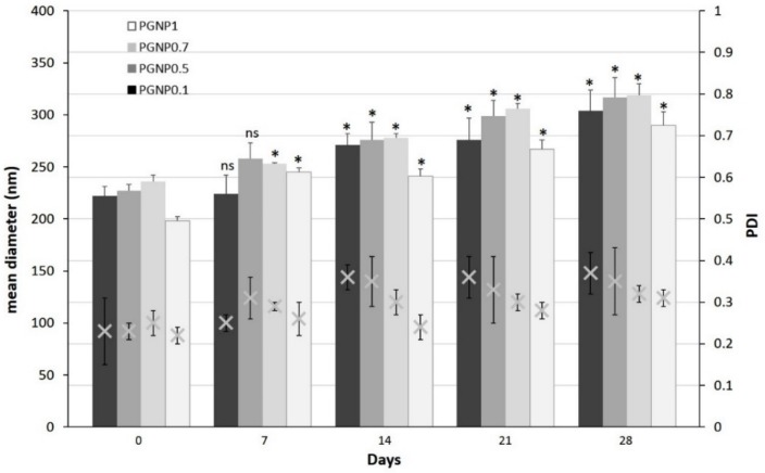
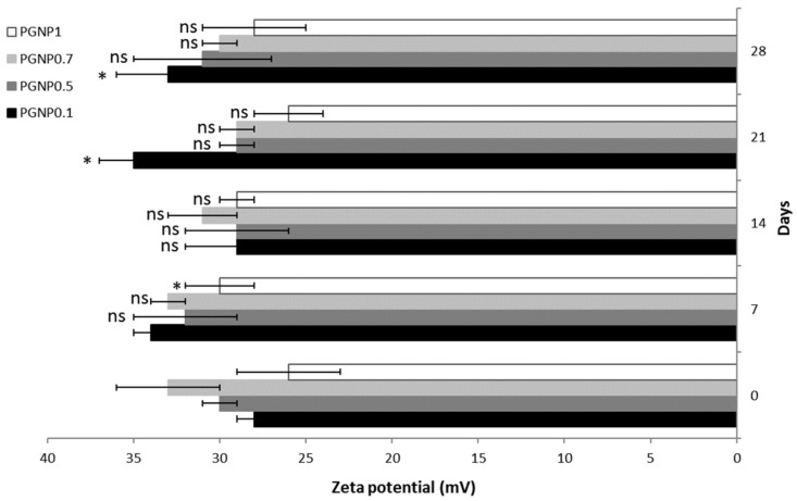
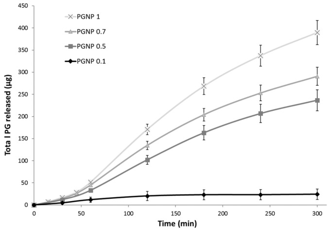
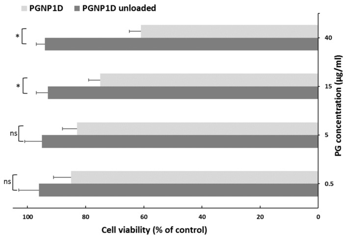
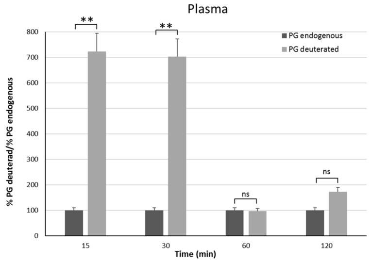
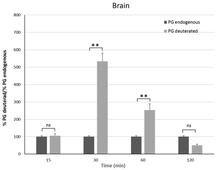
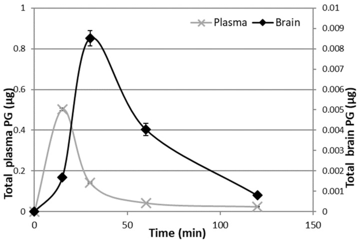
Progesterone is a sex hormone which shows neuroprotective effects in different neurodegenerative disorders, including Parkinson's disease, stroke, and Alzheimer's disease. However, the pharmacokinetic limitations associated with the peripheral administration of this molecule highlight the need for more efficient delivery approaches to increase brain progesterone levels. Since the nose-to-brain administration of mucoadhesive hydrogel nanoparticles is a non-invasive and convenient strategy for the delivery of therapeutics to the central nervous system, in this work, progesterone-loaded hydrogel nanoparticle formulations have been prepared, characterized, and tested in vivo. Nanoparticles, loaded with different progesterone concentrations, have been obtained by polyelectrolyte complex formation between trimethyl chitosan and sodium alginate, followed by ionotropic gelation with sodium tripolyphosphate as a cross-linking agent. All formulations showed a mean diameter ranging from 200 nm to 236 nm, a polydispersity index smaller than 0.23, and a high progesterone encapsulation efficiency (83-95%). The zeta potential values were all positive and greater than 28 mV, thus ensuring nanoparticles stability against aggregation phenomena as well as interaction with negative sialic residues of the nasal mucosa. Finally, in vivo studies on Sprague-Dawley male rats demonstrated a 5-fold increase in brain progesterone concentrations compared to basal progesterone level after 30 min of hydrogel nanoparticle inhalation.

Keywords: brain; hydrogel nanoparticles; progesterone; trimethyl chitosan.


Conflict of interest statement

The authors declare no conflict of interest.

Figures

Figure 1
Figure 1
Scanning electron microscopy image of freeze-dried PGNP1D nanoparticles.
Figure 2
Figure 2
Variation of mean diameter and polydispersity index (PDI) of PG-loaded nanoparticles during 28 days of storage at 4 °C. Results are expressed as means of three independent measurements ± standard deviations. For each formulation, statistical analysis was performed comparing the mean diameter value at each time point with respect to that at time zero. * = different from day 0 (p ≤ 0.05), ns indicates not significant data (p > 0.05).
Figure 3
Figure 3
Zeta potential of PG-loaded nanoparticles during 28 days of storage at 4 °C. Results are expressed as means of three independent measurements ± standard deviations. For each formulation, statistical analysis was performed comparing the zeta potential value at each time point with respect to that at time zero. * = different from day 0 (p ≤ 0.05), ns indicates not significant data (p > 0.05).
Figure 4
Figure 4
In vitro PG release in water/ethanol (40:60 v/v) solution from PG-loaded nanoparticles.
Figure 5
Figure 5
In vitro cytotoxicity studies of PGNP1D and relative unloaded formulations at different dilutions on the human nasal cell line RPMI 2650. The cell viability is recorded as percentage cell viability in comparison with non-treated control cells (100% viability). Results are represented as mean ± SD from three independent experiments. The asterisks indicate statistically significant data (* = p ≤ 0.05), ns indicates not significant data (p > 0.05).
Figure 6
Figure 6
Plasma endogenous and deuterated PG detected on male Sprague–Dawley rats 15 min, 30 min, 1 h, and 2 h after continuous PGNP1D inhalation. The asterisks indicate statistically significant data (** = p ≤ 0.01), ns indicates not significant data (p > 0.05).
Figure 7
Figure 7
Brain endogenous and deuterated PG detected on male Sprague–Dawley mice 15 min, 30 min, 1 h, and 2 h after continuous PGNP1D inhalation. The asterisks indicate statistically significant data (** = p ≤ 0.01), ns indicates not significant data (p > 0.05).
Figure 8
Figure 8
Deuterated PG in plasma and brain 15 min, 30 min, 1 h, and 2 h after continuous PGNP1D inhalation.

References

    1. Bourque M., Morissette M., Di Paolo T. Repurposing sex steroids and related drugs as potential treatment for Parkinson’s disease. Neuropharmacology. 2019;147:37–54. doi: 10.1016/j.neuropharm.2018.04.005. - DOI - PubMed
    1. Vegeto E., Villa A., Della Torre S., Crippa V., Rusmini P., Cristofani R., Galbiati M., Maggi A., Poletti A. The role of sex and sex Hormones in Neurodegenerative Diseases. Endocr. Rev. 2019 doi: 10.1210/endrev/bnz005. in press. - DOI - PMC - PubMed
    1. Hirsch L., Jette N., Frolkis A., Steeves T., Pringsheim T. The Incidence of Parkinson’s Disease: A Systematic Review and Meta-Analysis. Neuroepidemiology. 2016;46:292–300. doi: 10.1159/000445751. - DOI - PubMed
    1. Ascherio A., Chen H., Schwarzschild M.A., Zhang S.M., Colditz G.A., Speizer F.E. Caffeine, postmenopausal estrogen, and risk of Parkinson’s disease. Neurology. 2003;60:790–795. doi: 10.1212/01.WNL.0000046523.05125.87. - DOI - PubMed
    1. Giatti S., Calabrese D., Pesaresi M., Cermenati G., Mitro N., Viviani B., Garcia-Segura L.M., Caruso D. Levels and actions of progesterone and its metabolites in the nervous system during physiological and pathological conditions. Prog. Neurobiol. 2014;113:56–69. - PubMed

LinkOut - more resources