. 2019 Dec 6;11(12):1966.
doi: 10.3390/cancers11121966.

Induction of NK Cell Reactivity against B-Cell Acute Lymphoblastic Leukemia by an Fc-Optimized FLT3 Antibody

Affiliations

Induction of NK Cell Reactivity against B-Cell Acute Lymphoblastic Leukemia by an Fc-Optimized FLT3 Antibody

Bastian J Schmied et al. Cancers (Basel). .

Abstract

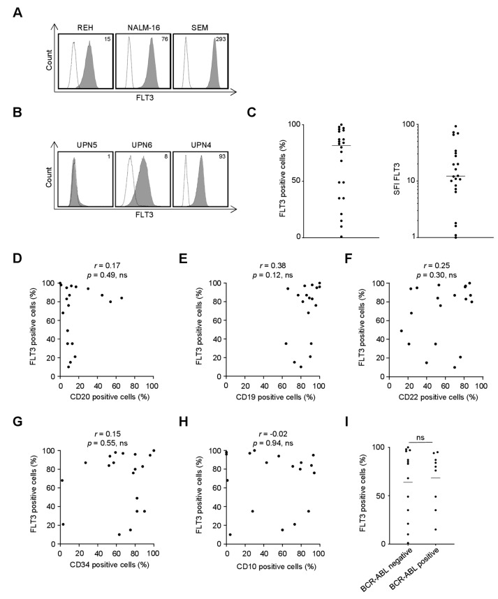
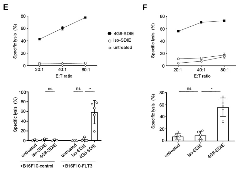
Antibody-dependent cellular cytotoxicity (ADCC) is a major mechanism by which antitumor antibodies mediate therapeutic efficacy. At present, we evaluate an Fc-optimized (amino acid substitutions S239D/I332E) FLT3 antibody termed 4G8-SDIEM (FLYSYN) in patients with acute myeloid leukemia (NCT02789254). Here we studied the possibility to induce NK cell ADCC against B-cell acute lymphoblastic leukemia (B-ALL) by Fc-optimized FLT3 antibody treatment. Flow cytometric analysis confirmed that FLT3 is widely expressed on B-ALL cell lines and leukemic cells of B-ALL patients. FLT3 expression did not correlate with that of CD20, which is targeted by Rituximab, a therapeutic monoclonal antibody (mAb) employed in B-ALL treatment regimens. Our FLT3 mAb with enhanced affinity to the Fc receptor CD16a termed 4G8-SDIE potently induced NK cell reactivity against FLT3-transfectants, the B-ALL cell line SEM and primary leukemic cells of adult B-ALL patients in a target-antigen dependent manner as revealed by analyses of NK cell activation and degranulation. This was mirrored by potent 4G8-SDIE mediated NK cell ADCC in experiments with FLT3-transfectants, the cell line SEM and primary cells as target cells. Taken together, the findings presented in this study provide evidence that 4G8-SDIE may be a promising agent for the treatment of B-ALL, particularly in CD20-negative cases.

Keywords: ADCC; B-ALL; CD135; FLT3; NK cells; acute lymphoblastic leukemia; antibody; immunotherapy.


Conflict of interest statement

H.-J.B. and G.J. were listed as inventors in a patent family for Fc-optimized FLT3 mAb, e.g., EP2516468B1; applicant was Tübingen University. There are no other conflicts of interest to declare.

Figures

Figure 1
Figure 1
Recognition of FLT3 expressed on the surface of B-cell acute lymphoblastic leukemia (B-ALL) cell lines and primary cells by mAb 4G8. B-ALL cell lines and primary leukemic cells of B-ALL patients were incubated with mouse anti-human FLT3 mAb clone 4G8 or murine IgG1 as isotype control (both 10 µg/mL) followed by a goat anti-mouse PE conjugate and subsequently analyzed by flow cytometry. (A) Exemplary data for FLT3 expression on REH, NALM-16 and SEM cells is shown (shaded peaks, anti-FLT3; open peaks, control). Numbers in the upper right corner depict specific fluorescence intensity (SFI) levels calculated as described in the method section. (B,C) Malignant cells within peripheral blood mononuclear cells of B-ALL patients (n = 22) were identified by counterstaining for CD34, CD10, CD19 or CD20 according to their pre-specified immunophenotype. (B) Exemplary data for patient cells with no (left), intermediate (middle) and high (right) surface expression of FLT3. (C) Combined analysis with FLT3 surface expression depicted as % FLT3+ B-ALL blasts (left) and SFI levels (right). (DI) Association of FLT3 surface expression (depicted as % positive cells) on primary B-ALL samples with expression of CD20 (D), CD19 (E), CD22 (F), CD34 (G), CD10 (H) and BCR-ABL (I). p: p-value; r: Pearson correlation coefficient; UPN: uniform patient number.
Figure 2
Figure 2
Production and characterization of 4G8-SDIE. (A) Schematic illustration of the structure of 4G8-SDIE. (B) Purified 4G8-SDIE was analyzed by size exclusion chromatography (left) and SDS-PAGE (right). Expected molecular weights, based on the amino acid sequence, were ~23, ~49 and ~145 kDa for light chain, heavy chain and full antibody, respectively. Dots represent the respective standards. M: marker; mAU: milli absorption unit; NR: non-reduced; R: reduced. (C) B16F10-FLT3 or control transfectants were incubated with 5 µg/mL 4G8-SDIE or iso-SDIE (isotype control antibody with similar characteristics, but irrelevant target specificity) followed by an anti-human phycoerythrin (PE) conjugate and analyzed by flow cytometry. Shaded peaks: 4G8-SDIE; open peaks: iso-SDIE. (D) Peripheral blood mononuclear cells (PBMC) of healthy donors were cultured with primary B-ALL cells in the presence or absence of iso-SDIE, chimeric 4G8 with wildtype Fc-part (4G8-WT) or 4G8-SDIE (all 10 µg/mL). B-ALL cell lysis was analyzed by 2 h Europium cytotoxicity assays. On the left, exemplary results obtained with cells from one healthy PBMC donor and one B-ALL patient are shown, on the right pooled data obtained with cells from two PBMC donors and B-ALL patients UPN4/6 at an E:T ratio of 80:1 are depicted. Bars and error bars represent means of results and standard deviations, respectively. (E) B16F10-FLT3 transfectants, the B-ALL cell lines SEM and NALM-16, and primary cells of two B-ALL patients (UPN 1 and 4) were incubated with increasing concentrations of 4G8-SDIE or iso-SDIE (10 µg/mL) followed by an anti-human PE conjugate and analyzed by flow cytometry. Malignant cells within PBMC of B-ALL patients were identified according to their pre-specified immunophenotype. Mean fluorescence intensities (MFI) are depicted. *: significant (p-value < 0.05); ns: not significant; UPN: uniform patient number.
Figure 3
Figure 3
Induction of natural killer (NK) cell reactivity against FLT3+ target cells. Peripheral blood mononuclear cells (PBMC) of healthy donors were cultured with or without B16F10-FLT3 or control transfectants (A,C,E) or the FLT3+ B-ALL cell line SEM (B,D,F) in the presence or absence of 4G8-SDIE/iso-SDIE (1 µg/mL). Top panels of each subfigure display exemplary data obtained with PBMC from one donor and B16F10-FLT3 or SEM cells. Bottom panels depict combined results from analyses with three to five independent PBMC donors. Bars and error bars represent means of results and standard deviations, respectively. (A,B) Cells were cultured at an effector to target (E:T) ratio of 2.5:1 for 24 h. Subsequently, activation of NK cells identified as CD19CD56+CD3 lymphocytes was determined by flow cytometric analysis of CD69. (C,D) Cells were cultured at an E:T ratio of 2.5:1 for 4 h in the presence of GolgiStop, GolgiPlug and an anti-human CD107a phycoerythrin (PE) conjugate. Subsequently, degranulation of NK cells (CD19CD56+CD3 lymphocytes) was determined by flow cytometric analysis of CD107a. (E,F) Target cell lysis was analyzed by 2 h Europium cytotoxicity assays. Combined analyses show data obtained at an E:T ratio of 80:1. ns: not significant; *: significant (p-value < 0.05).
Figure 3
Figure 3
Induction of natural killer (NK) cell reactivity against FLT3+ target cells. Peripheral blood mononuclear cells (PBMC) of healthy donors were cultured with or without B16F10-FLT3 or control transfectants (A,C,E) or the FLT3+ B-ALL cell line SEM (B,D,F) in the presence or absence of 4G8-SDIE/iso-SDIE (1 µg/mL). Top panels of each subfigure display exemplary data obtained with PBMC from one donor and B16F10-FLT3 or SEM cells. Bottom panels depict combined results from analyses with three to five independent PBMC donors. Bars and error bars represent means of results and standard deviations, respectively. (A,B) Cells were cultured at an effector to target (E:T) ratio of 2.5:1 for 24 h. Subsequently, activation of NK cells identified as CD19CD56+CD3 lymphocytes was determined by flow cytometric analysis of CD69. (C,D) Cells were cultured at an E:T ratio of 2.5:1 for 4 h in the presence of GolgiStop, GolgiPlug and an anti-human CD107a phycoerythrin (PE) conjugate. Subsequently, degranulation of NK cells (CD19CD56+CD3 lymphocytes) was determined by flow cytometric analysis of CD107a. (E,F) Target cell lysis was analyzed by 2 h Europium cytotoxicity assays. Combined analyses show data obtained at an E:T ratio of 80:1. ns: not significant; *: significant (p-value < 0.05).
Figure 4
Figure 4
Induction of NK cell reactivity against primary B-ALL cells. Peripheral blood mononuclear cells (PBMC) of healthy donors were cultured with or without FLT3+ B-ALL patient cells (UPN1/4/6/12/17/22, all blast count ≥86%) in the presence or absence of 4G8-SDIE/iso-SDIE (1 µg/mL). Left panels depict exemplary results obtained with cells from one healthy PBMC donor and one B-ALL patient; right panels show combined results obtained in multiple analyses with cells from different PBMC donors and B-ALL patients. Bars and error bars represent means of results and standard deviations, respectively. (A) Cells were cultured at an effector to target (E:T) ratio of 2.5:1 for 24 h. Subsequently, activation of NK cells identified as CD19CD56+CD3 lymphocytes was determined by flow cytometric analysis of CD69. Combined analyses show data obtained with cells from two healthy PBMC donors and four B-ALL patients. (B) Cells were cultured at an E:T ratio of 2.5:1 for 4 h in the presence of GolgiStop, GolgiPlug and an anti-human CD107a phycoerythrin (PE) conjugate. Subsequently, degranulation of NK cells (CD19CD56+CD3 lymphocytes) was determined by flow cytometric analysis of CD107a. Combined analyses show data obtained with cells from two healthy PBMC donors and six B-ALL patients. (C) B-ALL cell lysis was analyzed by 2 h Europium cytotoxicity assays. On the left exemplary data obtained with cells from one healthy PBMC donor and one B-ALL patient at different E:T ratios, on the right pooled data obtained with cells from three healthy PBMC donors and five B-ALL patients at an E:T ratio of 80:1 are shown. ns: not significant; *: significant (p-value < 0.05).

References

    1. Rothschilds A.M., Wittrup K.D. What, Why, Where, and When: Bringing Timing to Immuno-Oncology. Trends Immunol. 2019;40:12–21. doi: 10.1016/j.it.2018.11.003. - DOI - PubMed
    1. Arteaga C.L., Sliwkowski M.X., Osborne C.K., Perez E.A., Puglisi F., Gianni L. Treatment of HER2-positive breast cancer: Current status and future perspectives. Nat. Rev. Clin. Oncol. 2012;9:16–32. doi: 10.1038/nrclinonc.2011.177. - DOI - PubMed
    1. Keating G.M. Rituximab: A review of its use in chronic lymphocytic leukaemia, low-grade or follicular lymphoma and diffuse large B-cell lymphoma. Drugs. 2010;70:1445–1476. doi: 10.2165/11201110-000000000-00000. - DOI - PubMed
    1. Kellner C., Otte A., Cappuzzello E., Klausz K., Peipp M. Modulating Cytotoxic Effector Functions by Fc Engineering to Improve Cancer Therapy. Transfus. Med. Hemother. 2017;44:327–336. doi: 10.1159/000479980. - DOI - PMC - PubMed
    1. Weiner G.J. Rituximab: Mechanism of action. Semin. Hematol. 2010;47:115–123. doi: 10.1053/j.seminhematol.2010.01.011. - DOI - PMC - PubMed

LinkOut - more resources