. 2019 Dec 7;20(24):6179.
doi: 10.3390/ijms20246179.

Proteome Alterations in Equine Osteochondrotic Chondrocytes

Affiliations

Proteome Alterations in Equine Osteochondrotic Chondrocytes

Elisabetta Chiaradia et al. Int J Mol Sci. .

Abstract

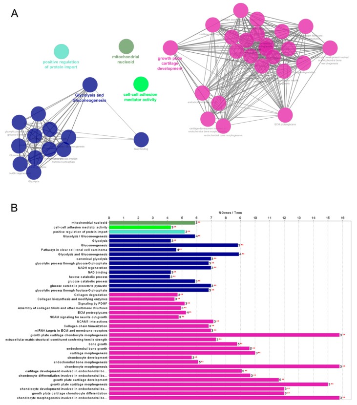
Osteochondrosis is a failure of the endochondral ossification that affects developing joints in humans and several animal species. It is a localized idiopathic joint disorder characterized by focal chondronecrosis and growing cartilage retention, which can lead to the formation of fissures, subchondral bone cysts, or intra-articular fragments. Osteochondrosis is a complex multifactorial disease associated with extracellular matrix alterations and failure in chondrocyte differentiation, mainly due to genetic, biochemical, and nutritional factors, as well as traumas. This study describes the main proteomic alterations occurring in chondrocytes isolated from osteochondrotic cartilage fragments. A comparative analysis performed on equine osteochondrotic and healthy chondrocytes showed 26 protein species as differentially represented. In particular, quantitative changes in the extracellular matrix, cytoskeletal and chaperone proteins, and in cell adhesion and signaling molecules were observed in osteochondrotic cells, compared to healthy controls. Functional group analysis annotated most of these proteins in "growth plate and cartilage development", while others were included in "glycolysis and gluconeogenesis", "positive regulation of protein import", "cell-cell adhesion mediator activity", and "mitochondrion nucleoid". These results may help to clarify some chondrocyte functional alterations that may play a significant role in determining the onset and progression of equine osteochondrosis and, being related, of human juvenile osteochondrosis.

Keywords: chondrocytes; endochondral ossification; growth plate and cartilage development; horse; human juvenile osteochondrosis; joint diseases; osteochondrosis; proteomics.


Conflict of interest statement

The authors declare no conflict of interest.

Figures

Figure 1
Figure 1
Representative image of a 2D gel of equine chondrocytes obtained using linear IPG (immobilized pH gradient) strip 3–10 and 12% T SDS-PAGE. The differentially represented protein spots (fold change > 2 and p < 0.05) were marked and numbered. Corresponding identified proteins are listed in Table 1. Spots found in OC and Control gels are marked in black and white circles, while spots found in OC gels only are marked in red circles.
Figure 2
Figure 2
(A) STRING Protein Protein Interaction (PPI) analysis of differentially expressed proteins in OC, nodes colors are in accordance with cell component categories: green = extracellular regions (GO:005576); red = mitochondrion (GO:005739); blue = cytosol (GO:005829); (B) PANTHER analysis classified the proteins identified according to the corresponding “Protein Class”.
Figure 3
Figure 3
ClueGO + CluePedia analyses of identified deregulated proteins in OC chondrocytes. Analysis was performed integrating biological processes, molecular functions, cellular component, Reactome, Kyoto Encyclopedia of Genes and Genomes (KEGG), and WikiPathways. (A) Functionally grouped network with terms as nodes linked based on their kappa score level (≥0.4), where only the label of the most significant term per group is shown; (B) GO/pathway terms specific for deregulated proteins. The bars represent the number of proteins associated with the terms. The percentage of genes per term is shown as bar label. Term/group resulted over significance ** p < 0.001.
Figure 4
Figure 4
Validation of a panel of five proteins found to be modulated in equine OC cells using different approaches. (A) Representative images of Wester Blotting of vimentin (VIM), annexin A2 and A1 (ANXA1-2), and collagen VI alpha 1 chain (COL6A1), as well as tubulin (TUB), which were used as housekeeping proteins. The bar graph shows normalized Western blot band densities. Images of independent blots were acquired and quantified using Quantity One Software. Data represent the mean ± SEM of three independent experiments. (* p = 0.01; # p = 0.0018; $ p = 0.001; § p = 0.0027). (B) Actin analysis by fluorescent phalloidine of Control and OC cells (scale bar 50 μm), seeded on glass slips. Data represent the mean ± SEM of 3 independent experiments. (C) Immunohistochemistry of equine cartilage samples (from the lateral trochlear ridge of femur) for COL6A1 (characterized by pericellular immunolabeling) (magnification 200×).
Figure 5
Figure 5
Representative images of healthy (Control) and OC equine joint. (A,B) Control and OC lateral trochlear ridge of the femur with fragment obtained during arthroscopy. (C,D) Lateromedial radiographic images of healthy (Control) and OC metacarpophalangeal joint with OC fragment of the dorsoproximal aspect of the proximal phalanx.

References

    1. Ytrehus B., Carlson C.S., Ekman S. Etiology and pathogenesis of osteochondrosis. Vet. Pathol. 2007;44:429–448. doi: 10.1354/vp.44-4-429. - DOI - PubMed
    1. Bates J.T., Jacobs J.C., Shea K.G., Oxford J.T. Emerging genetic basis of osteochondritis dissecans. Clin. Sports Med. 2014;33:199–220. doi: 10.1016/j.csm.2013.11.004. - DOI - PMC - PubMed
    1. Naccache F., Metzger J., Distl O. Genetic risk factors for osteochondrosis in various horse breeds. Equine Vet. J. 2018;50:556–563. doi: 10.1111/evj.12824. - DOI - PubMed
    1. Van Weeren P.R., Jeffcott L.B. Problems and pointers in osteochondrosis: Twenty years on. Vet. J. 2013;197:96–102. doi: 10.1016/j.tvjl.2013.03.048. - DOI - PubMed
    1. Van Weeren R. Fifty years of osteochondrosis. Equine Vet. J. 2018;50:554–555. doi: 10.1111/evj.12821. - DOI - PubMed

LinkOut - more resources