Review
. 2019 Dec 7;12(24):4096.
doi: 10.3390/ma12244096.

Magnetic Nanoparticles Supporting Bio-responsive T1/ T2 Magnetic Resonance Imaging

Affiliations
Review

Magnetic Nanoparticles Supporting Bio-responsive T1/ T2 Magnetic Resonance Imaging

Connor M Ellis et al. Materials (Basel). .

Abstract

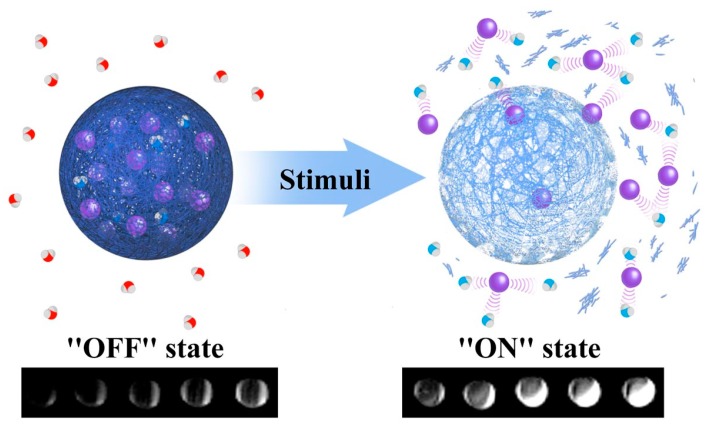
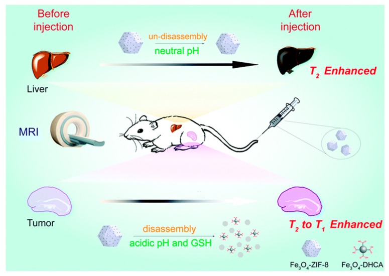
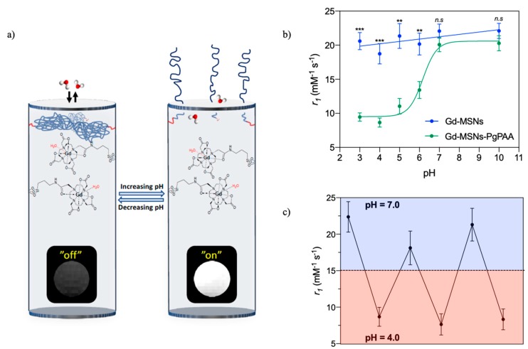
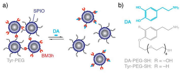
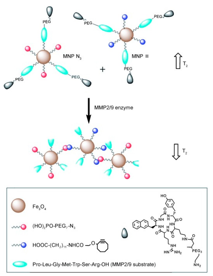
: The use of nanoparticulate systems as contrast agents for magnetic resonance imaging (MRI) is well-established and known to facilitate an enhanced image sensitivity within scans of a particular pathological region of interest. Such a capability can enable both a non-invasive diagnosis and the monitoring of disease progression/response to treatment. In this review, magnetic nanoparticles that exhibit a bio-responsive MR relaxivity are discussed, with pH-, enzyme-, biomolecular-, and protein-responsive systems considered. The ability of a contrast agent to respond to a biological stimulus provides not only enriched diagnostic capabilities over corresponding non-responsive analogues, but also an improved longitudinal monitoring of specific physiological conditions.

Keywords: Bio-responsive; Biomolecule-responsive; Diagnosis; Enzyme-responsive; Iron Oxide Nanoparticles; Magnetic Resonance Imaging; Mesoporous Silica Nanoparticles; Nanoparticle; Therapy; pH-responsive.


Conflict of interest statement

The authors declare no conflict of interest.

Figures

Figure 1
Figure 1
A schematic representing the impact of a generic stimulus on nanoparticle generated magnetic resonance imaging (MRI) contrast. The stimulus may be pH, enzyme activity, or temperature in nature, or may reflect the prescence of specific proteins/enzymes. In this example T1 contrast capabilities are switched “on”/“off” with a particular biological stimulus. The MR active moieties (purple spheres) are encapsulated within a responsive matrix and released into solution in the prescence of this particular stimulus (enhancing MR contrast by interacting with local water proton; red = non-enhanced relaxation, blue = enhanced relaxation). Adapted with permission from the authors of [13]. Copyright (2013) American Chemical Society.
Figure 2
Figure 2
A schematic showing how the incorporation of iron oxide nanoparticles (IONPs) into a pH-responsive zeolitic imidazole framework (ZIF-8 moiety) can engender a responsive contrast capability. At neutral pH, the nanocomposite remains intact, with T2 contrast exhibited. On decreasing pH, the ZIF-8 structure disassembles switching from T2 to T1 contrast capabilities. This framework was shown to be similarly responsive to glutathione (GSH). Reproduced from the work of [30] with permission from The Royal Society of Chemistry.
Figure 3
Figure 3
(a) Schematic representing a pH-responsive, reversible capping of the pore channels of a mesoporous silica nanoparticle (MSN) with poly(acrylic acid) (PAA) (blue polymer chains). The associated change in MR contrast is also shown. (b) A graph detailing the change in relaxivity with pH. (c) The reversible relaxivity switching with pH. Adapted from the work of [40] with permission from The Royal Society of Chemistry.
Figure 4
Figure 4
Dopamine sensitive IONPs. (a) The effect of dopamine on particle aggregation. In the absence of dopamine, nanocluster assemblies form owing to binding between protein (BM3h) and a dopamine analogue (Tyr-PEG). On addition of dopamine (DA), competitive binding inhibits self-assembly. (b) The structures of dopamine and tethered dopamine analogue. Reprinted (adapted) with permission from the authors of [11]. Copyright (2019) American Chemical Society.
Figure 5
Figure 5
A schematic representing the click coupling of IONPs functionalised with azide (red) groups and those functionalised with alkyne groups (blue). These particles expose azide/alkyne groups in the prescence of matrix metalloproteinase (MMP) enzymes, facilitating a click reaction mediated particle coupling. The effect on the T2 relaxation can be seen alongside both the individual IONPs (top) and the nanocluster (bottom). The particles also contain tumour specific peptide ligands and are functionalised with PEG to improve biocompatibility in vivo. Reprinted with permission from the authors of [52]. Copyright (2014), with permission from John Wiley and Sons.

References

    1. Shin T.-H., Choi Y., Kim S., Cheon J. Recent advances in magnetic nanoparticle-based multi-modal imaging. Chem. Soc. Rev. 2015;44:4501–4516. doi: 10.1039/C4CS00345D. - DOI - PubMed
    1. Ni D., Bu W., Ehlerding E.B., Cai W., Shi J. Engineering of inorganic nanoparticles as magnetic resonance imaging contrast agents. Chem. Soc. Rev. 2017;46:7438–7468. doi: 10.1039/C7CS00316A. - DOI - PMC - PubMed
    1. Gobbo O.L., Sjaastad K., Radomski M.W., Volkov Y., Prina-Mello A. Magnetic nanoparticles in cancer theranostics. Theranostics. 2015;5:1249. doi: 10.7150/thno.11544. - DOI - PMC - PubMed
    1. Singh A., Sahoo S.K. Magnetic nanoparticles: A novel platform for cancer theranostics. Drug Discov. Today. 2014;19:474–481. doi: 10.1016/j.drudis.2013.10.005. - DOI - PubMed
    1. Pellico J., Ellis C.M., Davis J.J. Nanoparticle-Based Paramagnetic Contrast Agents for Magnetic Resonance Imaging. Contrast Media Mol. Imaging. 2019;2019:13. doi: 10.1155/2019/1845637. - DOI - PMC - PubMed

LinkOut - more resources