Review
. 2020 Apr;26(2):135-146.
doi: 10.1177/1591019919891297. Epub 2019 Dec 9.

Vessel wall magnetic resonance imaging in intracranial aneurysms: Principles and emerging clinical applications

Affiliations
Review

Vessel wall magnetic resonance imaging in intracranial aneurysms: Principles and emerging clinical applications

Corrado Santarosa et al. Interv Neuroradiol. 2020 Apr.

Abstract

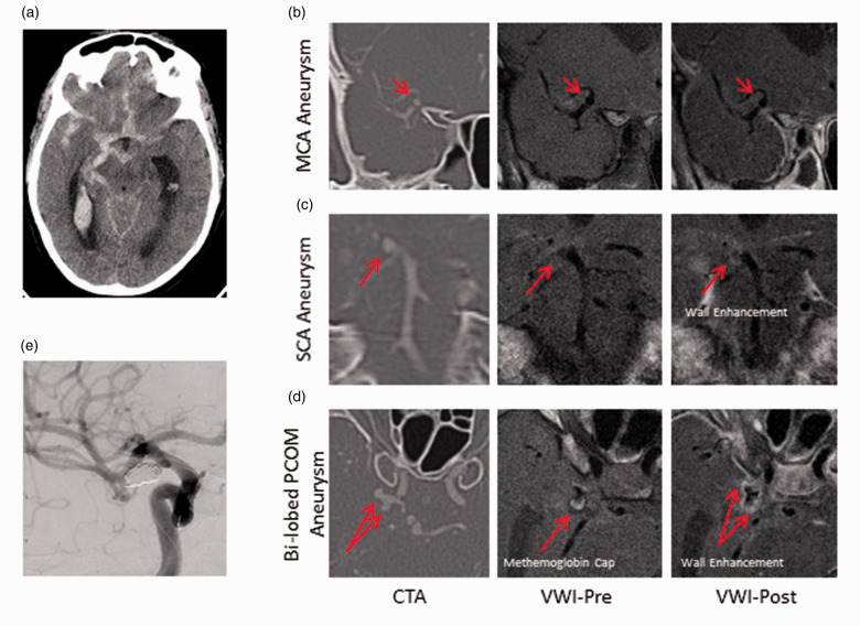
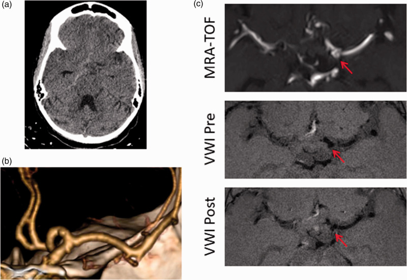
Intracranial high-resolution vessel wall magnetic resonance imaging is an imaging paradigm that complements conventional imaging modalities used in the evaluation of neurovascular pathology. This review focuses on the emerging utility of vessel wall magnetic resonance imaging in the characterization of intracranial aneurysms. We first discuss the technical principles of vessel wall magnetic resonance imaging highlighting methods to determine aneurysm wall enhancement and how to avoid common interpretive pitfalls. We then review its clinical application in the characterization of ruptured and unruptured intracranial aneurysms, in particular, the emergence of aneurysm wall enhancement as a biomarker of aneurysm instability. We offer our perspective from a high-volume neurovascular center where vessel wall magnetic resonance imaging is in routine clinical use.

Keywords: Intracranial aneurysm; magnetic resonance imaging; vessel wall imaging.


Figures

Figure 1.
Figure 1.
Pitfalls of vessel wall interpretation. (a) 3D TOF-MRA with left PCOM aneurysm (circle); (b) axial contrast-enhanced 3D MRA showing complete filling of the aneurysm showing the extent of the true lumen; (c) axial pre-contrast VW-MRI showing incomplete intraluminal suppression likely due to slow or turbulent flow asterisk, compared to (b); and (d) axial post-contrast VW-MRI with intraluminal enhancement due to incomplete suppression of blood signal (red asterisk) and normal enhancement of the surrounding cavernous dura which complicates interpretation (yellow asterisk).
Figure 2.
Figure 2.
Enhancement from adjacent veins. An incidental basilar aneurysm underwent VW-MRI. (a) and (d) Coronal and sagittal MRA MIP showing basilar tip aneurysm, (b) and (c) coronal VW-MRI images pre- and post-contrast demonstrate a focal area of “enhancement”, (e) and (f) adjacent axial VW-MRI sequences more clearly demonstrating the linear nature of this enhancing structure adjacent to the aneurysm (a vein).
Figure 3.
Figure 3.
Qualitative grading of vessel wall enhancement. Upper panel: MCA aneurysm with no enhancement. Middle panel: Basilar aneurysm with thin enhancement. Lower panel: PCOM aneurysm with thick enhancement. MIP 3D TOF MRA (first column), pre-contrast (second column), and post-contrast T1-weighted HR-MRA (third column). Aneurysm is denoted by circle or arrow. MRA: magnetic resonance angiography; VWI: vessel wall imaging.
Figure 4.
Figure 4.
Quantitative grading of vessel wall enhancement. (a) MIP 3D TOF MRA with right ophthalmic aneurysm (circle), (b) axial VW-MRI post-contrast T1 slice through the pituitary stalk with ROI (yellow circle), (c) axial VW-MRI post-contrast T1 slice through the body of the aneurysm with ROI on the aneurysm wall, and (d) corresponding axial VW-MRI pre-contrast T1 slice with matching ROI (green circle). Conceptually, the signal intensity of the wall is calculated by measuring the brightest part of the aneurysm wall on post-contrast sequences and subtracting the correlating measure from the wall on pre-contrast sequences. Normalization occurs by dividing this number by the highest intensity of the wall on pre-contrast images. Additional metrics can be derived by using brain parenchyma (not shown) or the pituitary stalk as additional normalizing factors.
Figure 5.
Figure 5.
Multiple enhancing aneurysms in a patient with SAH. A 58yF smoker presented with worst headache of life. (a) CT head with R > L basal cistern SAH and IVH. (b) to (d) CTA, VW-MRI pre/post-contrast images of a non-enhancing right MCA aneurysm (b), an enhancing right SCA aneurysm (c), and an enhancing, bi-lobed right PCOM aneurysm (d). Arrows denote the aneurysm in each slice. Note the presence of T1 bright signal at the tip of the posteriorly oriented lobe of the PCOM aneurysm which may represent methemoglobin. (e) Only the PCOM aneurysm was coiled as this was judged to be the site of rupture. CTA: computed tomography angiography; MCA: middle cerebral artery; PCOM: posterior communicating artery; SCA: superior cerebellar artery; VWI: vessel wall imaging.
Figure 6.
Figure 6.
Spontaneous SAH in a patient with an incidental, non-enhancing aneurysm. 22yF with sickle cell disease admitted in sickle crisis has sudden onset headache. (a) CT with right basal cistern SAH; (b) CTA reconstruction demonstrates a small left A1 aneurysm; and (c) Axial MRA (top), VW-MRI pre-contrast (middle), and VW-MRI post-contrast (bottom) images demonstrate no enhancement of the aneurysm. Arrow denotes aneurysm. This aneurysm was judged not to have ruptured and was not treated (incorrect hemorrhage pattern, non-enhancement of the aneurysm, alternative explanation). Patient has been followed for four years with aneurysm stability and no recurrent hemorrhage. MRA: magnetic resonance angiography; TOF: time of flight; VWI: vessel wall imaging.

References

    1. Vlak MH, Algra A, Brandenburg R, et al. Prevalence of unruptured intracranial aneurysms, with emphasis on sex, age, comorbidity, country, and time period: a systematic review and meta-analysis. Lancet Neurol 2011; 10: 626–636. - PubMed
    1. Etminan N, Rinkel GJ. Unruptured intracranial aneurysms: development, rupture and preventive management. Nat Rev Neurol 2016; 12: 699–713. - PubMed
    1. de Rooij NK, Linn FH, van der Plas JA, et al. Incidence of subarachnoid haemorrhage: a systematic review with emphasis on region, age, gender and time trends. J Neurol Neurosurg Psychiatry 2007; 78: 1365–1372. - PMC - PubMed
    1. Bijlenga P, Gondar R, Schilling S, et al. PHASES score for the management of intracranial aneurysm: a cross-sectional population-based retrospective study. Stroke 2017; 48: 2105–2112. - PubMed
    1. Wiebers DO, Whisnant JP, Huston J, 3rd, et al. Unruptured intracranial aneurysms: natural history, clinical outcome, and risks of surgical and endovascular treatment. Lancet 2003; 362: 103–110. - PubMed

MeSH terms

LinkOut - more resources