. 2019 Dec 9;11(1):80.
doi: 10.1186/s13073-019-0676-0.

Distinct patterns of complex rearrangements and a mutational signature of microhomeology are frequently observed in PLP1 copy number gain structural variants

Affiliations

Distinct patterns of complex rearrangements and a mutational signature of microhomeology are frequently observed in PLP1 copy number gain structural variants

Vahid Bahrambeigi et al. Genome Med. .

Abstract

Background: We investigated the features of the genomic rearrangements in a cohort of 50 male individuals with proteolipid protein 1 (PLP1) copy number gain events who were ascertained with Pelizaeus-Merzbacher disease (PMD; MIM: 312080). We then compared our new data to previous structural variant mutagenesis studies involving the Xq22 region of the human genome. The aggregate data from 159 sequenced join-points (discontinuous sequences in the reference genome that are joined during the rearrangement process) were studied. Analysis of these data from 150 individuals enabled the spectrum and relative distribution of the underlying genomic mutational signatures to be delineated.

Methods: Genomic rearrangements in PMD individuals with PLP1 copy number gain events were investigated by high-density customized array or clinical chromosomal microarray analysis and breakpoint junction sequence analysis.

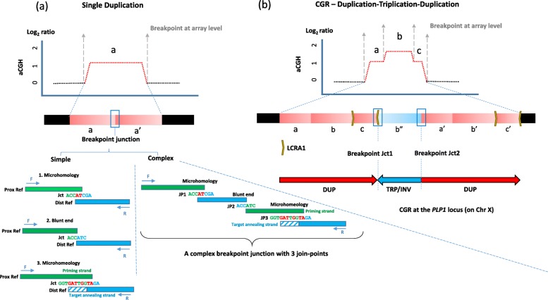
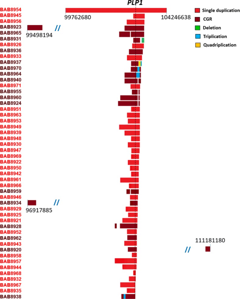
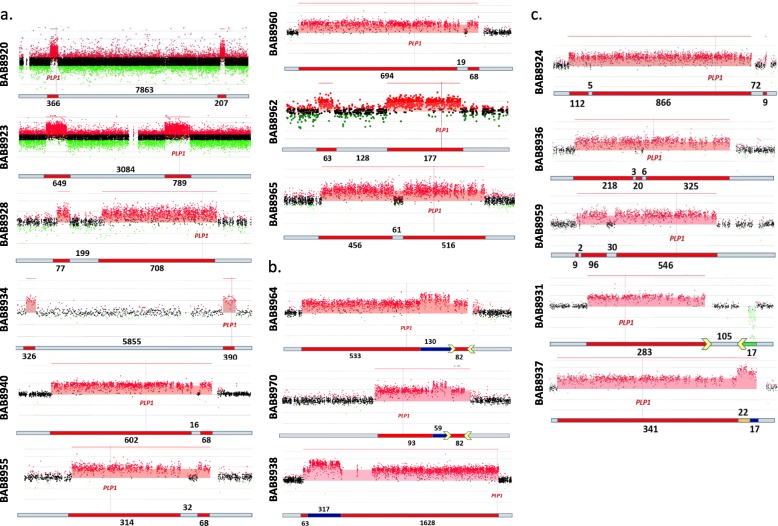
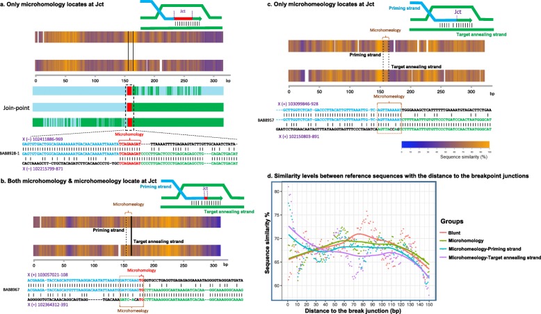
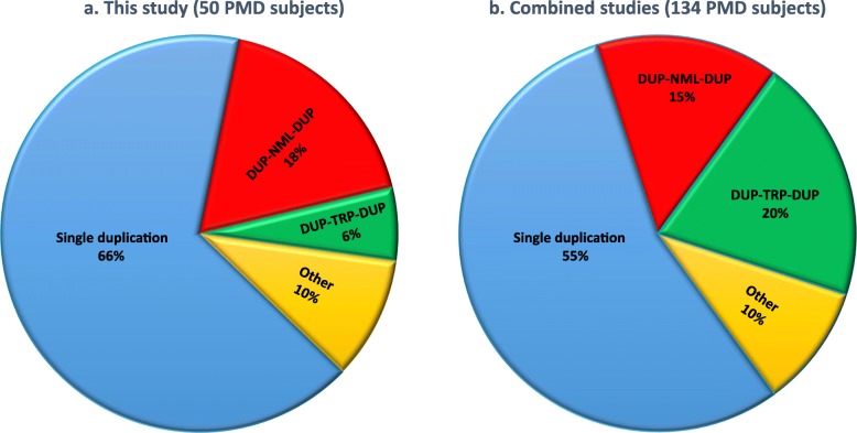
Results: High-density customized array showed that the majority of cases (33/50; ~ 66%) present with single duplications, although complex genomic rearrangements (CGRs) are also frequent (17/50; ~ 34%). Breakpoint mapping to nucleotide resolution revealed further previously unknown structural and sequence complexities, even in single duplications. Meta-analysis of all studied rearrangements that occur at the PLP1 locus showed that single duplications were found in ~ 54% of individuals and that, among all CGR cases, triplication flanked by duplications is the most frequent CGR array CGH pattern observed. Importantly, in ~ 32% of join-points, there is evidence for a mutational signature of microhomeology (highly similar yet imperfect sequence matches).

Conclusions: These data reveal a high frequency of CGRs at the PLP1 locus and support the assertion that replication-based mechanisms are prominent contributors to the formation of CGRs at Xq22. We propose that microhomeology can facilitate template switching, by stabilizing strand annealing of the primer using W-C base complementarity, and is a mutational signature for replicative repair.

Keywords: BIR; Duplication; Genome instability; Genomic rearrangements; HR; LCR; MMBIR; Microhomeology; PMD; RBM.


Conflict of interest statement

J.R.L. has stock ownership in 23andMe, is a paid consultant for Regeneron Pharmaceuticals, and is a co-inventor on multiple US and European patents related to molecular diagnostics for inherited neuropathies, eye diseases, and bacterial genomic fingerprinting. The Department of Molecular and Human Genetics at Baylor College of Medicine derives revenue from the chromosomal microarray analysis (CMA) and clinical exome sequencing offered in the Baylor Genetics Laboratory (BMGL: http://www.bmgl.com/BMGL/Default.aspx). The remaining authors declare that they have no competing interests.

Figures

Fig. 1
Fig. 1
Genomic rearrangements with different levels of complexity. At the array-resolution level, genomic rearrangements with the PLP1 gain can be apparently simple as a a single duplication or b a CGR. In aCGH figures, transitions of copy number alterations from copy neutral regions (black dots) to copy number gains (red dots) are demonstrated by gray vertical dashed arrows (breakpoints). At the nucleotide sequence level as shown in a, in the simplest case scenario, a single duplication has a breakpoint junction with only one join-point (a—left), a product of one TS by NHEJ (for blunt end), or microhomology and/or microhomeology-mediated rearrangement. Or, a breakpoint junction can contain several join-points (a—right). Such breakpoint junctions are products of iterative TS by different rearrangement mechanisms such as NHEJ or MMBIR. Bases indicated in red are in both the proximal and distal reference sequences. Rectangle with diagonal lines indicates a region of imperfect match between proximal and distal reference sequences. In addition to the iterative TS that lead to the appearance of complex breakpoints, iterative TS can result in copy number transitions of large genomic segments and formation of more complex genomic structures. b As a representative of such complex genomic structures, a schematic figure of a CGR with DUP-TRP/INV-DUP pattern resulted from two TSs creating breakpoint junctions Jct1 and Jct2, as shown. The horizontal bar below the aCGH depicts the rearrangement product. Duplications are represented in red and triplication in blue; yellow arrows represent inverted low copy repeats that mediate the TS in Jct1. Positions of the genomic segments are denoted as a, b, and c, duplicated segments as a′, b′, and c′, and the triplicated segment as b″. The TS between low copy repeats forming Jct1 switched the direction of replication resulting in an inversion of the TRP segment, and the second TS forming Jct2 switched the direction of the replication again resulting in directly oriented DUP segments. The Y-axis on the aCGH plots represents expected log2 ratios in male using a gender-matched control and that PLP1 maps to chromosome X. Jct: junction; JP: join-point
Fig. 2
Fig. 2
An overview of genomic rearrangements as seen on aCGH in 50 individuals with PMD. Genomic rearrangements at Xq22 vary in size and genomic positions. The largest duplication (~ 4.5 Mb) is found in individual BAB8954. Three individuals show additional duplications distant from the duplicated PLP1 locus (individuals BAB8920, BAB8923, and BAB8934). The black numbers refer to genomic coordinates on chromosome X. The left column lists the 50 subjects studied. Slash lines indicate a break in numbering for genomic coordinates. The location of PLP1 is indicated by a black vertical broken line
Fig. 3
Fig. 3
CGRs detected by aCGH at the PLP1 locus. a Two duplications separated by CNRs were detected on aCGH in 9 individuals with PMD. The distance between the two duplications differs among these individuals, ranging from 16 to 7863 kb. In the schematic figure below each array, duplications are depicted in red and CNRs in gray. Three cases (BAB8940, BAB8955, and BAB8960) could be single duplications on the H2 inversion haplotype or could be two duplications with one TS involving reversal of the direction of replication between IRs LCRA1a and LCRA1b (Additional file 1: Figure S9); three (BAB8923, BAB8928, and BAB8965) have directly oriented DUP-NML-DUP structures (Additional file 1: Figures S6–1, S6–2 and S6–3); one has two tandem head to tail duplications (BAB8962; Additional file 1: Figure S6–4); and two (BAB8920, BAB8934) have DUP-NML-INV/DUP structures (Additional file 1: Figure S7). b A DUP-TRP-DUP pattern of rearrangement was detected on aCGH in three individuals with PMD (Additional file 1: Figure S10). Breakpoint junction analyses indicated that one of these individuals (BAB8964) probably has the previously reported DUP-TRP/INV-DUP pattern of rearrangement with inversion mediated by a TS between inverted repeats LCRA1a and LCRA1b. Based on aCGH data, BAB8970 probably has the same structure, although breakpoint junctions were not resolved (Additional file 1: Figures S10–1 and S10–2). Breakpoint junction analysis indicates that BAB8939 also carries a DUP-TRP/INV-DUP, but the inversion was not mediated by LCRA1a and LCRA1b (Additional file 1: Figure S10–3). Duplications are indicated in red, triplications in blue, and LCR blocks (LCRA1a and LCRA1b) in yellow. c Additional CGR patterns at the PLP1 locus were identified on aCGH. DUP-NML-DUP-NML-DUP rearrangement pattern in which duplications are separated by short CNRs (BAB8924, BAB8936, and BAB8959). In BAB8924, based on the sequenced breakpoint junction, this case may have two tandem head to tail duplications on the H2 haplotype that has an inversion within LCRA1a and LCRA1b (Additional file 1: Figure S12–1a) or may have three duplications with one TS between LCRA1a and LCRA1b resulting in an inversion (not shown). We were not able to resolve any breakpoint junctions in BAB8936 (Additional file 1: Figure S12–1b). Breakpoint junction sequencing in BAB8959 showed that the CGR based on aCGH may not have occurred during the same cell division (Additional file 1: Figures S12–2). One individual, BAB8931, exhibited DUP-NML-DEL pattern of rearrangement with a ~ 283-kb duplication (breakpoint junction in LCRA1a) followed by ~ 106 kb of CNR and then a ~ 16-kb deletion (breakpoint junction in LCRA1b). The most complex rearrangement in this study was observed in individual BAB8937 with a DUP-QUAD-TRP rearrangement pattern. In this case, duplication is followed by a quadruplication and then a triplication. The possible mechanism for such rearrangements is shown in Additional file 1: Figure S11. Duplications are indicated in red, CNRs in gray, deletion in green, triplication in blue, quadruplication in orange, and LCR blocks in yellow in the horizontal bar under each array
Fig. 4
Fig. 4
Representative similarity plots (heat maps) between reference sequences surrounding CNV breakpoint junctions containing a only microhomology (> 2 bp of nucleotide similarity) flanked by solid vertical lines), b both microhomeology and microhomology, and c only microhomeology. We present here an example for each type of the observed junctional sequences using heat map (top) and the sequence alignment at a nucleotide level (bottom). Reference sequences were aligned using the Needleman-Wunsch algorithm, as described in the “Methods” section. The 5′ reference sequence is indicated in blue color and 3′ reference sequence is indicated in green. In the upper panel of heat map plot, the 5′ reference sequence was plotted as a rectangle on the top while the 3′ was on the bottom. The heat map shading indicates the sequence similarity level of a 20-bp moving window: orange-high similarity, blue-low similarity, and white-gap. Schematic figures in b and c indicate microhomeology-mediated priming strand (blue) invasion to the target annealing strand (green). Microhomology is shown in red. d An aggregative plot showing the change of similarity levels between reference sequences along an increase in the distance to the breakpoint junctions. We compared such patterns among four junction categories: blunt junctions (red), junctions containing a microhomology only (green), and the priming sides (blue) and target annealing sides (purple) of junctions containing a microhomeology
Fig. 5
Fig. 5
An overview of genomic rearrangements with gain at the PLP1 locus. a Genomic rearrangements in the present cohort with 50 PMD individuals (Table 1). b Meta-analysis of combined results from six previously published studies (Additional file 2: Table S3a). Genomic rearrangements involving triplications are the most frequent CGRs at the PLP1 locus

References

    1. Lupski JR. Genomic disorders: structural features of the genome can lead to DNA rearrangements and human disease traits. Trends Genet. 1998;14:417–422. doi: 10.1016/S0168-9525(98)01555-8. - DOI - PubMed
    1. Stankiewicz P, Lupski JR. Genome architecture, rearrangements and genomic disorders. Trends Genet. 2002;18:74–82. doi: 10.1016/S0168-9525(02)02592-1. - DOI - PubMed
    1. Carvalho CM, Lupski JR. Mechanisms underlying structural variant formation in genomic disorders. Nat Rev Genet. 2016;17:224–238. doi: 10.1038/nrg.2015.25. - DOI - PMC - PubMed
    1. Ellis D, Malcolm S. Proteolipid protein gene dosage effect in Pelizaeus-Merzbacher disease. Nat Genet. 1994;6:333–334. doi: 10.1038/ng0494-333. - DOI - PubMed
    1. Inoue Ken. PLP1-related inherited dysmyelinating disorders: Pelizaeus-Merzbacher disease and spastic paraplegia type 2. Neurogenetics. 2004;6(1):1–16. doi: 10.1007/s10048-004-0207-y. - DOI - PubMed

Publication types

Substances