. 2020 Jan;27(1):96-105.
doi: 10.1016/j.acra.2019.09.014.

Augmented Radiologist Workflow Improves Report Value and Saves Time: A Potential Model for Implementation of Artificial Intelligence

Affiliations

Augmented Radiologist Workflow Improves Report Value and Saves Time: A Potential Model for Implementation of Artificial Intelligence

Huy M Do et al. Acad Radiol. 2020 Jan.

Abstract

Rationale and objectives: Our primary aim was to improve radiology reports by increasing concordance of target lesion measurements with oncology records using radiology preprocessors (RP). Faster notification of incidental actionable findings to referring clinicians and clinical radiologist exam interpretation time savings with RPs quantifying tumor burden were also assessed.

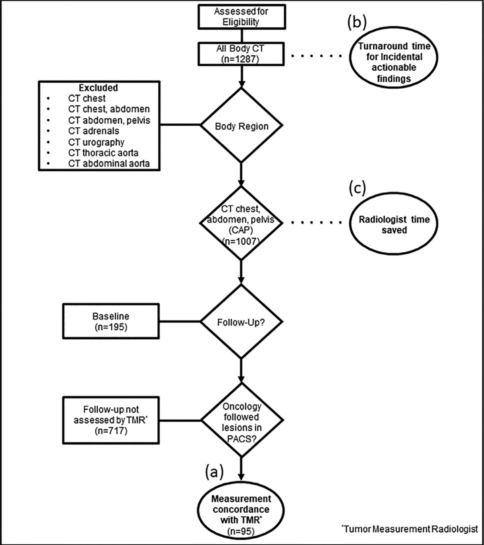
Materials and methods: In this prospective quality improvement initiative, RPs annotated lesions before radiologist interpretation of CT exams. Clinical radiologists then hyperlinked approved measurements into interactive reports during interpretations. RPs evaluated concordance with our tumor measurement radiologist, the determinant of tumor burden. Actionable finding detection and notification times were also deduced. Clinical radiologist interpretation times were calculated from established average CT chest, abdomen, and pelvis interpretation times.

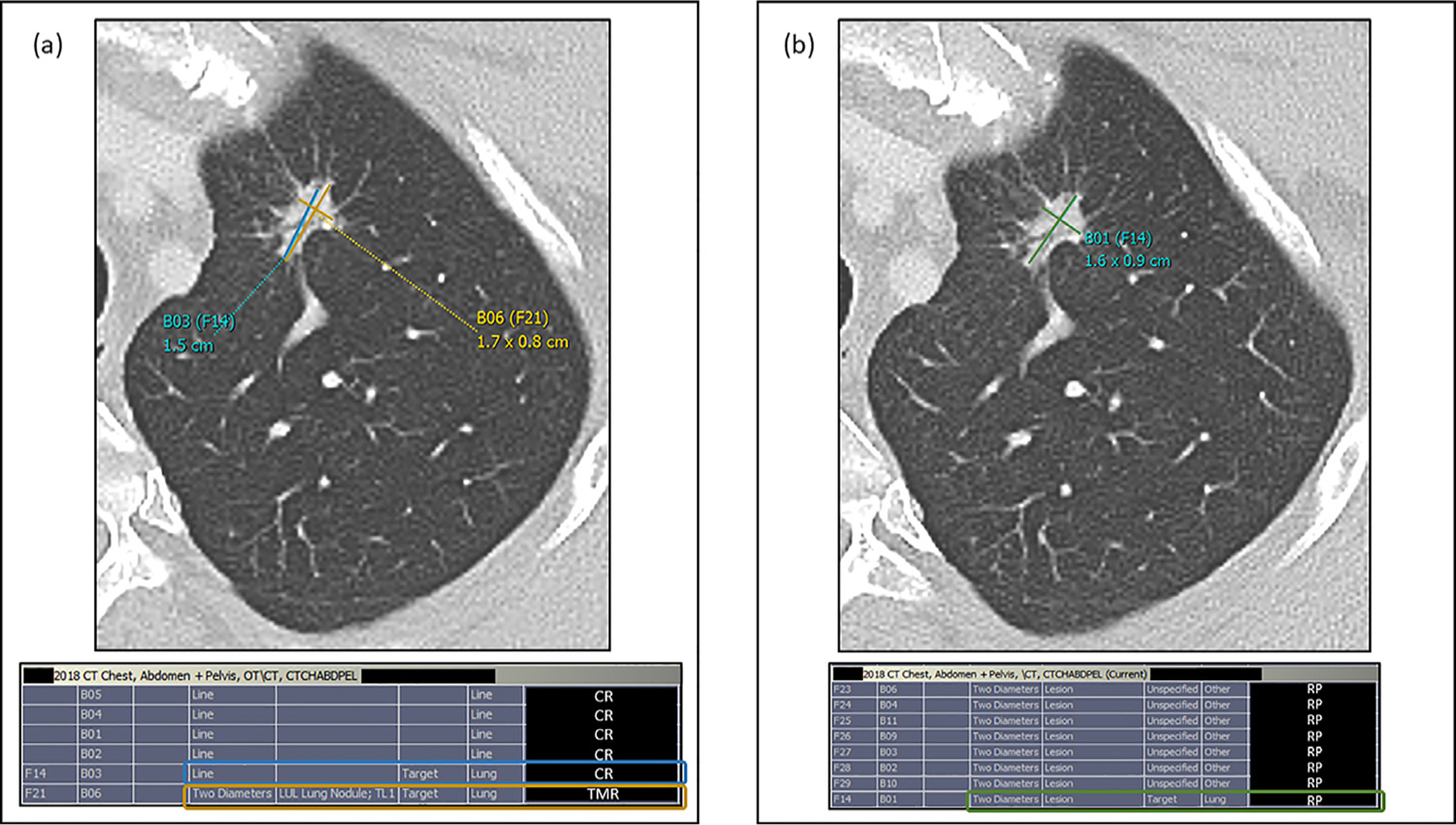
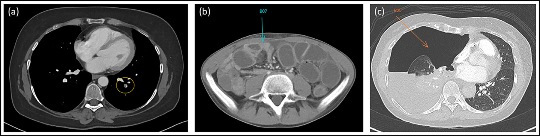
Results: RPs assessed 1287 body CT exams with 812 follow-up CT chest, abdomen, and pelvis studies; 95 (11.7%) of which had 241 verified target lesions. There was improved concordance (67.8% vs. 22.5%) of target lesion measurements. RPs detected 93.1% incidental actionable findings with faster clinician notification by a median time of 1 hour (range: 15 minutes-16 hours). Radiologist exam interpretation times decreased by 37%.

Conclusions: This workflow resulted in three-fold improved target lesion measurement concordance with oncology records, earlier detection and faster notification of incidental actionable findings to referring clinicians, and decreased exam interpretation times for clinical radiologists. These findings demonstrate potential roles for automation (such as AI) to improve report value, worklist prioritization, and patient care.

Keywords: Actionable findings; Artificial intelligence; Cancer clinical trials; Radiology preprocessors; Tumor quantification.


Conflict of interest statement

DECLARATION OF INTEREST

None declared.

CONFLICT OF INTEREST

Dr. Huy Do and Dr. Les Folio were associate investigators in a corporate research agreement with Carestream Health (Rochester, NY), the PACS used in this initiative.

Figures

Figure 1.
Figure 1.
Study inclusion flowchart with exclusion criteria, depicting the three variables assessed in this study: (a) Lesion measurement concordance with the tumor measurement radiologist (TMR); (b) turn-around notification times of incidental actionable findings to referring oncology teams; and (c) clinical radiologist time savings.
Figure 2.
Figure 2.
(a) Example target lesion measurement discrepancy on prior CT CAP exam with corresponding measured lesions in PACS bookmark table for a patient with lung cancer. In this standard workflow (status quo, no radiology preprocessor [RP] involvement), the clinical radiologist measured a spiculated left apical lung nodule (designated target lesion according to clinical trial protocol) seen above using the single line tool (B03(F14), blue) in our PACS, which was remeasured by our tumor measurement radiologist (TMR) using our PACS two diameter tool (B06 (F21), yellow) and recorded as a measurement discrepancy for the purposes of this study. This discrepancy was also confirmed using the bookmark table as evidenced by two different measurements of the same target lesion, initially measured by the clinical radiologist (CR, blue rectangle) and remeasured by the TMR (yellow rectangle). (b) Example target lesion measurement concordance on follow-up CT CAP exam for the same patient. In the RP augmented workflow, the RP measurement of the same left apical lung nodule target lesion using the two diameter tool (B01(F14), green) was accepted by the TMR as it was not modified and saved as a key image in our PACS. RP measurement concordance with the TMR was confirmed using the bookmark table as the RP measurement of the target lesion (RP, green rectangle) was not remeasured or modified by the TMR. CAP; chest, abdomen, pelvis; PACS, Picture Archiving and Communication System.
Figure 3.
Figure 3.
Examples of three incidental actionable findings (from CT exams of three different patients) successfully detected and annotated by RPs in the RP augmented workflow. (a) Left basal segmental pulmonary arterial filling defect consistent with pulmonary embolism (PE, yellow oval). (b) Significant distention of small bowel loops with corresponding air-fluid levels on abdominal X-ray (not shown) and transition point of obstruction (blue arrow) consistent with small bowel obstruction (SBO). (c) Large amount of free air in right thoracic cavity (orange arrow) with associated collapse of the right lung and leftward mediastinal shift consistent with tension pneumothorax.

References

    1. Eisenhauer EA, Therasse P, Bogaerts J, Schwartz LH, Sargent D, Ford R, et al. New response evaluation criteria in solid tumours: revised RECIST guideline (version 1.1). Eur J Cancer 2009; 45(2):228–247. doi:10.1016/j.ejca.2008.10.026. - DOI - PubMed
    1. Padhani AR, Ollivier L. The RECIST (Response Evaluation Criteria in Solid Tumors) criteria: implications for diagnostic radiologists. Br J Radiol 2001; 74(887):983–986. doi:10.1259/bjr.74.887.740983. - DOI - PubMed
    1. Levy MA, Rubin DL. Tool support to enable evaluation of the clinical response to treatment. AMIA Annu Symp Proc 2008: 399–403. - PMC - PubMed
    1. Machado LB, Apolo AB, Steinberg SM, Folio LR. Radiology Reports With Hyperlinks Improve Target Lesion Selection and Measurement Concordance in Cancer Trials. AJR Am J Roentgenol 2017; 208(2). doi:10.2214/ajr.16.16845. W31–w7. - DOI - PMC - PubMed
    1. Folio LR, Choi MM, Solomon JM, Schaub NP. Automated registration, segmentation, and measurement of metastatic melanoma tumors in serial CT scans. Acad Radiol 2013; 20(5):604–613. doi:10.1016/j.acra.2012.12.013. - DOI - PubMed

Publication types