Review
. 2020 Jul 1;12(7):a036756.
doi: 10.1101/cshperspect.a036756.

Genetic and Epigenetic Control of Heart Development

Affiliations
Review

Genetic and Epigenetic Control of Heart Development

Brynn N Akerberg et al. Cold Spring Harb Perspect Biol. .

Abstract

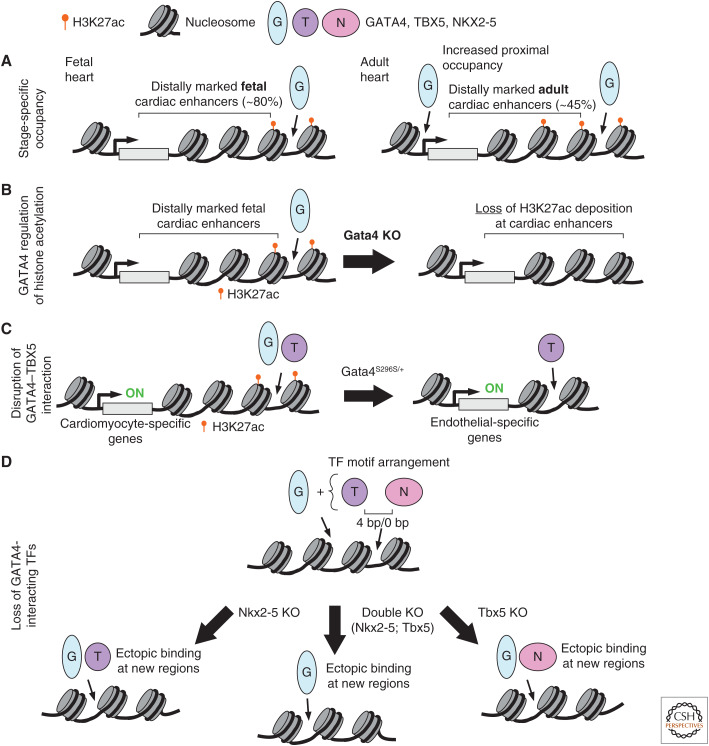
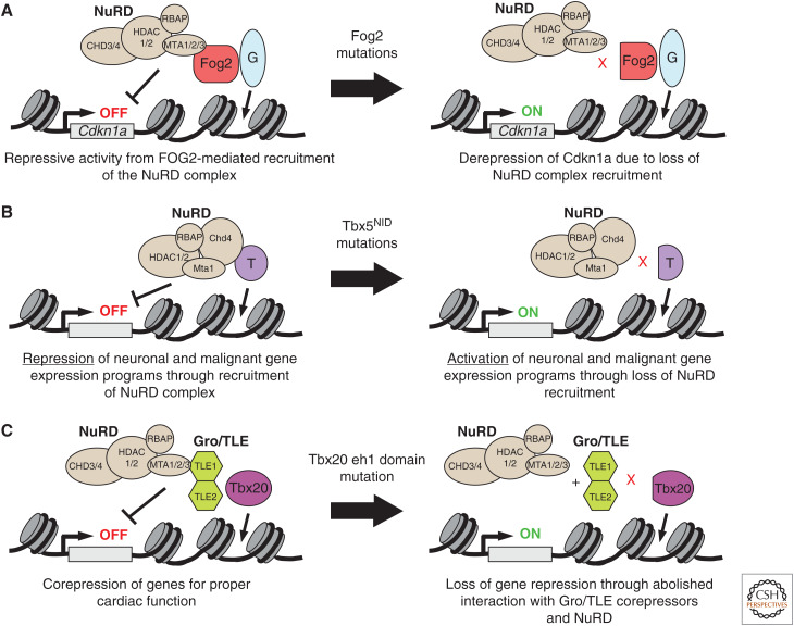
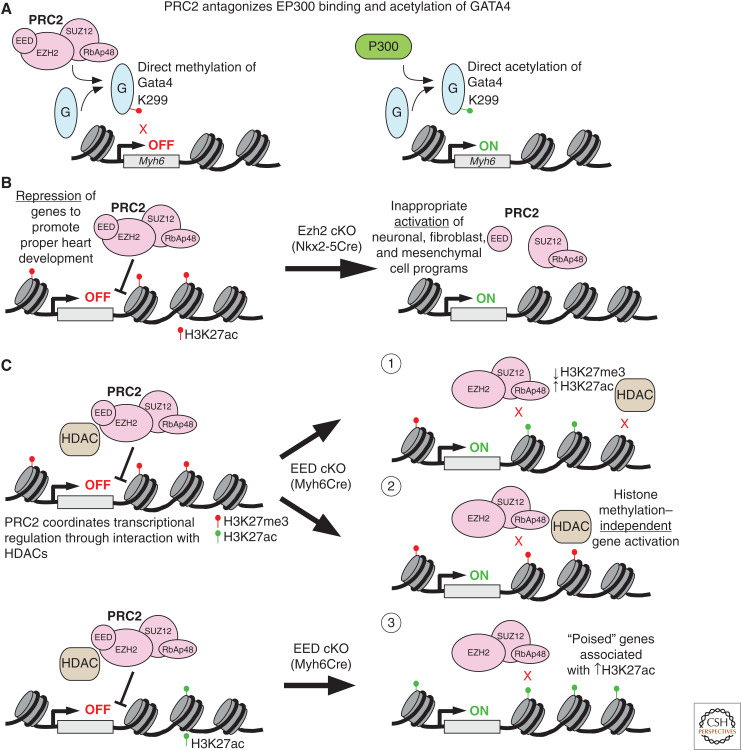
A transcriptional program implemented by transcription factors and epigenetic regulators governs cardiac development and disease. Mutations in these factors are important causes of congenital heart disease. Here, we review selected recent advances in our understanding of the transcriptional and epigenetic control of heart development, including determinants of cardiac transcription factor chromatin occupancy, the gene regulatory network that regulates atrial septation, the chromatin landscape and cardiac gene regulation, and the role of Brg/Brahma-associated factor (BAF), nucleosome remodeling and histone deacetylation (NuRD), and Polycomb epigenetic regulatory complexes in heart development.


Figures

Figure 1.
Figure 1.
Transcription factor (TF) interactions. (A) GATA-binding protein 4 (GATA4) occupies regions in a stage-specific manner. GATA4 bioChIP-Seq data revealed that GATA4 predominantly binds distal (>2 kb from transcription start sites [TSSs]) enhancers (H3K27ac) in fetal heart. The majority of fetal binding sites were not occupied by GATA4 in adult heart, and overall a greater proportion of GATA4 occupancy was proximal to the TSSs. (B) Depletion of GATA4 in the embryonic mouse heart resulted in loss of H3K27ac epigenetic marks at candidate cardiac enhancers. (C) GATA4 and T-Box 5 (TBX5) coordinately regulate transcription of cardiac genes through binding enhancers, which are often marked by H3K27ac. A GATA4 point mutation found in a family with septal defects, and cardiomyopathy abolishes physical interaction with TBX5 and leads to dysregulated gene expression, in part through ectopic TBX5 occupancy at endothelial-related genes. (D) A subset of sides co-occupied by NKX2-5 and TBX5 revealed a distinct binding motif orientation and spacing (0 bp or 4 bp) and frequent co-occupancy by GATA4. Loss of either NKX2-5 or TBX5 resulted in ectopic binding (binding to regions in mutant that were not found in wild type) of the other TF partner in combination with GATA4. Similarly, in cells with double knockout of both NKX2-5 and TBX5, GATA4 was found ectopically bound to new genomic regions.
Figure 2.
Figure 2.
Nucleosome remodeling and histone deacetylation (NuRD) complex and cardiac gene regulation. (A) The NuRD complex interacts with FOG2, a direct modulator of GATA4 transcriptional activity. One target of GATA4–FOG2–NuRD repression was Cdkn1a. (B) TBX5 directly interacts with NuRD complex subunits CHD4, HDAC2, and MTA1 to repress gene expression programs not normally expressed during heart development. TBX5 mutations in its NuRD interaction domain (NID) abolished direct interaction with the NuRD complex, leading to expression of normally repressed, ectopic gene programs. (C) Physical interaction between TBX20 and the NuRD complex is mediated through Gro/TLE corepressors. Specifically, the TBX20 eh1 domain is essential for proper transcriptional regulation by the Gro/TLE–NuRD complexes.
Figure 3.
Figure 3.
Polycomb repressive complex 2 (PRC2). (A) PRC2 binds to directly methylate K299 residue on GATA4 to attenuate transcriptional activation of downstream targets such as Myh6. This repressive activity of PRC2 is balanced by the EP300, which competes with PRC2 for GATA4 binding and directly acetylates GATA4 at the same residue, resulting in target gene activation. (B) EZH2, the catalytic subunit of the PRC2 complex, is required for proper heart development. Conditional knockout (cKO) of EZH2 by Nkx2-5Cre in cardiac progenitors globally decreased H3K27me3 levels and inappropriately derepressed neuronal, fibroblast, and mesenchymal gene programs in embryonic mouse hearts. (C) Myh6-specific deletion of EED, a core PRC2 component, leads to derepression of many genes. These derepressed genes fall into three categories based on their associated epigenetic marks: (1) increased H3K27me3 and increased H3K27ac, consistent with loss of EED-dependent histone deacetylase (HDAC) activity; (2) retained H3K27me3, consistent with histone methylation-independent gene regulation; and (3) little H3K27me3 in control and increased H3K27ac in EEDKO, suggestive of “poised” genes that are activated by relief of PRC2 and HDAC repression.

References

    1. Ai S, Peng Y, Li C, Gu F, Yu X, Yue Y, Ma Q, Chen J, Lin Z, Zhou P, et al. 2017a. EED orchestration of heart maturation through interaction with HDACs is H3K27me3-independent. eLife 6: e24570 10.7554/eLife.24570 - DOI - PMC - PubMed
    1. Ai S, Yu X, Li Y, Peng Y, Li C, Yue Y, Tao G, Li C, Pu WT, He A. 2017b. Divergent requirements for EZH1 in heart development versus regeneration. Circ Res 121: 106–112. 10.1161/CIRCRESAHA.117.311212 - DOI - PMC - PubMed
    1. Akerberg BN, Sarangam ML, Stankunas K. 2015. Endocardial Brg1 disruption illustrates the developmental origins of semilunar valve disease. Dev Biol 407: 158–172. 10.1016/j.ydbio.2015.06.015 - DOI - PMC - PubMed
    1. Akerberg BN, Gu F, VanDusen NJ, Zhang X, Dong R, Li K, Zhang B, Zhou B, Sethi I, Ma Q, et al. 2019. A reference map of murine cardiac transcription factor chromatin occupancy identifies dynamic and conserved enhancers. Nat Commun 10: 4907 10.1038/s41467-019-12812-3 - DOI - PMC - PubMed
    1. Ang YS, Rivas RN, Ribeiro AJS, Srivas R, Rivera J, Stone NR, Pratt K, Mohamed TMA, Fu J-D, Spencer CI, et al. 2016. Disease model of GATA4 mutation reveals transcription factor cooperativity in human cardiogenesis. Cell 167: 1734–1749.e22. 10.1016/j.cell.2016.11.033 - DOI - PMC - PubMed

Publication types

MeSH terms

LinkOut - more resources