. 2019 Dec 26;116(52):26846-26852.
doi: 10.1073/pnas.1907081116. Epub 2019 Dec 9.

CRISPR/Cas9-based targeted genome editing for correction of recessive dystrophic epidermolysis bullosa using iPS cells

Affiliations

CRISPR/Cas9-based targeted genome editing for correction of recessive dystrophic epidermolysis bullosa using iPS cells

Joanna Jacków et al. Proc Natl Acad Sci U S A. .

Abstract

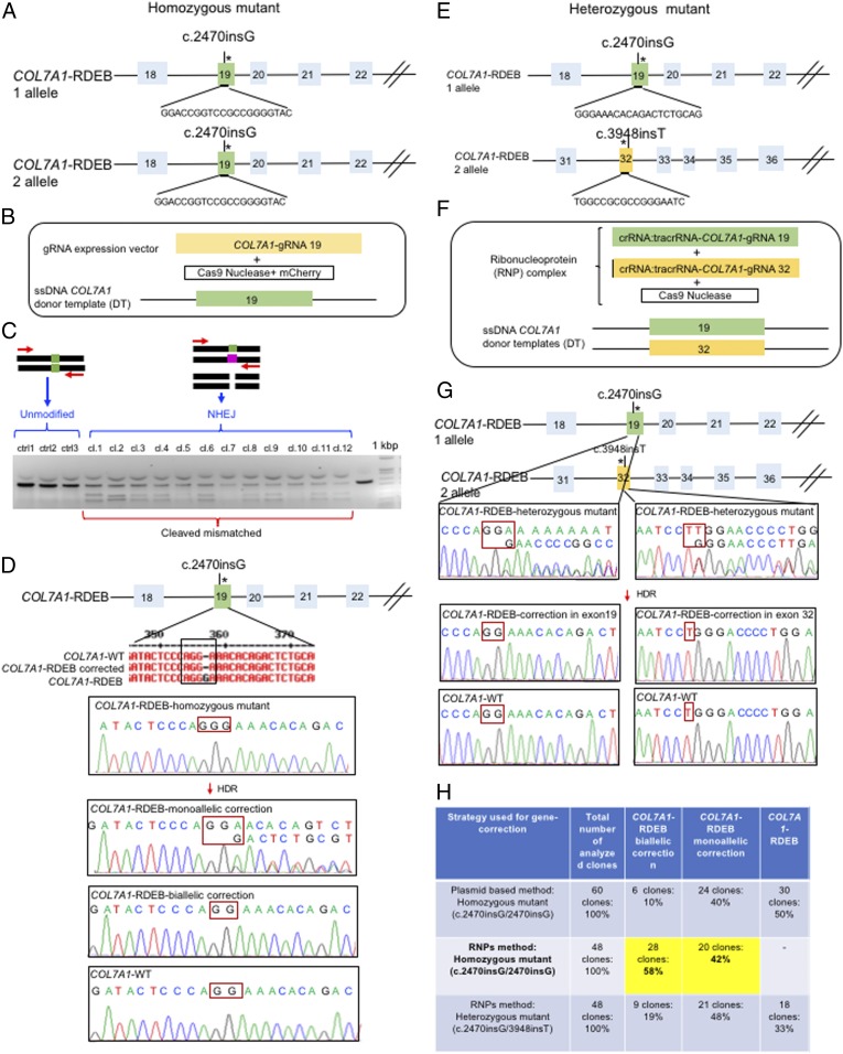
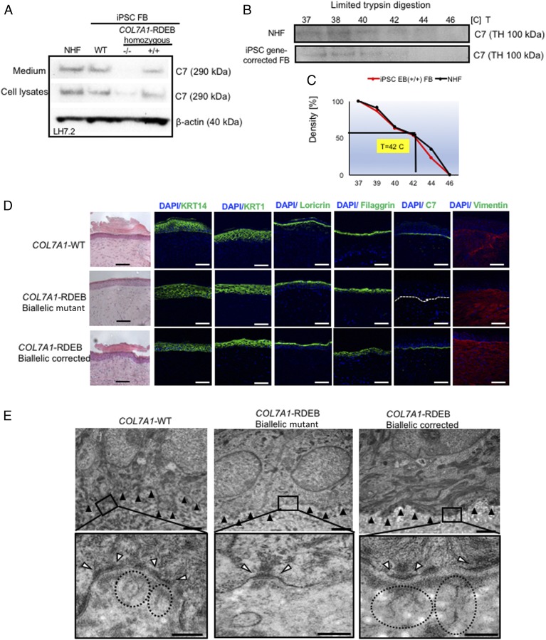
Recessive dystrophic epidermolysis bullosa (RDEB) is a severe inherited skin disorder caused by mutations in the COL7A1 gene encoding type VII collagen (C7). The spectrum of severity depends on the type of mutation in the COL7A1 gene. C7 is the major constituent of anchoring fibrils (AFs) at the basement membrane zone (BMZ). Patients with RDEB lack functional C7 and have severely impaired dermal-epidermal stability, resulting in extensive blistering and open wounds on the skin that greatly affect the patient's quality of life. There are currently no therapies approved for the treatment of RDEB. Here, we demonstrated the correction of mutations in exon 19 (c.2470insG) and exon 32 (c.3948insT) in the COL7A1 gene through homology-directed repair (HDR). We used the clustered regulatory interspaced short palindromic repeats (CRISPR) Cas9-gRNAs system to modify induced pluripotent stem cells (iPSCs) derived from patients with RDEB in both the heterozygous and homozygous states. Three-dimensional human skin equivalents (HSEs) were generated from gene-corrected iPSCs, differentiated into keratinocytes (KCs) and fibroblasts (FBs), and grafted onto immunodeficient mice, which showed normal expression of C7 at the BMZ as well as restored AFs 2 mo postgrafting. Safety assessment for potential off-target Cas9 cleavage activity did not reveal any unintended nuclease activity. Our findings represent a crucial advance for clinical applications of innovative autologous stem cell-based therapies for RDEB.

Keywords: CRISPR/Cas9 gene editing; iPSCs; recessive dystrophic epidermolysis bullosa; type VII collagen.


Conflict of interest statement

The authors declare no competing interest.

Figures

Fig. 1.
Fig. 1.
Evaluation of CRISPR/Cas9 gene-correction efficiency using plasmid- and protein-based methods in iPS cells. (A) Schematic representation of the CRISPR target site for the homozygous (c.2470insG) mutation in exon 19 of COL7A. (B) Components used for plasmid-based gene-correction strategy. (C) T7E1 assay after gene editing targeting exon 19 of the COL7A1 gene shows that the Cas9-mCherry nuclease had high cleavage efficiency, demonstrated by the cleavage products of genome editing. (D) Sanger sequencing confirmed various genotypes from targeted colonies in COL7A1-RDEB homozygous mutant iPSCs. (E) Schematic representation of CRISPR target site for heterozygous (c.2470insG/c.3948insT) mutations in exon 19 of COL7A. (F) Components used for protein-based gene-correction strategy. (G) Sanger sequencing confirmed various genotypes from targeted colonies in COL7A1-RDEB heterozygous mutant iPSCs. (H) Summary of the detected genotypes and efficacy after CRISPR/Cas9 gene editing.
Fig. 2.
Fig. 2.
Functional verification of gene-corrected RDEB patient iPSC-derived FBs and HSEs. (A) Western blot (WB) analysis assessing type VII collagen (C7) protein expression and secretion in normal human FBs, wild type iPSC-derived FBs (iPSC WT FB), COL7A1-RDEB homozygous mutant (−/−), and corrected (+/+) iPSCs differentiated to FBs. Conditioned medium was collected and directly probed for C7, using polyclonal rabbit LH7.2 antibody (kindly provided by Dr. A. Nystrom). (B) Thermal stability of C7 was analyzed and quantified by (C) limited trypsin digestion of medium from normal human FBs and iPSC gene-corrected RDEB-derived FBs at increasing temperatures, as previously reported. Triple helices (TH) of C7 were analyzed by immunoblot, using polyclonal rabbit NC2 antibody against C7 (kindly provided by Dr. A. Nystrom). (D) Generation of 3D HSEs using gene-corrected RDEB patient iPSC-derived KCs and FBs, which were then grafted onto nude mice are histologically comparable to those generated using iPSC WT KCs/FBs, 2 mo postgrafting. H&E staining revealed normal epidermal and dermal morphology. C7 deposition is demonstrated by immunofluorescence (IF) staining (green signal) 2 mo after grafting, using LH7.2 antibody. Additional IF staining was performed for keratin 14, keratin 10, loricrin, filaggrin, and vimentin on corrected, mutant, and WT xenografts. (E) Transmission electron microscopy was performed on positive iPSC WT KCs/FBs skin grafts, negative COL7A1-RDEB homozygous mutant and COL7A1-RDEB gene-corrected KCs/FBs skin grafts, and gene-corrected RDEB skin grafts, 2 mo postgrafting. (Top) Lower-magnification pictures; black boxes indicate where the higher magnification pictures were taken from. (Scale bar, 1 μm.) (Bottom) High-magnification pictures with an emphasis on the anchoring fibrils (AF), indicated by the dotted circles and white arrowheads point to the hemidesmosomes. (Scale bar, 200 nm.) The BMZ is indicated by black arrowheads.

Comment in

  • 26147

References

    1. Hovnanian A., et al. , Characterization of 18 new mutations in COL7A1 in recessive dystrophic epidermolysis bullosa provides evidence for distinct molecular mechanisms underlying defective anchoring fibril formation. Am. J. Hum. Genet. 61, 599–610 (1997). - PMC - PubMed
    1. Rashidghamat E., McGrath J. A., Novel and emerging therapies in the treatment of recessive dystrophic epidermolysis bullosa. Intractable Rare Dis. Res. 6, 6–20 (2017). - PMC - PubMed
    1. Kim E. J., Kang K. H., Ju J. H., CRISPR-Cas9: A promising tool for gene editing on induced pluripotent stem cells. Korean J. Intern. Med. 32, 42–61 (2017). - PMC - PubMed
    1. Cox D. B., Platt R. J., Zhang F., Therapeutic genome editing: Prospects and challenges. Nat. Med. 21, 121–131 (2015). - PMC - PubMed
    1. Moreno A. M., Mali P., Therapeutic genome engineering via CRISPR-Cas systems. Wiley Interdiscip. Rev. Syst. Biol. Med. 9, e1380 (2017). - PubMed

LinkOut - more resources