. 2020 Jan 17;295(3):717-728.
doi: 10.1074/jbc.RA119.011274. Epub 2019 Dec 8.

The Arabidopsis At GCD3 protein is a glucosylceramidase that preferentially hydrolyzes long-acyl-chain glucosylceramides

Affiliations

The Arabidopsis At GCD3 protein is a glucosylceramidase that preferentially hydrolyzes long-acyl-chain glucosylceramides

Guang-Yi Dai et al. J Biol Chem. .

Abstract

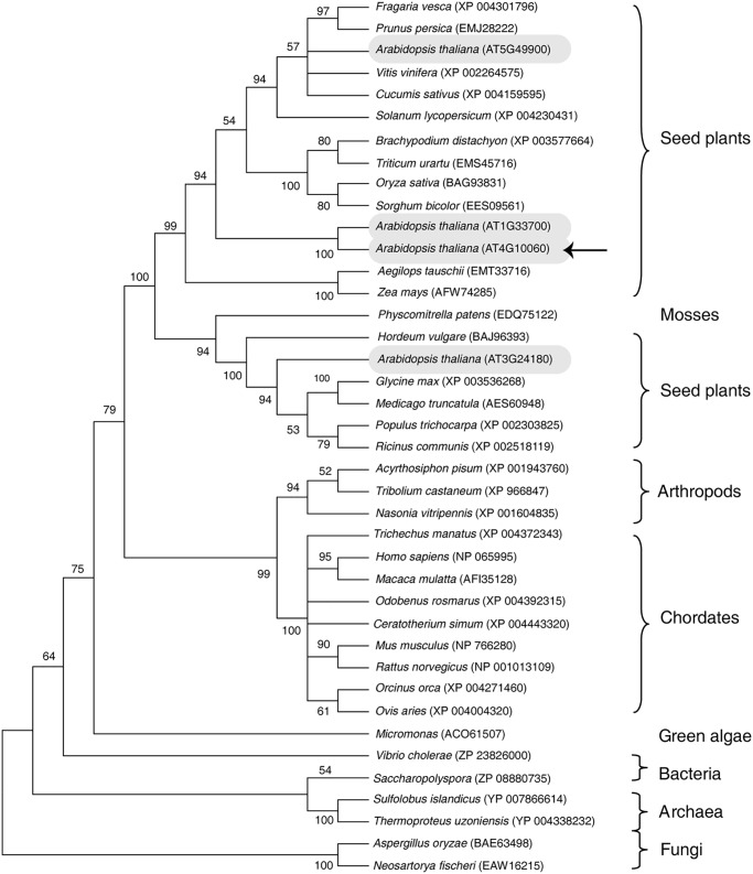
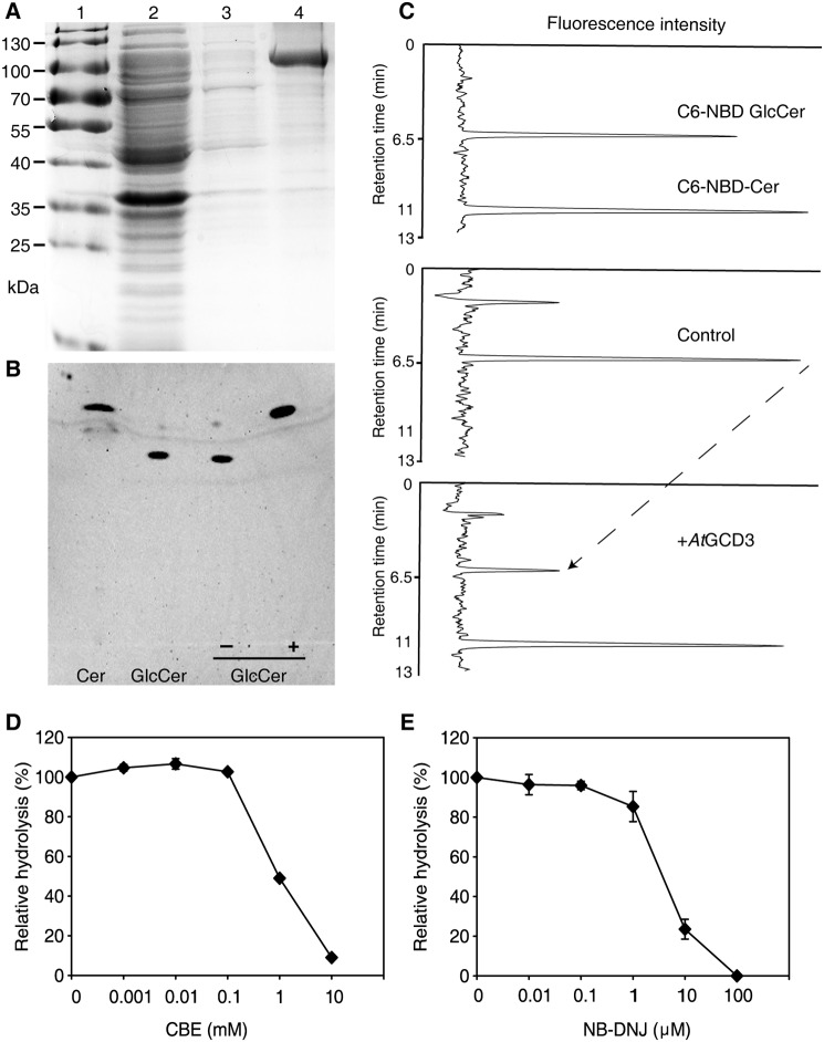
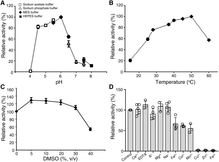
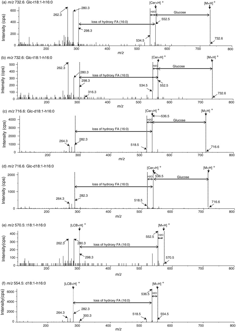
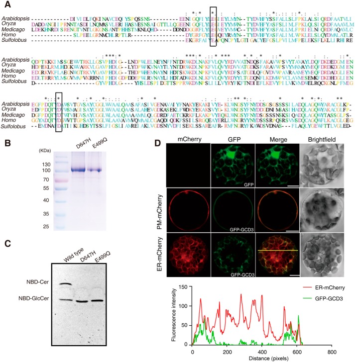
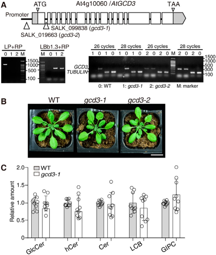
Cellular membranes contain many lipids, some of which, such as sphingolipids, have important structural and signaling functions. The common sphingolipid glucosylceramide (GlcCer) is present in plants, fungi, and animals. As a major plant sphingolipid, GlcCer is involved in the formation of lipid microdomains, and the regulation of GlcCer is key for acclimation to stress. Although the GlcCer biosynthetic pathway has been elucidated, little is known about GlcCer catabolism, and a plant GlcCer-degrading enzyme (glucosylceramidase (GCD)) has yet to be identified. Here, we identified AtGCD3, one of four Arabidopsis thaliana homologs of human nonlysosomal glucosylceramidase, as a plant GCD. We found that recombinant AtGCD3 has a low Km for the fluorescent lipid C6-NBD GlcCer and preferentially hydrolyzes long acyl-chain GlcCer purified from Arabidopsis leaves. Testing of inhibitors of mammalian glucosylceramidases revealed that a specific inhibitor of human β-glucosidase 2, N-butyldeoxynojirimycin, inhibits AtGCD3 more effectively than does a specific inhibitor of human β-glucosidase 1, conduritol β-epoxide. We also found that Glu-499 and Asp-647 in AtGCD3 are vital for GCD activity. GFP-AtGCD3 fusion proteins mainly localized to the plasma membrane or the endoplasmic reticulum membrane. No obvious growth defects or changes in sphingolipid contents were observed in gcd3 mutants. Our results indicate that AtGCD3 is a plant glucosylceramidase that participates in GlcCer catabolism by preferentially hydrolyzing long-acyl-chain GlcCers.

Keywords: Arabidopsis thaliana; cell signaling; ceramide; fatty acid; glucosylceramidase; lipid metabolism; plant biology; sphingolipid; stress adaption.


Conflict of interest statement

The authors declare that they have no conflicts of interest with the contents of this article

Figures

Figure 1.
Figure 1.
Phylogenic analysis of AtGCD3. The phylogenetic tree of GBA2-type proteins was constructed by the maximum likelihood method using MEGA 6. The leaves of the tree are labeled with the organism genus and species and a database accession number. GBA2-type glucosylceramidases occur across Archaea, plants, and chordates. Four glucosylceramidase homologs (AT5G49900, AT1G33700, AT4G10060, and AT3G24180) in A. thaliana are shown in gray ovals. The arrow indicates AtGCD3, which we investigated in this study.
Figure 2.
Figure 2.
Glucosylceramidase activity of purified, recombinant AtGCD3. A, purification of recombinant AtGCD3. Each fraction was subjected to SDS-PAGE and stained with Coomassie Brilliant Blue. Lane 1, protein marker; lane 2, flow-through fraction; lane 3, wash-through fraction; lane 4, purified AtGCD3. B, TLC demonstrating that the fluorescent ceramide was released from C6-NBD GlcCer by AtGCD3. In each reaction, 100 pmol of C6-NBD GlcCer was incubated with (+) or without (−) recombinant AtGCD3 for 60 min and dissolved in MeOH as described under “Experimental procedures.” C, HPLC showing the C6-NBD-Cer released from C6-NBD GlcCer by AtGCD3. Enzyme assays were conducted as described under “Experimental procedures.” The reaction was stopped by adding 4 volumes of acetonitrile, and the retention times of standard C6-NBD GlcCer and C6-NBD Ceramide were 6.5 and 11 min, respectively. Note that when AtGCD3 was added, the peak at 11-min retention time increased, and the peak at 6.5 min (the dashed line) decreased. D and E, inhibitory effects of CBE and NB-DNJ on AtGCD3. Aliquots of 100 pmol of C6-NBD GlcCer were incubated with 1 μg of AtGCD3 and various concentrations of CBE (D) and NB-DNJ (E). After 1 h, samples were loaded onto TLC plates. Fluorescence was detected with a Typhoon Trio+ scanner. Values represent the mean from at least three independent experiments, with S.D. (error bars).
Figure 3.
Figure 3.
General properties of the recombinant AtGCD3. A, pH dependence of glucosylceramidase activity of purified AtGCD3. To vary the pH, 50 mm sodium acetate buffer (pH 4.0–5.5), MES (pH 5.5–6.5), sodium phosphate buffer (pH 6.5–7.5), or HEPES buffer (pH 7.5–8.0) were used. B, optimal temperature. Assay mixtures were incubated at 15–60 °C. C, effects of DMSO on AtGCD3 activity. D, metal ion dependence of AtGCD3 activity. Assay mixtures were incubated with a 1 mm concentration of the indicated metal ions. Data represent the mean ± S.D. (error bars) (n ≥ 3) of independent experiments.
Figure 4.
Figure 4.
Substrate specificity of recombinant AtGCD3. A and B, the recombinant AtGCD3 hydrolyzes C16 long-chain fatty acid GlcCer to C16 long-chain fatty acid hydroxyceramide in naturally occurring lipid substrates extracted from Arabidopsis leaves. Natural glucosylceramides were extracted from Arabidopsis, incubated with (B) or without (A) recombinant AtGCD3 at 30 °C for 24 h, and then subjected to HPLC ESI-MS/MS analysis. Each peak (a, b, c, d, e, and f) was further subjected to MS/MS, as shown in Fig. 5. C and D, quantitative analysis of hydrolysis using Arabidopsis leaf lipids. Naturally occurring glucosylceramides (C) were hydrolyzed to hydroxyceramides (D). The inset in D indicates that Glc-d18:0-h16:0 was hydrolyzed to d18:0-h16:0. E and F, the recombinant AtGCD3 preferentially hydrolyzes long-acyl-chain GlcCer. 500 nm commercially available GlcCers with different fatty acid chain length were incubated with (+) or without (−) AtGCD3 at 30 °C for 13 h, respectively, and analyzed by HPLC ESI-MS/MS. Commercial glucosylceramides (E) were hydrolyzed to ceramides (F). G, the recombinant AtGCD3 has no activity on galactosylceramide and MGDG. 500 nm Gal-d18:1–16:0 or MGDG was incubated with (+) or without (−) AtGCD3 at 30 °C for 13 h and analyzed by HPLC ESI-MS/MS. Data represent the mean ± S.D. (error bars) (n = 3) of independent experiments. The statistical significance of differences was determined with (+) or without (−) AtGCD3 in each GlcCer by Student's t test; p values are indicated where p < 0.05.
Figure 5.
Figure 5.
Mass spectra of glucosylceramides and the product hydroxyceramides. a–d, mass spectra of purified natural GlcCer containing Glc-t18:1-h16:0 (a and b: m/z 732.6) and Glc-d18:1-h16:0 (c and d: m/z 716.6) with or without recombinant AtGCD3, as shown in Fig. 4. e and f, MS/MS spectra showing that C16 hydroxyceramide was generated through hydrolysis of relevant GlcCers with recombinant AtGCD3. t18:1-h16:0 (e, m/z 570.5) and d18:1-h16:0 (f, m/z 554.5) were hydrolyzed products of Glc-t18:1-h16:0 (b) and Glc-d18:1-h16:0 (d), respectively. The two-way arrows indicate neutral loss of sphingolipids, and one-way arrows show the product ion.
Figure 6.
Figure 6.
Alignment of the AtGCD3 amino acid sequence with nonlysosomal glucosylceramidase and analysis of catalytic residues of AtGCD3. A, multiprotein alignment of the AtGCD3 amino acid sequence with other glucosylceramidases. Invariant residues are indicated with an asterisk; increased levels of conservation are indicated with a colon and a dot. The residues corresponding to the nucleophile Glu-499 and acid/base Asp-647 of AtGCD3 are boxed. Arabidopsis, gi|332657434 (AT4g10060) from A. thaliana; Oryza, gi|215704397 from Oryza sativa; Medicago, gi|355479745 from Medicago truncatula; Homo, NP_065995.1 from Homo sapiens; Sulfolobus, NC_002754.1 from S. solfataricus P2. B, SDS-PAGE of the purified E499Q and D647H mutant proteins. The two mutant enzymes were produced by site-directed mutagenesis, as shown in A (boxes), and were expressed and purified as described under “Experimental procedures.” C, TLC showing the hydrolysis of NBD-GlcCer by the WT and mutant AtGCD3. Note that AtGCD3 mutant proteins E499Q and D647H were completely inactive. D, subcellular localization of GFP-AtGCD3 fusion protein. GFP-AtGCD3 was co-transformed with organelle markers (mCherry) in WT Arabidopsis protoplasts. After incubation at 23 °C for 16 h under the light, fluorescence was observed by laser confocal microscopy. Row 3 panels are shown in three-dimensional maximum projection (eight sections at 0.84-μm step size), and others are single-plane. Free GFP was used as control (row 1 panels). The graph below shows the overlap of fluorescence intensity peaks along profiles as indicated in the merged micrograph. Bars, 10 μm.
Figure 7.
Figure 7.
Characterization of T-DNA insertion mutants of AtGCD3. A, schematic diagram of the GCD3 locus. Gray boxes, UTR; white boxes, exons. Thin lines between exons represent introns. T-DNA was inserted in the promoter region of AtGCD3 in SALK_019663 (gcd3-2) and was inserted in the first intron of AtGCD3 in SALK_099838 (gcd3-1). The triangles indicate the insertion sites. The T-DNA insertion mutants gcd3-1 and gcd3-2 were homozygotes, as verified by two-round PCR. Primer LBb1.3 and RP indicated the existence of the T-DNA insertion, and primer LP and RP showed that both are homozygotes. RT-PCR confirmed that gcd3-1 was a genuine null mutant. TUBULIN was used as an internal control. B, normal phenotype of a 32-day-old gcd3-1 plant. Bar, 2 cm. C, sphingolipid profiles of 3-week-old rosette leaves of WT and gcd3-1 plants. The contents of sphingolipid in WT and gcd3-1 plants were quantified after extraction, separation, and identification by HPLC ESI-MS/MS as described under “Experimental procedures.” All data are available in Table S1. The relative values indicate the level of sphingolipids in gcd3-1 relative to that found in WT (set as 1). The values represent the mean ± S.D. (error bars) (n = 9 independent experiments). Significance was determined using Student's t test.

Similar articles

Cited by

References

    1. Markham J. E., Lynch D. V., Napier J. A., Dunn T. M., and Cahoon E. B. (2013) Plant sphingolipids: function follows form. Curr. Opin Plant Biol. 16, 350–357 10.1016/j.pbi.2013.02.009 - DOI - PubMed
    1. Pata M. O., Hannun Y. A., and Ng C. K. Y. (2010) Plant sphingolipids: decoding the enigma of the Sphinx. New Phytol. 185, 611–630 10.1111/j.1469-8137.2009.03123.x - DOI - PMC - PubMed
    1. Bi F.-C., Liu Z., Wu J.-X., Liang H., Xi X.-L., Fang C., Sun T.-J., Yin J., Dai G.-Y., Rong C., Greenberg J. T., Su W.-W., and Yao N. (2014) Loss of ceramide kinase in Arabidopsis impairs defenses and promotes ceramide accumulation and mitochondrial H2O2 bursts. Plant Cell 26, 3449–3467 10.1105/tpc.114.127050 - DOI - PMC - PubMed
    1. Yoshida S., and Uemura M. (1986) Lipid composition of plasma membranes and tonoplasts isolated from etiolated seedlings of mung bean (Vigna radiata L.). Plant Physiol. 82, 807–812 10.1104/pp.82.3.807 - DOI - PMC - PubMed
    1. Sperling P., Franke S., Lüthje S., and Heinz E. (2005) Are glucocerebrosides the predominant sphingolipids in plant plasma membranes? Plant Physiol. Biochem. 43, 1031–1038 10.1016/j.plaphy.2005.10.004 - DOI - PubMed

Publication types

MeSH terms