. 2020 Feb 14;295(7):1781-1791.
doi: 10.1074/jbc.RA119.011612. Epub 2019 Dec 9.

The human protein haptoglobin inhibits IsdH-mediated heme-sequestering by Staphylococcus aureus

Affiliations

The human protein haptoglobin inhibits IsdH-mediated heme-sequestering by Staphylococcus aureus

Jakob H Mikkelsen et al. J Biol Chem. .

Abstract

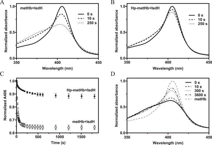
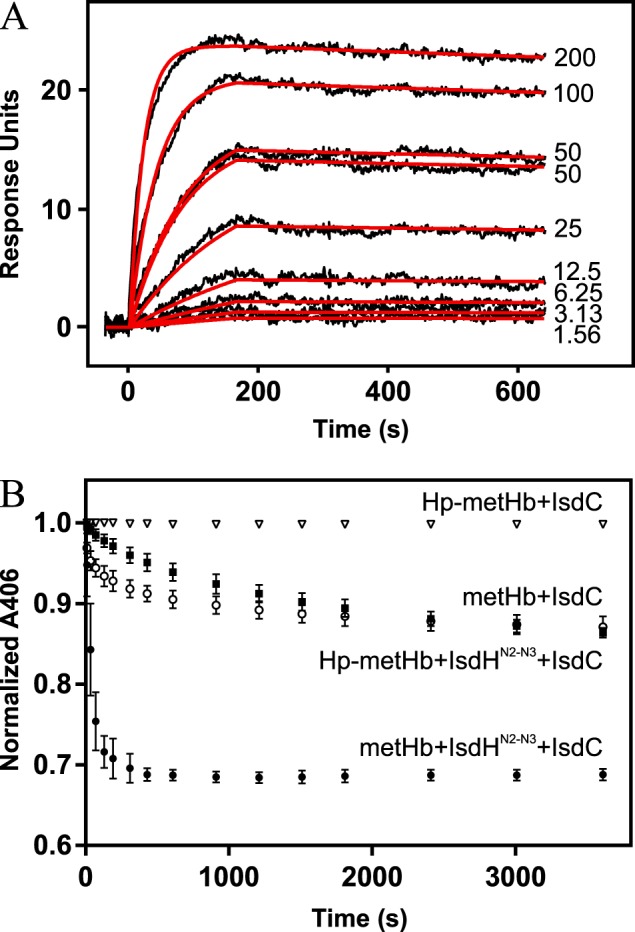
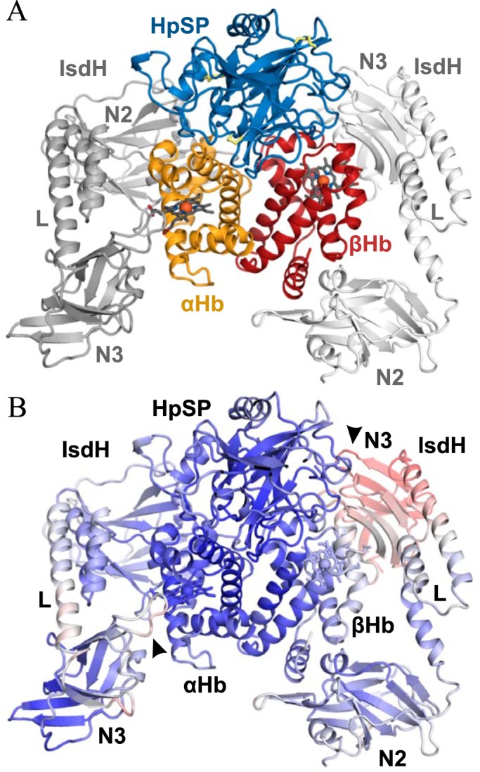
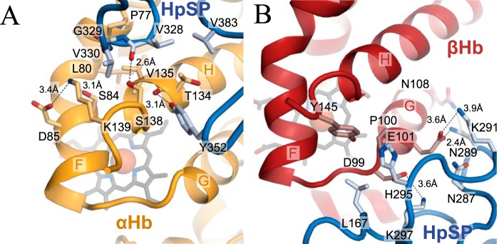
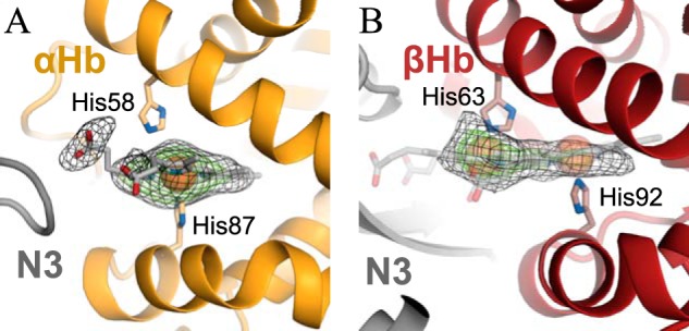
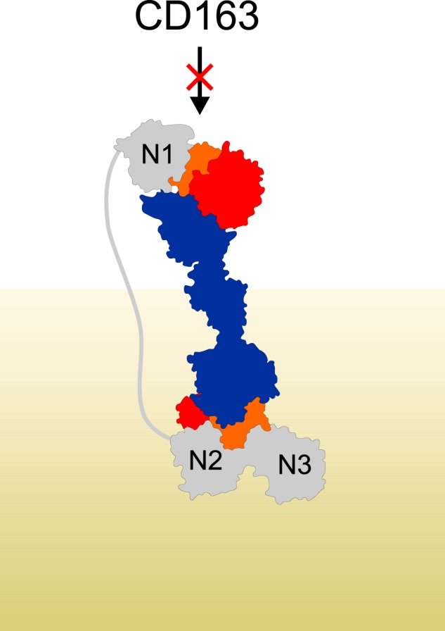
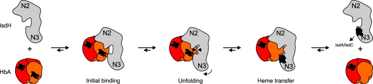
Iron is an essential nutrient for all living organisms. To acquire iron, many pathogens have developed elaborate systems to steal it from their hosts. The iron acquisition system in the opportunistic pathogen Staphylococcus aureus comprises nine proteins, called iron-regulated surface determinants (Isds). The Isd components enable S. aureus to extract heme from hemoglobin (Hb), transport it into the bacterial cytoplasm, and ultimately release iron from the porphyrin ring. IsdB and IsdH act as hemoglobin receptors and are known to actively extract heme from extracellular Hb. To limit microbial pathogenicity during infection, host organisms attempt to restrict the availability of nutrient metals at the host-pathogen interface. The human acute phase protein haptoglobin (Hp) protects the host from oxidative damage by clearing hemoglobin that has leaked from red blood cells and also restricts the availability of extracellular Hb-bound iron to invading pathogens. To investigate whether Hp serves an additional role in nutritional immunity through a direct inhibition of IsdH-mediated iron acquisition, here we measured heme extraction from the Hp-Hb complex by UV-visible spectroscopy and determined the crystal structure of the Hp-Hb-IsdH complex at 2.9 Å resolution. We found that Hp strongly inhibits IsdH-mediated heme extraction and that Hp binding prevents local unfolding of the Hb heme pocket, leaving IsdH unable to wrest the heme from Hb. Furthermore, we noted that the Hp-Hb binding appears to trap IsdH in an initial state before heme transfer. Our findings provide insights into Hp-mediated IsdH inhibition and the dynamics of IsdH-mediated heme extraction.

Keywords: Haptoglobin; Hemoglobin; IsdH; Staphylococcus aureus; Staphylococcus aureus (S. aureus); hemoglobin; infection; inhibitor; iron.


Conflict of interest statement

The authors declare that they have no conflicts of interest with the contents of this article.

Figures

Figure 1.
Figure 1.
Heme transfer from metHb and Hp–metHb to IsdHN2-N3. A and B, spectral changes over time for metHb+IsdHN2-N3 (A) and Hp–metHb+IsdHN2-N3 (B). C, absorbances at 406 nm for metHb + IsdHN2-N3 and Hp–metHb + IsdhN2-N3 followed for 30 min. Normalized absorbances at 406 nm are represented as mean values ± S.D. (n = 3). D, spectral changes over time for metHb preincubated with IsdHN2-N3. Hp was added at time point 0. After 1 h, no further spectral change was observed indicating that the system reached equilibrium.
Figure 2.
Figure 2.
Hp–Hb binding kinetics and spontaneous release of heme from Hp–metHb and metHb. A, surface plasmon resonance determination of Hp1-1 affinity for Hb. Sensogram data are shown as black curves. Concentrations (in nm) of the injected Hb are shown next to the curves. A 1:1 Langmuir model was fitted to the sensogram (red curves). B, absorbances at 406 nm followed for 60 min. Normalized absorbances at 406 nm are represented as mean values ± S.D. (n = 3).
Figure 3.
Figure 3.
Structure of the HpSP–αβHb–IsdHN2-N3 complex. A, Cartoon representation of the 2.9 Å resolution crystal structure of HpSP–αβHb–IsdHN2-N3 complex. Hp is shown in blue, αHb is in orange, βHb is in red, and IsdHN2-N3 is light and dark gray. N2 and N3 denotes IsdHN2 and IsdHN3, respectively. L denotes the IsdHlinker domain. Heme groups are represented by dark gray sticks, and iron atoms are represented by red spheres. Disulfides are shown as yellow sticks. B, B-factors of the determined structure of Hp–Hb–IsdHN2-N3 are plotted on a cartoon representation of the structure with blue representing low B-factor, white representing intermediate, and red representing high. The interfaces αHb–IsdHN3 and βHb-IsdhN3 are marked with arrows.
Figure 4.
Figure 4.
Interactions between the IsdHlinker and Hb in Hb–IsdHN2-N3versus HpSP–αβHb–IsdHN2-N3. A, structural comparison of IsdHN2-N3 bound to αHb in Hb–IsdHN2-N3 (light color scheme) (PDB entry 4XS0) (9) and HpSP–αβHb–IsdHN2-N3 (dark color scheme). The structures are aligned on IsdHN2. DynDom analysis reveal that the two structures are related by a 5° rotation of the IsdHlinker and IsdHN3. The axis of rotation is marked by a black rod. Residues that are rotated as a rigid body are shown in light blue, residues in a fixed position are shown in red, and flexible residues are shown in green. B, interface between the αHb F-helix and IsdHlinker in the structure of Hb–IsdHN2-N3 (PDB entry 4XS0) (9). C, interface between the αHb F-helix and IsdHlinker in the structure of HpSP–αβHb–IsdHN2-N3. D, structural comparison of αHb–IsdHN2-N3 and βHb–IsdHN2-N3 in the structure of HpSP–αβHb–IsdHN2-N3. The structures are aligned on IsdHN2. The globin chains of αHb and βHb are structurally distinct, which is emphasized by differences in the position of the F-helix. In βHb, the position of the F-helix is closer to the IsdHlinker. E, interactions between βHb and IsdHlinker. In contrast to αHb, we observe direct interactions between IsdHlinker and the βHb F-helix. Hydrogen bond and salt bridges are shown as dashed black lines.
Figure 5.
Figure 5.
HpSP interactions with the αHb and βHb heme-binding pockets. A and B, selected interactions between HpSP and the regions surrounding the heme-binding pockets of αHb (A) and βHb (B) in the structure of HpSP–αβHb-IsdhN2-N3. Hydrogen bond and salt bridges are shown as dashed black lines.
Figure 6.
Figure 6.
Heme-binding sites in HpSP–αβHb–IsdHN2-N3. A and B, Electron density maps (mFo − DFc, heme omit maps contoured at 2.5 σ (black mesh) and 5.0 σ (green mesh)) of the heme-groups bound to αHb (A) and βHb (B). Well-defined electron density for the heme-group is observed at αHb at the canonical site with the heme iron coordinated by the proximal histidine (His-87). At βHb, the electron density is not well-defined and indicates that the heme group can be in two alternative positions, either at the canonical site or in a novel position with the heme iron coordinated by the distal histidine (His-63).
Figure 7.
Figure 7.
IsdHN1 blocks interaction between Hp–Hb and CD163. Shown is a model of IsdHN1-N3 binding to Hp–Hb. The model was generated based on the structures of HpSP–αβHb–IsdHN2-N3 and Hp–Hb–IsdHN1 (PDB entry 4WJG) (36). The distance between IsdHN1 C terminus and IsdHN2 N terminus is ∼180 Å. IsdH is shown in gray, Hp is in blue, αHb is in orange, and βHb is in red. The peptidoglycan layer on the surface of the bacterium is indicated in khaki.
Figure 8.
Figure 8.
Suggested mechanism of IsdH mediated heme extraction. The structure of HpSP–αβHb–IsdHN2-N3 possibly represents a novel conformational state of IsdHN2-N3 after Hb binding (initial binding) but prior to Hb unfolding and heme transfer. The proposed mechanism is obtained by combining our results with the previous structural analysis of Hb–IsdHN2-N3 (37).

References

    1. Nairz M., Schroll A., Sonnweber T., and Weiss G. (2010) The struggle for iron: a metal at the host–pathogen interface. Cell. Microbiol. 12, 1691–1702 10.1111/j.1462-5822.2010.01529.x - DOI - PubMed
    1. Conroy B. S., Grigg J. C., Kolesnikov M., Morales L. D., and Murphy M. E. P. (2019) Staphylococcus aureus heme and siderophore-iron acquisition pathways. Biometals 32, 409–424 10.1007/s10534-019-00188-2 - DOI - PubMed
    1. Maresso A. W., and Schneewind O. (2006) Iron acquisition and transport in Staphylococcus aureus. Biometals 19, 193–203 10.1007/s10534-005-4863-7 - DOI - PubMed
    1. Torres V. J., Attia A. S., Mason W. J., Hood M. I., Corbin B. D., Beasley F. C., Anderson K. L., Stauff D. L., McDonald W. H., Zimmerman L. J., Friedman D. B., Heinrichs D. E., Dunman P. M., and Skaar E. P. (2010) Staphylococcus aureus fur regulates the expression of virulence factors that contribute to the pathogenesis of pneumonia. Infect Immun. 78, 1618–1628 10.1128/IAI.01423-09 - DOI - PMC - PubMed
    1. Torres V. J., Pishchany G., Humayun M., Schneewind O., and Skaar E. P. (2006) Staphylococcus aureus IsdB is a hemoglobin receptor required for heme iron utilization. J. Bacteriol. 188, 8421–8429 10.1128/JB.01335-06 - DOI - PMC - PubMed

MeSH terms