. 2019 Dec 9;10(12):934.
doi: 10.1038/s41419-019-2155-3.

Hypoxia regulates the mitochondrial activity of hepatocellular carcinoma cells through HIF/HEY1/PINK1 pathway

Affiliations

Hypoxia regulates the mitochondrial activity of hepatocellular carcinoma cells through HIF/HEY1/PINK1 pathway

David Kung-Chun Chiu et al. Cell Death Dis. .

Abstract

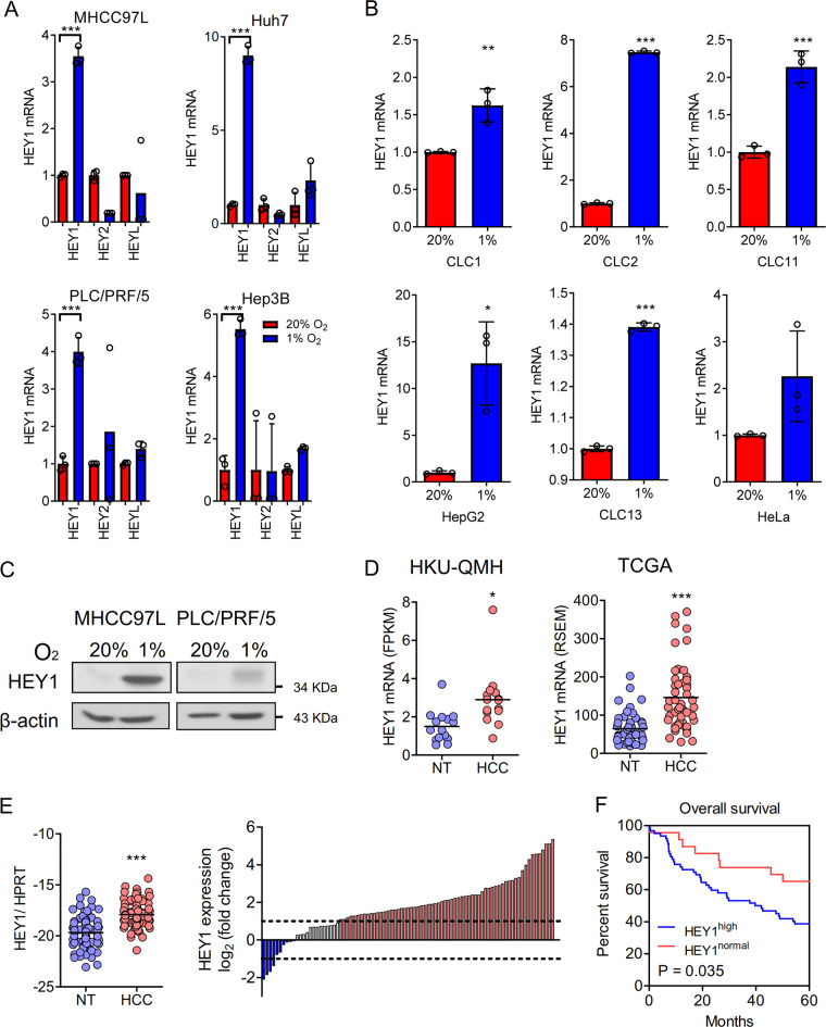
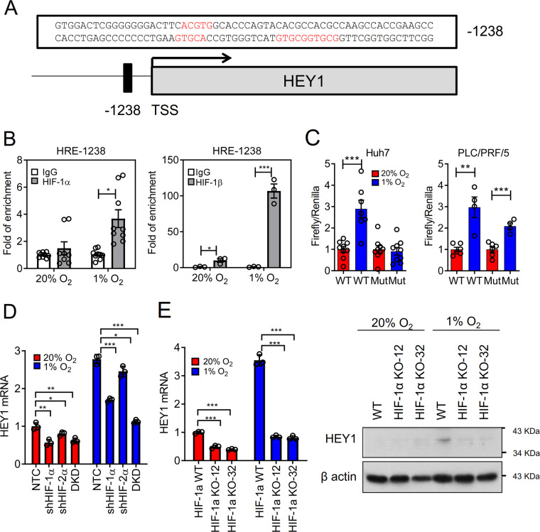
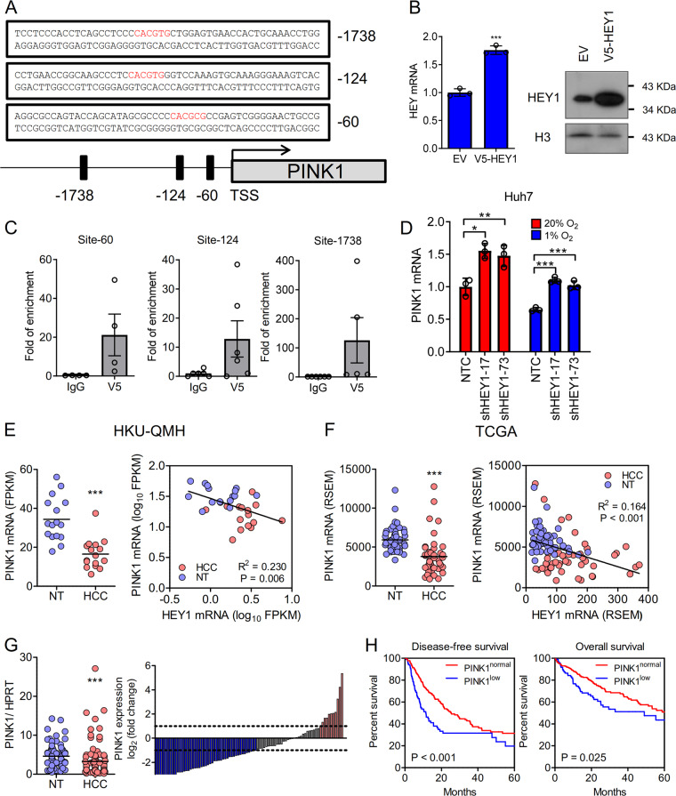
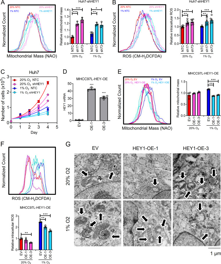
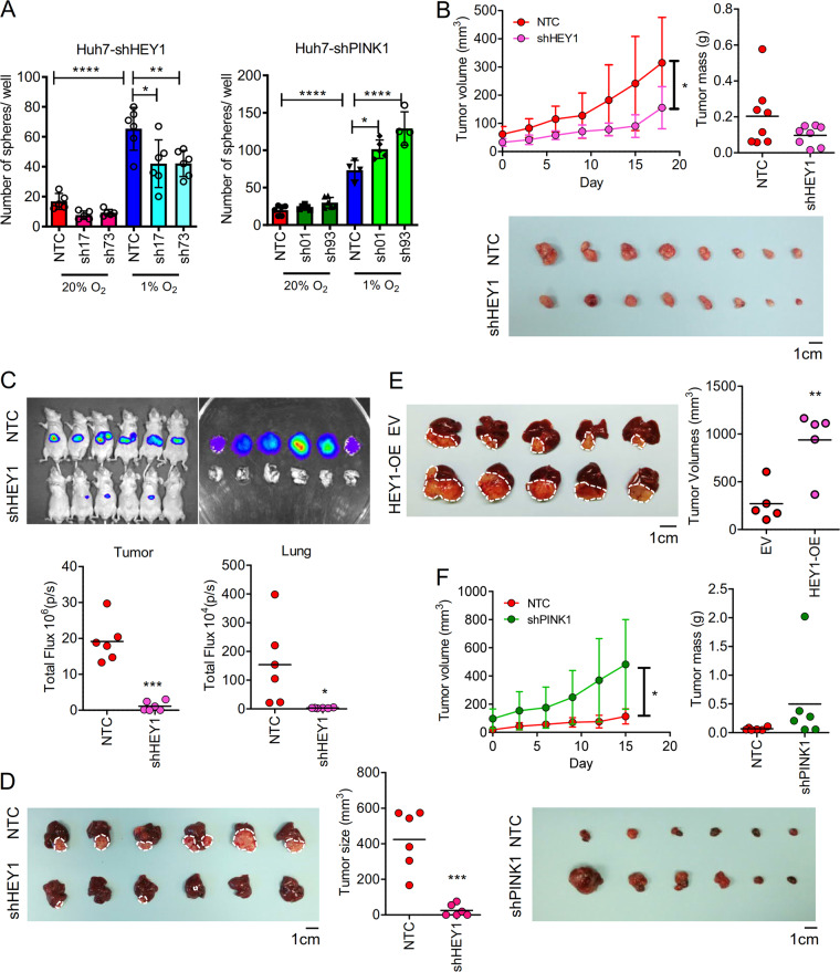
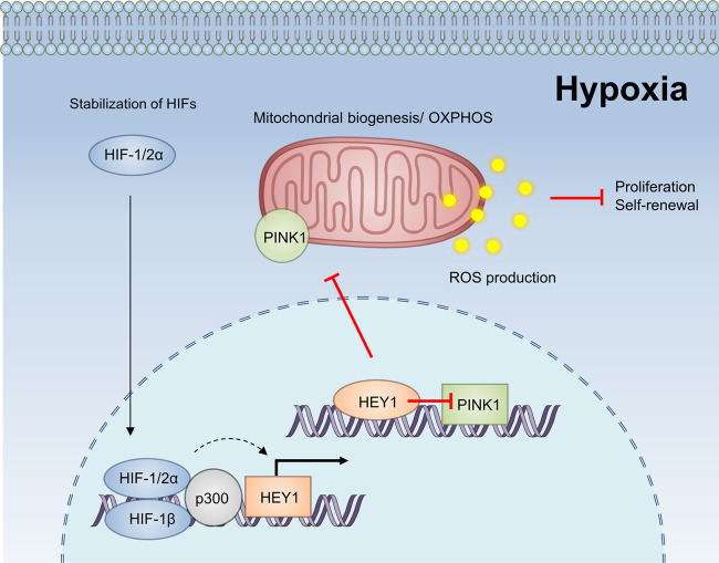
Hypoxia is commonly found in cancers. Hypoxia, due to the lack of oxygen (O2) as the electron recipient, causes inefficient electron transfer through the electron transport chain at the mitochondria leading to accumulation of reactive oxygen species (ROS) which could create irreversible cellular damages. Through hypoxia-inducible factor 1 (HIF-1) which elicits various molecular events, cells are able to overcome low O2. Knowledge about the new molecular mechanisms governed by HIF-1 is important for new therapeutic interventions targeting hypoxic tumors. Using hepatocellular carcinoma (HCC) as a model, we revealed that the HIF-1 and the Notch signaling pathways cross-talk to control mitochondrial biogenesis of cancer cells to maintain REDOX balance. From transcriptome sequencing, we found that HEY1, a transcriptional repressor, in the NOTCH pathway was consistently induced by hypoxia in HCC cell lines. We identified a strong hypoxia response element (HRE) in HEY1 by chromatin immunoprecipitation (ChIP) and luciferase reporter assays. Transcriptome and ChIP sequencing further identified PINK1, a gene essential for mitochondrial biogenesis, as a novel transcriptional target of HEY1. HCC cells with HEY1 knockdown re-expressed PINK1. HEY1 and PINK1 expressions inversely correlated in human HCC samples. Overexpression of HEY1 and under-expression of PINK1 were detected in human HCC and associated with poor clinical outcomes. Functionally, we found that overexpression of HEY1 or knockdown of PINK1 consistently reduced mitochondrial cristae, mitochondrial mass, oxidative stress level, and increased HCC growth.


Conflict of interest statement

The authors declare that they have no conflict of interest.

Figures

Fig. 1
Fig. 1. Expressions of HEY family members in human HCC cell lines and clinical samples.
a RT-qPCR of HEY1, HEY2, HEYL mRNA expression levels in 4 human HCC cell lines (MHCC97L, Huh7, PLC/PRF/5, Hep3B) exposed to 20 and 1% O2 for 24 h. b RT-qPCR confirmed HEY1 mRNA expressions in 5 human HCC cell lines (CLC1, CLC2, CLC11, HepG2, CLC13) and 1 human cervical cancer cell line (HeLa). Data were normalized to the corresponding values in 20% O2 and house keeping gene, 18 S. c HEY1 protein expression in MHCC97L and PLC/PRF/5 exposed to 20 and 1% O2 for 48 h. d HEY1 mRNA expression in (middle) in-house 16 pairs and (right) 49 pairs from TCGA database of human HCC tissues and corresponding non-tumorous liver tissues (NT) was detected by transcriptome sequencing. e Left: HEY1 mRNA expression was determined by qRT-PCR in 87 cases of in-house (HKU-QMH) human HCC and NT tissues. Right: Waterfall plot shows that HEY1 was over-expressed in 72.41% (63/87) of HCC patients by at least 2 fold. f HEY1 overexpression ( ≥ 2 fold) was closely associated with poor overall survival in HCC patients. Data are presented as mean ± s.d. (Student’s t-test, *P < 0.05, **P < 0.01, ***P < 0.001).
Fig. 2
Fig. 2. HEY1 is a transcriptional target of HIF-1.
a 4 copies of HREs (red) were found in 1238 bp promixal to transcription start site (TSS) of HEY1. b ChIP assay was performed with HIF-1α, HIF-1β, and IgG control antibodies in MHCC97L cells exposed to 20 and 1% O2 for 24 h. c Huh7 and PLC/PRF/5 cells were transfected with p2.1 Firefly luciferase reporter encompassing wildtype (WT) or mutated (Mut) HEY1 HREs and Renilla luciferase control plasmids. Transfected cells were exposed to 20 and 1% O2 for 24 h for dual luciferase activities quantification. d HEY1 mRNA expression in MHCC97L-shNTC, -shHIF-1α, -shHIF-2α, and HIF-1α and HIF-2α double knockdown (DKD) subclones exposed to 20 and 1% O2 for 24 h. e HEY1 mRNA (left) and protein (right) expressions in MHCC97L-WT and -HIF-1αKO subclones (established by TALEN) exposed to 20 and 1% O2 for 24 (mRNA) and 48 (protein) hours. For qRT-PCR, values were normalized to house keeping gene, 18 S. Data are presented as mean ± s.d. (Student’s t-test, *P < 0.05, **P < 0.01, ***P < 0.001).
Fig. 3
Fig. 3. HEY1 represses PINK1.
a 3 putative E boxes (red) were located upstream to the TSS of PINK1. b, c ChIP assay was performed in Hey1 over-expressing HCC cells (Huh7-V5-HEY1) with V5 and IgG control antibodies. d PINK1 mRNA expression in Huh7-NTC, -shHEY1-17, shHEY-73 cells that were exposed to 20 and 1% O2 for 24 and 48 h, respectively. e Left: transcriptome sequencing revealed that PINK1 was down-regulated in human HCC. Right: PINK1 and HEY1 expressions were inversely correlated in our in-house 16 cases of HCC and NT tissues. f Left: PINK1 mRNA expression in HCC and NT tissues in 49 HCC patients from the TCGA database. Right: PINK1 and HEY1 expressions were inversely correlated in HCC and NT tissues from TCGA data base. g Left: RT-qPCR confirmed that PINK1 was down-regulated in an expanded cohort of 87 HCC patients. Waterfall plot shows that PINK1 was under-expressed in 47/87 (54.02%) HCC patients by at least 1 fold. h PINK1 was defined as down-regulated with Z-score < -1 otherwise normal. PINK1 down-regulation was associated with shorter disease-free and overall survivals in HCC patients. RSEM: RNA-Seq by Expectation-Maximization. For qRT-PCR, values were normalized to house keeping gene, 18 S or HPRT. Data are presented as mean ± s.d. (Student’s t-test, *P < 0.05, **P < 0.01, ***P < 0.001).
Fig. 4
Fig. 4. PINK1 increases mitochondrial mass and ROS and inhibits HCC proliferation.
a Mitochondrial mass, b ROS in Huh7-NTC and shPINK1 subclones exposed to 20 and 1% O2 for 24 h. c Huh7-NTC, -shPINK1-01, shPINK1-93 cells were exposed to 20 and 1% O2 for 24 h. Cell images under transmission electron microscope (TEM). d Proliferation of Huh7-NTC and shPINK1 subclones exposed to 20 and 1% O2 for 4 days. Data are presented as mean ± s.d. (Student’s t-test, *P < 0.05, **P < 0.01, ***P < 0.001).
Fig. 5
Fig. 5. HEY1 decreases mitochondrial mass and ROS and promotes HCC proliferation.
a Mitochondrial mass b ROS in Huh7-NTC and shHEY1 subclones exposed to 20 and 1% O2 for 24 h. c Proliferation of Huh7-NTC and shHEY1 subclones exposed to 20 and 1% O2 for 4 days. d HEY1 mRNA expression in MHCC97L-HEY1-OE cells established by CRISPR-dCas9-SAM system. e Mitochondrial mass and f ROS in MHCC97L-EV and –HEY1-OE subclones exposed to 20 and 1% O2 for 24 h. g Cell images of HEY1-OE cells under TEM. Arrows point to mitochondria. Data are presented as mean ± s.d. (Student’s t-test, *P < 0.05, **P < 0.01, ***P < 0.001).
Fig. 6
Fig. 6. HEY1 promotes whereas PINK1 represses HCC growth in vivo.
a Sphere formation assay. The number of spheres derived from Huh7-NTC and –shHEY1/PINK1 cells exposed to 20 and 1% O2 were counted. b In total 1 × 106 Huh7-NTC and -shHEY1-17 cells were injected into BALB/c nude mice subcutaneously. Tumor volume was calculated based on the 3 dimensions of the tumors measured by caliper and tumor mass was measured when mice were sacrificed at day 18. In total 1 × 106 luciferase-labelled MHCC97L-NTC and -shHEY1-17 cells were injected into the left lobes of the livers of BALB/c nude mice. (n = 8 for each group). c Bioluminescence of (left) tumors and (right) lung metastases in mice after 6 weeks of orthotopic implantation. d Livers were harvested 6 weeks post-injection and tumor size was measured. (n = 6 for each group). e 2 × 106 MHCC97L-EV and -HEY1-OE cells were injected into BALB/c nude mice orthotopically. Livers were harvested 6 weeks post-injection and tumor size was measured. (n = 6 for each group). f 1 × 106 Huh7-NTC and –shPINK1 cells were injected into BALB/c nude mice subcutaneously. Tumor volume was calculated based on the 3 dimensions of the tumors measured by caliper and tumor mass was measured when mice were killed at day 15. (n = 6 for each group) Data are presented as mean ± s.d. (one-way ANOVA, Turkey’s multiple comparison test in A for comparison of multiple groups, Student’s t-test in b, c, e, f for comparison between two groups, *P < 0.05, **P < 0.01, ***P < 0.001, ***P < 0.0001).
Fig. 7
Fig. 7. Summary.
HIF-1 transcriptionally activates HEY1. HEY1 in turn transcriptionally represses PINK1. PINK1 is important to mitochondrial biogenesis. During hypoxia, ROS accumulates at the mitochondria due to inefficient electron transfer through the electron transport chain in the mitochondria, HIF-1 overcomes the oxidative stress through elevating HEY1 to reduce PINK1-mediated mitochondrial biogenesis, making HCC cells less dependent on mitochondria to reduce mitochondrial ROS production. Therefore, the HIF-1/HEY1 pathway confers survival advantages to HCC cells which frequently experience hypoxia.

References

    1. Cheng, A. L. et al. Efficacy and safety of sorafenib in patients in the Asia-Pacific region with advanced hepatocellular carcinoma: a phase III randomised, double-blind, placebo-controlled trial. Lancet Oncol.10, 25–34 (2009). - PubMed
    1. Llovet, J. M. et al. Sorafenib in advanced hepatocellular carcinoma. N. Engl. J. Med.359, 378–390 (2008). - PubMed
    1. Bruix J. Regorafenib shows significant survival gains in refractory liver cancer. European Society for Medical Oncology (EMSO) Congress, World Congress on Gastrointestical Cancer, 2016.
    1. El-Khoueiry A. B., et al. Nivolumab in patients with advanced hepatocellular carcinoma (CheckMate 040): an open-label, non-comparative, phase 1/2 dose escalation and expansion trial. Lancet389, 2492–2502 (2017). - PMC - PubMed
    1. Wong C. C. KAK, Ng I. O. The impact of hypoxia in hepatocellular carcinoma metastasis. Front. Med. 8, 33–41 (2014). - PubMed

MeSH terms

Substances