. 2019 Dec 9;9(1):18600.
doi: 10.1038/s41598-019-55079-w.

Neonatal obstructive nephropathy induces necroptosis and necroinflammation

Affiliations

Neonatal obstructive nephropathy induces necroptosis and necroinflammation

Bastian Popper et al. Sci Rep. .

Abstract

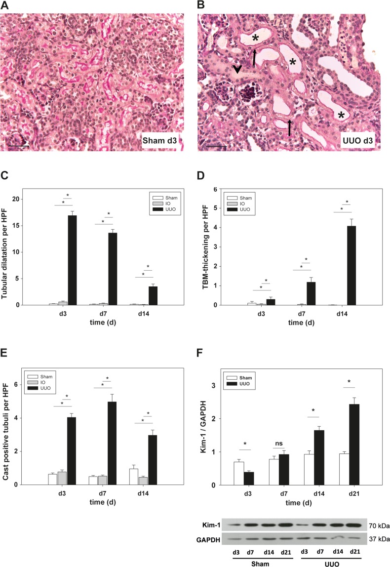
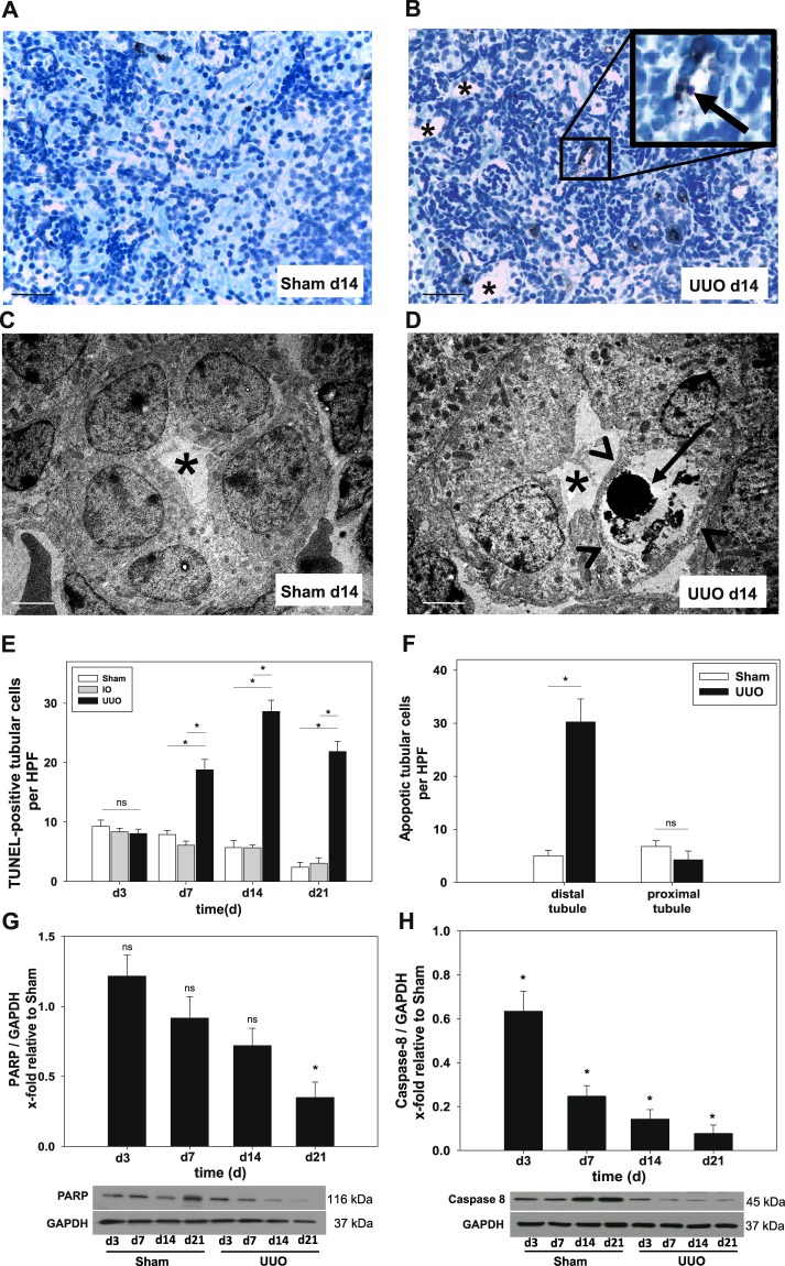
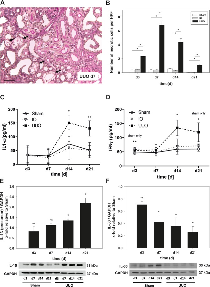
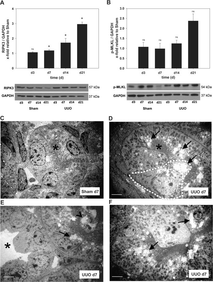
Urinary tract obstruction during kidney development causes tubular apoptosis, tubular necrosis, and interstitial inflammation. Necroptosis is a subtype of programmed necrosis mediated by the receptor-interacting serine/threonine-protein kinase-3 (RIPK3) and the pseudokinase mixed lineage kinase domain-like (MLKL). Necrosis induces inflammation and stimulates cell death in an autoamplification loop named necroinflammation. Here, we studied necroptosis and necroinflammation in obstructive nephropathy induced by unilateral ureteral obstruction (UUO) in neonatal C57Bl/6J mice. Ureteral obstruction induced tubular dilatation, tubular basement membrane thickening, cast formation, and increased expression of kidney injury molecule-1 (KIM-1). Morphological investigations showed either apoptotic or necrotic cells in the tubular compartment. Biochemical analysis revealed increased caspase-8 activity and upregulation of RIPK3 as well as phosphorylated-MLKL in UUO-kidneys. Pro-inflammatory cytokines (IL-1α, INF-γ, TNF-α) were upregulated following UUO. Taken together we show that necroptosis and necroinflammation are accompanied phenomena in neonatal kidneys with obstruction. These findings may help to develop novel strategies to treat congenital obstructive nephropathy.


Conflict of interest statement

The authors declare no competing interests.

Figures

Figure 1
Figure 1
Histological investigation of PAS-stained kidney sections and Western blot analysis to detect renal injury following unilateral ureteral obstruction (UUO) in neonatal WT mice or sham-operated controls (sham) as well as intact opposite kidneys (IO). UUO surgery was performed on the second day of life (day 2). (AC) Tubular dilatation increased within one day after UUO (asterisks) in comparison to sham-operated controls. Quantification revealed a significant increase at all time points investigated (p < 0.05). (D) UUO-induced thickening of the tubular basement membrane (arrows) which reached statistical significance at day 3 and peaked on day 14 in comparison to controls and IO kidneys. (E) Cast formation was quantified in UUO mice and controls. A significant increase in obstructed kidneys could be determined at all time points investigated. F. Whole kidneys were processed for Western blot analysis as described under Methods (n = 3/group). UUO induced protein expression of Kidney injury molecule (KIM-1) at day 14 and day 21 of life (p < 0.05). Bar = 100 µm. Magnification of 400x; *p < 0.05, ns = not significant, n = 8/group. Data are presented as mean + SEM.
Figure 2
Figure 2
Morphometric, ultrastructural and western blot analysis to detect tubular apoptosis after UUO in neonatal WT mice. UUO was performed on the second day of life. (A,B) Apoptotic cells were detected by TUNEL staining in sections of sham-operated, IO- and UUO-kidneys at days 3, 7, 14 and 21 and were analyzed at x400 magnification. TUNEL-positive tubular epithelial cells (arrow) appeared in distal tubules. Representative pictures at day 14. Dilatated tubules are indicated by asterisks. (C,D) Transmission electron micrographs of sham-operated and UUO-kidneys showed chromatin condensation (arrow) whilst cell membranes appear intact (arrow heads) in distal tubules (asterisks). (E) Quantification revealed a significant increase in the number of TUNEL-positive cells in UUO kidneys compared to controls. (F) In depth analysis of the tubular compartment showed a preference of distal tubular epithelial cells to undergo apoptosis. (G) Whole kidneys were processed for Western blot analysis as described under Methods (n = 3/group). UUO induced cleavage of PARP and Caspase 8 (H) indexed as x-fold relative to sham-operated controls. A/B Bar 100 µm, C/D Bar 20 µm; *p < 0.05, ns = not significant, n = 8/group. Data are presented as mean + SEM.
Figure 3
Figure 3
Morphometric, multiplex ELISA and western blot analysis were performed to detect tubular necrosis and necroinflammation after UUO in neonatal WT mice. Surgery (UUO) was performed on the second day of life. (A) PAS-staining of kidney sections of WT mice after UUO to detect tubular necrosis (representative picture at day 7 - arrows). (B) UUO induced significant increase of necrotic tubular cells with a maximum 5 days after ligation (d7 of life). (C,D) Supernatants of whole kidney lysates (n = 8/group) were analyzed by multiplex ELISA. Protein levels of pro-inflammatory cytokines IL-1α (C) and INF-γ (D) increased significantly in neonatal mice after UUO. (E,F) Whole kidneys were processed for Western blot analysis as described under Methods (n = 3/group). Results are indicated as x-fold relative to sham-operated controls. E. Expression of precursor form of IL-1β increased after UUO and peaked at day 21. F. Expression of IL-33 decreased after UUO. Bar 100 µm; *p < 0.05, ns = not significant. Data are presented as mean + SEM or +/− SD.
Figure 4
Figure 4
Western blot and ultrastructural analysis to detect necroptosis after UUO. Neonatal WT-mice were subjected to UUO or sham operation on the second day of life. (A,B) Whole kidneys were processed for Western blot analysis as described under Methods (n = 3/group). Expression of necrosome core proteins (RIPK3 and phospho(p)-MLKL) increased after ureteral ligation. UUO induced increased expression of RIPK3 (A) and p-MLKL (B) in comparison to controls. (CF) Transmission electron microscopy analysis of sham- and UUO-kidneys. (C) Normal morphology of proximal tubular cells (asterisks indicates intact brush border). (D) Proximal tubular cells increase in size (dashed line) and protrude into the tubular lumen. Marked vacuolization of the apical cytoplasm is seen in almost all cells of the proximal tubule (arrow). (E/F) Distal tubular segments remain almost unaffected (asterisks) whilst neighboring proximal segments contain luminal cell detritus (arrow head) and vacuolized tubular cells with massive cell swelling (arrows) and chromatin condensation in the nucleus (# in F). Bars 20 µm; *p < 0.05, **p < 0.01; n = 8/group. Data are presented as mean + SEM.

References

    1. Ingraham SE, McHugh KM. Current perspectives on congenital obstructive nephropathy. Pediatr Nephrol. 2011;26:1453–1461. doi: 10.1007/s00467-011-1799-8. - DOI - PubMed
    1. Lange-Sperandio, B. In Pediatric Nephrology. (ed. Harmon, E. W, Avner, D. E., Niaudet, P., Yoshikawa, N., Emma, F., Goldstein, L. S., editors.) pp. 1749–1777 (2016).
    1. Cachat F, et al. Ureteral obstruction in neonatal mice elicits segment-specific tubular cell responses leading to nephron loss. Kidney Int. 2003;63:564–575. doi: 10.1046/j.1523-1755.2003.00775.x. - DOI - PubMed
    1. Lange-Sperandio B, et al. Leukocytes induce epithelial to mesenchymal transition after unilateral ureteral obstruction in neonatal mice. The American journal of pathology. 2007;171:861–871. doi: 10.2353/ajpath.2007.061199. - DOI - PMC - PubMed
    1. Vanden Berghe T, Linkermann A, Jouan-Lanhouet S, Walczak H, Vandenabeele P. Regulated necrosis: the expanding network of non-apoptotic cell death pathways. Nat Rev Mol Cell Biol. 2014;15:135–147. doi: 10.1038/nrm3737. - DOI - PubMed