. 2020 Jan;21(1):54-64.
doi: 10.1038/s41590-019-0550-7. Epub 2019 Dec 9.

Ptpn6 inhibits caspase-8- and Ripk3/Mlkl-dependent inflammation

Affiliations

Ptpn6 inhibits caspase-8- and Ripk3/Mlkl-dependent inflammation

Mary Speir et al. Nat Immunol. 2020 Jan.

Abstract

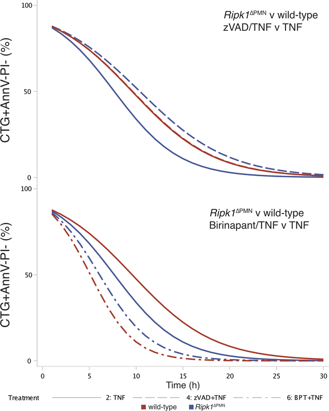
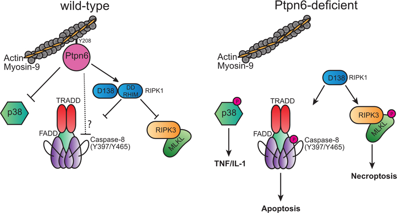
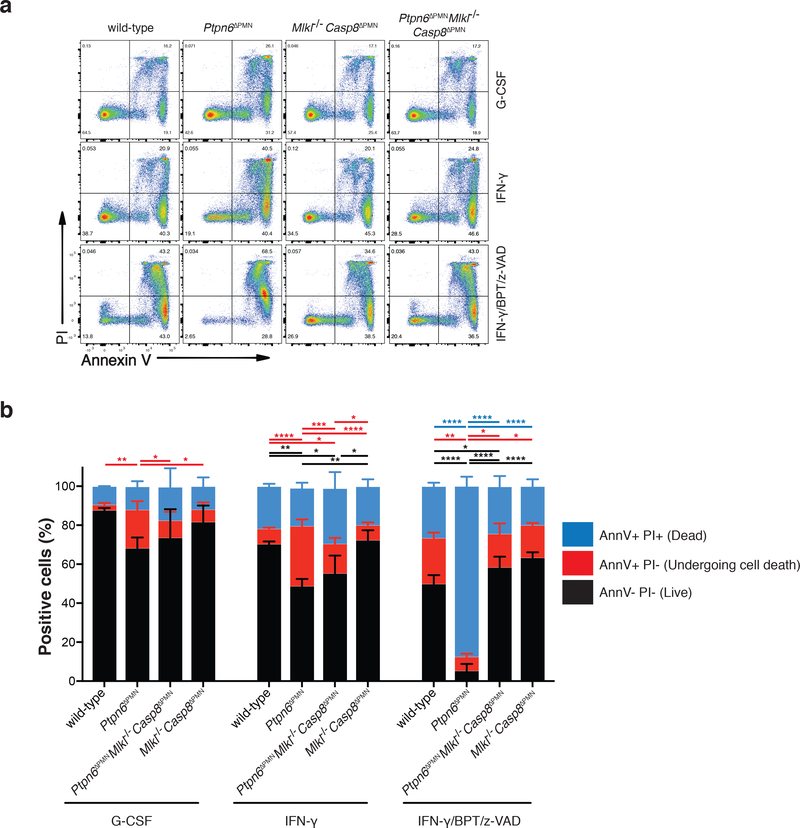
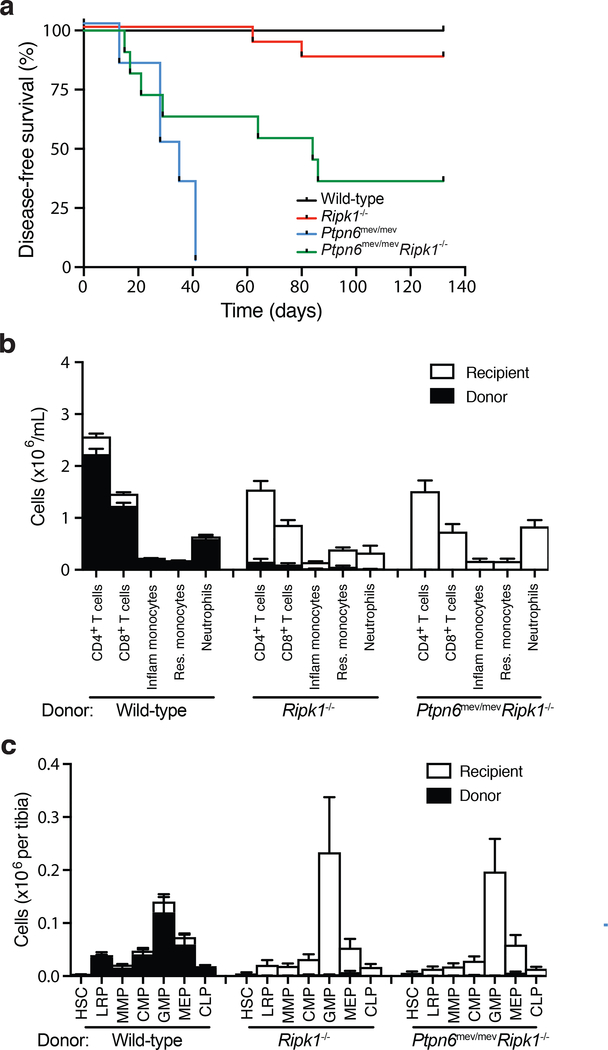
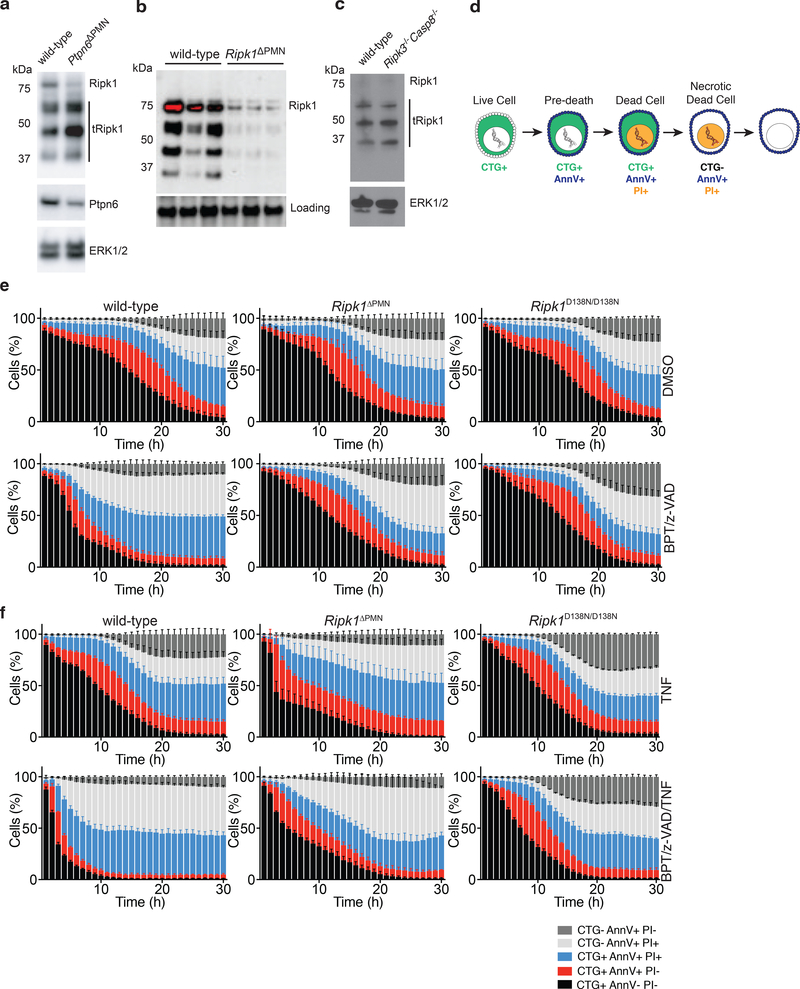
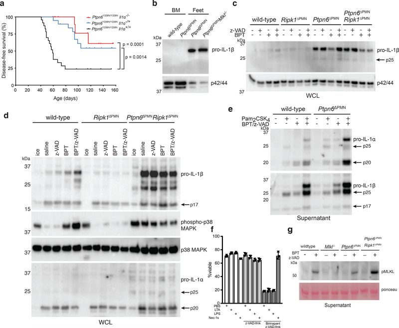
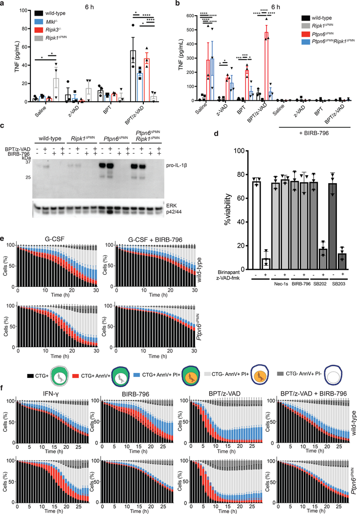
Ptpn6 is a cytoplasmic phosphatase that functions to prevent autoimmune and interleukin-1 (IL-1) receptor-dependent, caspase-1-independent inflammatory disease. Conditional deletion of Ptpn6 in neutrophils (Ptpn6∆PMN) is sufficient to initiate IL-1 receptor-dependent cutaneous inflammatory disease, but the source of IL-1 and the mechanisms behind IL-1 release remain unclear. Here, we investigate the mechanisms controlling IL-1α/β release from neutrophils by inhibiting caspase-8-dependent apoptosis and Ripk1-Ripk3-Mlkl-regulated necroptosis. Loss of Ripk1 accelerated disease onset, whereas combined deletion of caspase-8 and either Ripk3 or Mlkl strongly protected Ptpn6∆PMN mice. Ptpn6∆PMN neutrophils displayed increased p38 mitogen-activated protein kinase-dependent Ripk1-independent IL-1 and tumor necrosis factor production, and were prone to cell death. Together, these data emphasize dual functions for Ptpn6 in the negative regulation of p38 mitogen-activated protein kinase activation to control tumor necrosis factor and IL-1α/β expression, and in maintaining Ripk1 function to prevent caspase-8- and Ripk3-Mlkl-dependent cell death and concomitant IL-1α/β release.


Conflict of interest statement

Disclosure of Conflicts of Interest

The authors declare no competing financial or non-financial conflicts of interest.

Figures

Extended Data 1.
Extended Data 1.. Ptpn6 prevents an inflammatory signature in bone marrow neutrophils.
a) Gene Set Enrichment Analysis of RNA-Seq transcriptomic data derived from bone marrow neutrophils of female Ptpn6ΔPMNcaspase-8ΔPMNRipk3−/−Mlkl−/− mice and controls. b) Age-dependent effects on the neutrophil transcriptome in Ptpn6ΔPMNcaspase-8ΔPMNRipk3−/−Mlkl−/− mice and controls.
Extended Data 2.
Extended Data 2.. Flow cytometry gating strategy for hematopoietic stem and progenitor cells (HSPC).
For gating of HSPC in Figure 3, bone marrow cells were sorted after enrichment for hematopoietic progenitor cells by magnetic bead-based depletion of lineage-positive hematopoietic cells. (a) Definition and gating strategy for lineage restricted progenitors (LRP), multi-potent progenitors (MPP), and hematopoietic stem cells (HSC), (b) common myeloid progenitors (CMP), megakaryocyte erythroid progenitors (MEP), granulocyte macrophage progenitors (GMP), and (c) common lymphoid progenitors. Lineage-negative cells and gating were defined using isotype control antibodies.
Extended Data 3.
Extended Data 3.. Loss of RIPK1 sensitizes neutrophils to TNF-mediated cell death.
a-b) Live-cell imaging of CTG-labeled wild-type, Ripk1ΔPMN, and Ripk1D138N/D138N neutrophils treated with 2 μM BPT and/or 10 μM z-VAD-fmk +/− 100 ng/mL TNFα. PI and Annexin V were used to monitor changes in viability. Mean and SEM, n=3 biologically independent samples, and triplicate fields of view per independent biological sample. c) Live-cell imaging of CTG-labeled wild-type, Ptpn6ΔPMN, Ripk1ΔPMN, and Ptpn6ΔPMNRipk1ΔPMN neutrophils treated with saline or 100 ng/mL TNFα. PI and Annexin V were used to monitor changes in viability. Mean and SEM of technical replicates shown. Data are representative of two independent experiments.
Extended Data 4.
Extended Data 4.. TNF induces caspase activation in the absence of Ripk1 in neutrophils.
Live-cell imaging of CellTracker Orange-labeled wild-type, Ripk3−/−, Ripk1ΔPMN, and Ptpn6ΔPMNRipk1ΔPMN neutrophils treated with 2 μM BPT and/or 10 μM z-VAD-fmk +/− 100 ng/mL TNFα. CellEvent caspase-3/7 Green Detection Reagent and Draq7 were used to monitor changes in caspase activation and viability. Mean and SEM, n=3 technical triplicate samples from triplicate fields of view.
Extended Data 5.
Extended Data 5.. Generalized linear mixed effects models (logit link).
Comparison of CTG-positive proportion of a) z-VAD-fmk and b) birinapant treatment of TNF-stimulated Ripk1ΔPMN and wild-type neutrophils. Red curves indicate predicted profiles for wild-type, blue for Ripk1ΔPMN, with line patterns indicating predicted profile for treatment.
Extended Data 6.
Extended Data 6.. A model illustrating the role of Ptpn6 in regulation of cell death signaling in neutrophils.
Ptpn6 function is controlled in part by Y208-dependent anchoring to the actin-myosin-9 cytoskeleton. In the absence of Ptpn6, the negative regulatory functions of Ripk1 are lost but the kinase domain remains active to influence apoptotic and necroptotic cell death. In Ptpn6ΔPMN neutrophils lacking RIPK1 kinase activity or RIPK1, necroptotic and apoptotic cell death proceed unabated.
Figure 1.
Figure 1.. Loss of Ptpn6 sensitizes neutrophils to necroptotic cell death.
a-b) Flow cytometric analysis of wild-type, Ptpn6ΔPMN, Caspase-8ΔPMNMlkl−/− and Ptpn6ΔPMNCaspase-8ΔPMNMlkl−/− neutrophils stained with PI and Annexin V (Ann V) 12 h after addition of the necroptotic stimulus birinapant (BPT, 2 μM) + z-VAD-fmk (zVAD, 10 μM). Cells were first primed with 100 ng/mL G-CSF or 100 ng/mL IFN-γ. Mean ± SEM, n=3 biologically independent experiments. *p<0.05 ** p<0.01 ***p<0.001 ****p<0.0001, analyzed using one-way ANOVA with Tukey’s multiple comparison test.
Figure 2.
Figure 2.. Caspase-8 and Ripk3/Mlkl drives footpad inflammation in Ptpn6 mutant mice
a) Immunoblot for cleaved caspase-8 and Ptpn6 from freshly-isolated wild-type and Ptpn6ΔPMN bone marrow neutrophils. Luminescence was quantified using a ChemiDoc Gel Imaging System and ImageLab software. Mean ± SEM, n=4 independent neutrophil samples from Ptpn6ΔPMN or wild-type littermate controls. Data analyzed by Student’s t test. p values are shown. b) Kaplan-Meier plots showing disease-free survival of Ptpn6ΔPMN mice (n=145) compared with Ptpn6ΔPMNRipk3−/− (n=15), Ptpn6ΔPMNRipk3−/−caspase-8ΔPMN (n=45) mice, Ptpn6ΔPMNMlkl−/− (n=10), Ptpn6ΔPMNcaspase-8ΔPMNMlkl−/− (n=35) mice, Ptpn6ΔPMNcaspase-8ΔPMN (n=49), Ptpn6ΔPMNcaspase-8ΔPMNRipk3−/−Mlkl−/− (n=34), Ptpn6ΔPMNRipk1ΔPMN (n=9) and Ptpn6ΔPMNRipk1ΔPMN/D138N (n=8) mice. Survival curves were analyzed using a log-rank (Mantel-Cox) test compared to Ptpn6ΔPMN mice. Each panel shares the disease-free survival data of Ptpn6ΔPMN mice. c) RNA-Seq transcriptomic analysis of bone marrow neutrophils from female Ptpn6ΔPMN mice lacking Ripk3, Mlkl, and/or Casp8. Values are gene-wise z-scores of counts using the variance stabilized transformation from DESeq2.
Figure 3.
Figure 3.. RIPK1-deficiency prevents lethal inflammatory disease in Ptpn6 mutant mice by impairing hematopoiesis.
a) Kaplan-Meier plot comparing disease-free survival of lethally-irradiated wild-type mice transplanted with 106 Ripk1−/−, Ptpn6mev/mevRipk1−/−, Ptpn6mev/mev or wild-type fetal liver cells. Survival curves were analyzed using a log-rank (Mantel-Cox) test. p<0.0001, wild-type v Ptpn6mev/mev; p=0.0025, wild-type v Ptpn6mev/mevRipk1−/−; p=0.0454, Ptpn6mev/mev v Ptpn6mev/mevRipk1−/−; p<0.0001, Ptpn6mev/mev v Ptpn6mev/mevRipk1−/−. b-c) Characterization of the (b) hematopoietic compartment in peripheral blood 12 weeks post-transplant and (c) progenitor cells in the bone marrow of recipient mice 20 weeks post-transplant with 106 wild-type, Ripk1−/− or Ptpn6mev/mevRipk1−/− fetal liver cells. In (c), counts of hematopoietic stem cells (HSC), lineage-restricted progenitors (LRP), multipotent progenitors (MPP), common myeloid progenitors (CMP), granulocyte-macrophage progenitors (GMP), megakaryocyte-erythroid progenitors (MEP), and common lymphoid progenitors (CLP) from the bone marrow of lethally-irradiated recipients 20 weeks post-transplant, mean ± SEM, n = 4. p<0.05 for all comparisons of Ptpn6mev/mevRipk1−/− and Ripk1−/− to wild-type.
Figure 4.
Figure 4.. Loss of RIPK1 sensitizes neutrophils to TNF-mediated cell death.
a) Immunoblot showing Ripk1 and Ptpn6 in peripheral blood neutrophils isolated from wild-type and Ptpn6ΔPMN mice. ERK p42/44 is a loading control. Neutrophils pooled from 10–11 independent mice. Representative of 3 biologically independent experiments. b) Immunoblot for Ripk1 in lysates of bone marrow neutrophils from three independent Ripk1ΔPMN mice. c) Immunoblot for full-length and truncated Ripk1 (tRipk1) in lysates of wild-type and Ripk3−/−Casp8−/− bone marrow neutrophils. Representative data of 2 biologically independent experiments. d) Cell transition states identified by live cell imaging during regulated cell death of neutrophils. e-f) Live-cell imaging of CTG-labeled wild-type, Ripk1ΔPMN, and Ripk1D138N/D138N neutrophils treated with DMSO (e, top row), BPT/z-VAD (e, bottom row), TNF (f, top row), or BPT/z-VAD/TNF (f, bottom row) (drug concentrations: 2 μM BPT, 10 μM z-VAD-fmk, 100 ng/mL TNF). PI and Annexin V were used to monitor changes in viability. Mean and SEM, n=3 biologically independent experiments, and triplicate fields of view per biological replicate. BPT: birinapant; z-VAD: z-VAD-fmk; CTG: Cell Tracker Green; PI: propidium iodide.
Figure 5.
Figure 5.. Ptpn6-deficient neutrophils produce high levels of IL-1α and IL-1β in the absence of Ripk1.
a) Kaplan-Meier plot comparing disease-free survival of Ptpn6Y208N/Y208NIl1a+/+ (n=23) mutant mice with Ptpn6Y208N/Y208NIl1a−/− (n=23) and Ptpn6Y208N/Y208NIl1a+/− (n=19) mice. Survival curves were analyzed using a log-rank (Mantel-Cox) test compared to Ptpn6ΔPMNIl1a+/+ mice. p values are indicated. b) Immunoblot of IL-1β in neutrophils isolated from the bone marrow of wild-type and Ptpn6ΔPMN mice, or neutrophils pooled from the feet of three inflamed Ptpn6ΔPMN and two inflamed Ptpn6ΔPMNMlkl−/− mice. Representative of 3 biologically independent experiments. c) Immunoblot for IL-1β from whole cell lysates of wild-type, Ptpn6ΔPMN, Ripk1ΔPMN, and Ptpn6ΔPMN Ripk1ΔPMN bone marrow neutrophils primed for 1h with 100ng/mL IFNγ, and treated for 4 h with 2 μM BPT and/or 10 μM z-VAD-fmk, Representative of 3 biologically independent experiments. d) Immunoblot for IL-1β, IL-1α, p38 MAPK, and phospho-p38 MAPK from whole cell lysates of wild-type, Ripk1ΔPMN, and Ptpn6ΔPMN Ripk1ΔPMN neutrophils primed for 1h with 100ng/mL IFNγ, and treated for 4 h with 2 μM BPT and/or 10 μM z-VAD-fmk. Representative of 3 biologically independent experiments. ERK p42/44 was used as a loading control. e) Western blot showing IL-1α and IL-1β in the supernatant of wild-type and Ptpn6ΔPMN neutrophils treated overnight with the necroptotic stimuli BPT (2 μM) + z-VAD-fmk (10 μM) and/or 10 ng/mL Pam2CSK4, Representative of 3–4 independent experiments. f) Bone marrow neutrophil viability measured by PI uptake of neutrophils treated with 1 μg/mL lipotechoic acid, 100 ng/mL LPS or 10 μM Nec-1s in the presence of 10 μM z-VAD-fmk +/− 2 μM BPT. Mean ± SEM, n = 3 independent samples. LPS: lipopolysaccharide; LTA: lipoteichoic acid; Nec-1s: necrostatin-1s. g) Immunoblot for phospho-Mlkl in supernatant of neutrophils treated with BPT/z-VAD overnight. Ponceau staining is used as a loading control. Representative of 3 independent experiments.
Figure 6.
Figure 6.. p38 signaling drives TNF and IL-1 production by Ptpn6-deficient neutrophils in the absence of Ripk1.
a-b) Wild-type, Mlkl−/−, Ripk3−/−, Ripk1ΔPMN, Ptpn6ΔPMN, and Ptpn6ΔPMNRipk1ΔPMN neutrophils were primed for 1 h with 100 ng/mL IFN-γ before addition of 2 μM BPT and/or 10 μM z-VAD-fmk +/− 20 μM BIRB-796. TNF-α in the supernatant was measured after 6 h by ELISA. Mean and SEM, n=3 biologically independent experiments *p<0.05, **p<0.01, ***p<0.001, ****p<0.0001, analyzed using one-way ANOVA with Tukey’s multiple comparison test. c) Immunoblot of IL-1β in whole cell lysates after 4 h incubation. ERK p42/44 was used as a loading control. n = 3 biologically independent experiments. BPT: birinapant; z-VAD: z-VAD-fmk. d) Bone marrow neutrophils were treated with 2 μM birinapant and 10 μM z-VAD-fmk for 15h in the presence or absence of necrostatin-1s (Nec-1s), or the p38 inhibitors SB202190, SB203580 and BIRB-796. Viability was assessed by flow cytometry using propidium iodide (PI). Mean and SEM, n=3 biologically independent experiments. e-f) Live-cell imaging of CellTracker Green (CTG)-labeled wild-type and Ptpn6ΔPMN neutrophils treated with 100 ng/mL G-CSF, or 100 ng/mL IFN-γ, 2 μM birinapant, 10 μM z-VAD-fmk, +/− 20 μM BIRB-796. PI and Annexin V were used to monitor changes in viability. Mean and SEM, n=3 biologically independent experiments. BPT: birinapant; z-VAD: z-VAD-fmk
Figure 7.
Figure 7.. Ptpn6 contact with the actin-myosin cytoskeleton is mediated by Y208.
a) The structure of the Ptpn6 C-terminal SH2 domain bound to a phosphopeptide derived from the Natural Killer Group 2A (NKG2A) protein is displayed (PDB ID: 2YU7). The solvent accessible surface of the Ptpn6 SH2 domain (white) is shown overlaid on a ribbon diagram of the domain. The bound NKG2A phosphopeptide is shown in black with the phosphotyrosine residue indicated in stick representation. Tyrosine 208 is located at the C-terminal end of the BG loop. It is solvent accessible and does not contact the specificity-determining pocket (+3 pocket), nor the phosphotyrosine binding pocket. b) Coomassie staining for proteins associated with the wild-type and Y208N mutant Ptpn6 C-SH2 domains following immunoprecipitation from 108 neutrophils. Representative data from two biologically independent experiments. Peptides isolated from gel slices in (A) and (B) are described in Supplementary Table 2. c) Immunoblot for Myosin-9 in 5 × 10⁶ neutrophils following immunoprecipitation with the biotinylated peptides corresponding to the wild-type and Y208N mutant Ptpn6 C-SH2 domain. Representative data from three biologically independent experiments. d) Isothermal titration calorimetry (ITC) analysis comparing the interaction of FcγR2b phosphopeptide with wild-type and Y208N mutant recombinant C-terminal SH2 domain. Representative data from two biologically independent experiments.

References

    1. Uihlein LC, Brandling-Bennett HA, Lio PA & Liang MG Sweet syndrome in children. Pediatr Dermatol 29, 38–44 (2012). - PubMed
    1. Prat L, Bouaziz JD, Wallach D, Vignon-Pennamen MD & Bagot M Neutrophilic dermatoses as systemic diseases. Clin Dermatol 32, 376–388 (2014). - PubMed
    1. Going JJ, Going SM, Myskow MW & Beveridge GW Sweet’s syndrome: histological and immunohistochemical study of 15 cases. J Clin Pathol 40, 175–179 (1987). - PMC - PubMed
    1. Bourke JF, Jones JL, Fletcher A & Graham-Brown RA An immunohistochemical study of the dermal infiltrate and epidermal staining for interleukin 1 in 12 cases of Sweet’s syndrome. Br J Dermatol 134, 705–709 (1996). - PubMed
    1. Giasuddin AS, El-Orfi AH, Ziu MM & El-Barnawi NY Sweet’s syndrome: is the pathogenesis mediated by helper T cell type 1 cytokines? J Am Acad Dermatol 39, 940–943 (1998). - PubMed

METHODS-ONLY REFERENCES

    1. Pao LI et al. B cell-specific deletion of protein-tyrosine phosphatase Shp1 promotes B-1a cell development and causes systemic autoimmunity. Immunity 27, 35–48 (2007). - PubMed
    1. Shultz LD, Coman DR, Bailey CL, Beamer WG & Sidman CL ‘Viable motheaten,’ a new allele at the motheaten locus. I. Pathology. Am J Pathol 116, 179–192 (1984). - PMC - PubMed
    1. Kelliher MA et al. The death domain kinase RIP mediates the TNF-induced NF-kappaB signal. Immunity 8, 297–303 (1998). - PubMed
    1. Dannappel M et al. RIPK1 maintains epithelial homeostasis by inhibiting apoptosis and necroptosis. Nature 513, 90–94 (2014). - PMC - PubMed
    1. Polykratis A et al. Cutting edge: RIPK1 Kinase inactive mice are viable and protected from TNF-induced necroptosis in vivo. J Immunol 193, 1539–1543 (2014). - PMC - PubMed

Publication types

MeSH terms