. 2020 Jan;16(1):50-59.
doi: 10.1038/s41589-019-0424-1. Epub 2019 Dec 9.

CPSF3-dependent pre-mRNA processing as a druggable node in AML and Ewing's sarcoma

Affiliations

CPSF3-dependent pre-mRNA processing as a druggable node in AML and Ewing's sarcoma

Nathan T Ross et al. Nat Chem Biol. 2020 Jan.

Erratum in

  • Author Correction: CPSF3-dependent pre-mRNA processing as a druggable node in AML and Ewing's sarcoma.
    Ross NT, Lohmann F, Carbonneau S, Fazal A, Weihofen WA, Gleim S, Salcius M, Sigoillot F, Henault M, Carl SH, Rodríguez-Molina JB, Miller HR, Brittain SM, Murphy J, Zambrowski M, Boynton G, Wang Y, Chen A, Molind GJ, Wilbertz JH, Artus-Revel CG, Jia M, Akinjiyan FA, Turner J, Knehr J, Carbone W, Schuierer S, Reece-Hoyes JS, Xie K, Saran C, Williams ET, Roma G, Spencer M, Jenkins J, George EL, Thomas JR, Michaud G, Schirle M, Tallarico J, Passmore LA, Chao JA, Beckwith REJ. Ross NT, et al. Nat Chem Biol. 2020 Apr;16(4):479. doi: 10.1038/s41589-020-0508-y. Nat Chem Biol. 2020. PMID: 32139909

Abstract

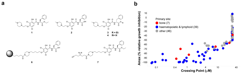
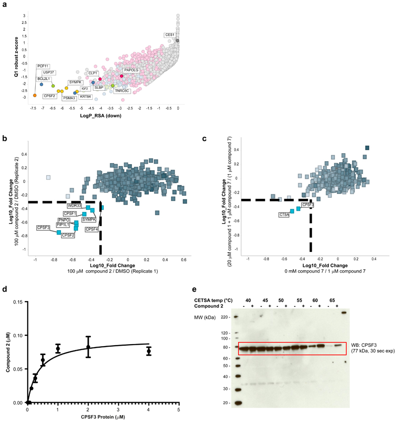
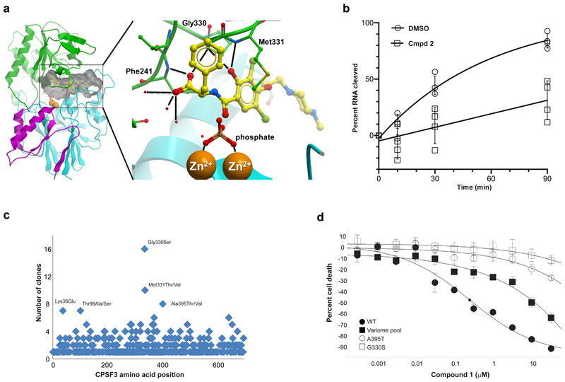
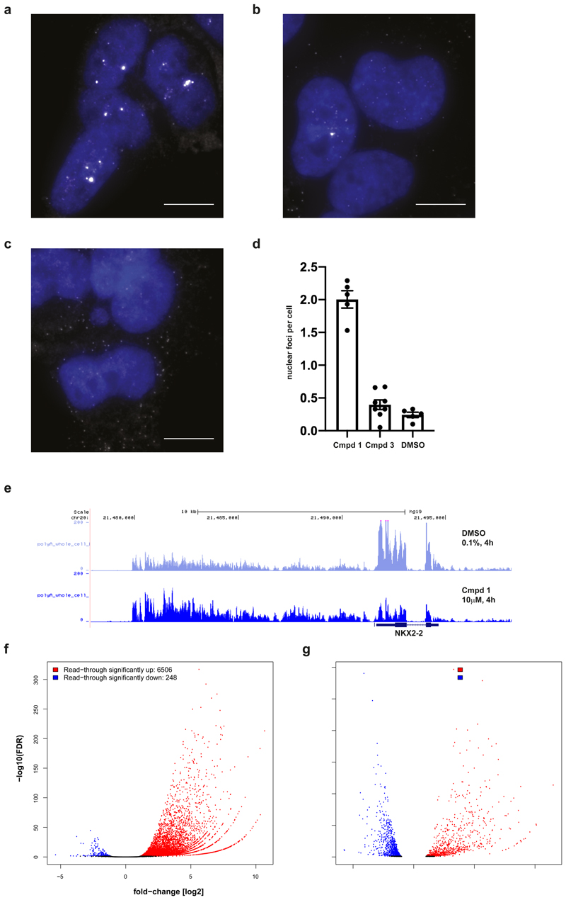
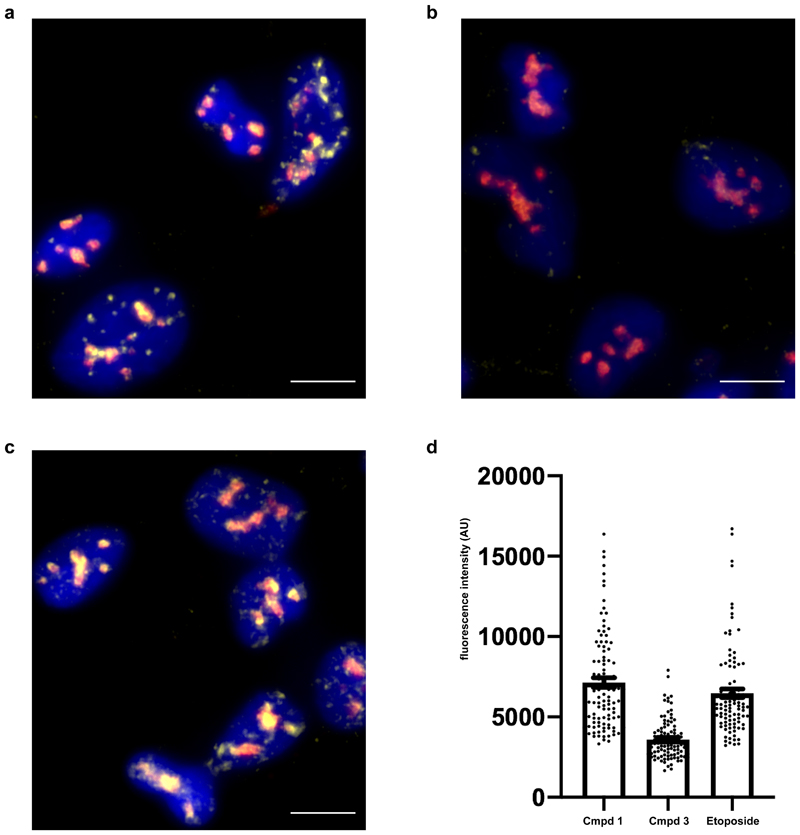
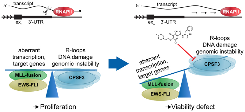
The post-genomic era has seen many advances in our understanding of cancer pathways, yet resistance and tumor heterogeneity necessitate multiple approaches to target even monogenic tumors. Here, we combine phenotypic screening with chemical genetics to identify pre-messenger RNA endonuclease cleavage and polyadenylation specificity factor 3 (CPSF3) as the target of JTE-607, a small molecule with previously unknown target. We show that CPSF3 represents a synthetic lethal node in a subset of acute myeloid leukemia (AML) and Ewing's sarcoma cancer cell lines. Inhibition of CPSF3 by JTE-607 alters expression of known downstream effectors in AML and Ewing's sarcoma lines, upregulates apoptosis and causes tumor-selective stasis in mouse xenografts. Mechanistically, it prevents the release of newly synthesized pre-mRNAs, resulting in read-through transcription and the formation of DNA-RNA hybrid R-loop structures. This study implicates pre-mRNA processing, and specifically CPSF3, as a druggable target providing an avenue to therapeutic intervention in cancer.


Conflict of interest statement

Competing Financial Interests Statement

All authors (except otherwise noted) were employees of Novartis Institutes for BioMedical Research at the time of their involvement in this study and may hold stock in Novartis. Correspondence should be addressed to R.E.J.B. (rohan.beckwith@novartis.com).

Figures

Figure 1
Figure 1. In vitro growth of AML and Ewing’s sarcoma cancer cell lines is sensitive to compound 1.
a, Chemical probes developed for study. b, Cell viability profile of compound 1 against a panel of 92 cancer cell lines, derived via CellTiter Glo (CTG) read-out after 72h of compound treatment in 11-point dose-response from 30 µM to 0.1 nM final concentration. Crossing Point was defined as the concentration at which the fitted curve of compound 1 for a given cell line achieved 50% of the positive control compound MG132. Amax was defined as the maximal inhibition observed with compound 1 for a given cell line over the range of tested concentrations. Cancer cell lines are color-coded by tumor primary site for bone (red, 7 cell lines), hematopoietic and lymphoid (blue, 39 cell lines), or other (grey, 46 cell lines). See Supplementary Table 1 for list of all 92 lines tested detailing primary site, lineage and compound sensitivity.
Figure 2
Figure 2. Chemical genetics and proteomics studies combine to identify mRNA processing and CPSF3 as the target of compound 2.
a, Full genome siRNA plus compound 1 synergy screen in A-673 cells, with cell viability read-out via CellTiter Glo assay. Per siRNA, the differential effect between DMSO-treated versus compound 1-treated cells was calculated. Plotting gene-level redundant siRNA activity (RSA statistical model, one-sided, down) as a measure of significance of effect versus Q1 Z-score as a measure of magnitude of effect identified siRNA-targeted genes whose knock-down sensitized cells to 0.4 µM compound 1 treatment. The siRNA library was tested as one replicate with each gene represented by n=8 siRNAs on average. Top screening hits, including known regulators of mRNA processing, are highlighted and named. b, Chemical proteomics studies using 6 in NOMO-1 cells. Proteins identified as specifically competed are highlighted and named. c, Photo-affinity labeling with PAL probe 7 in A-673 cells. Specifically enriched proteins are highlighted and named. d, Affinity binding measurement of compound 2 to human CPSF3 (Kd = 370 nM) as determined by SEC-TID. Error bars represent standard deviation of the mean (n=2). e, Western blot against CPSF3 following Cellular Thermal Shift Assay (CETSA) with compound 2 (+) or DMSO (-) treatment using NOMO-1 cell lysates (n=1).
Figure 3
Figure 3. Compound 2 binds directly to CPSF3, inhibiting its endonuclease activity and conferring growth inhibitory effects.
a, Co-crystal structure of CPSF3 and 2. Ribbon representation of the CPSF3 metallo-β-lactamase domain in cyan (residues 7 to 208) and magenta (396 to 459), and the β-CASP domain in green (209 to 395). Two zinc ions at the active site (orange spheres) are coordinated by a phosphate. 2 is shown as yellow ball-andstick and the interfacial cavity outlined by a grey surface. Insert highlights polar interactions between 2 and CPSF3. The carboxylate group of 2 forms bifurcated hydrogen bonds to the backbone NH of Phe241 and Gly330, respectively. Three ordered water molecules contact the carboxylate. The hydroxyl group is in hydrogen bonding distance to the backbone NH groups of Gly330 and Met331, respectively. b, In vitro cleavage reaction monitored over time using yeast recombinant 8-subunit core CPF complex, cleavage factors (CFIA and CFIB) and a Cyc1 model RNA substrate in the presence of 1% DMSO or 100 µM compound 2. The data was fitted to an exponential plateau model (solid line), error bars represent standard deviation of the mean (n=4). c, Missense mutation distribution in CPSF3 that desensitize to compound 1 in cell viability assay as identified from variomics studies in A-673. d, Cell viability assessment of A-673 cells expressing in trans either wild-type (WT) CPSF3 encoding cDNA or dominant mutant versions of human CPSF3 as derived from variomics study in response to treatment with compound 1. Small black circle on fitted curve for WT condition indicates IC50 value. Center values represent mean, error bars represent standard deviation (n=3).
Figure 4
Figure 4. Compound 1 induces transcript accumulation and RNA Pol II read-through.
a,b,c, Images of A-673 cells that were treated with compound 1 (a), compound 3 (b) or DMSO (c) for 4 h prior to fixation and probed for NKX2-2 mRNAs (white) using FISH probes targeted to the coding sequence of the transcript. Nuclei (blue) were stained with DAPI. Images are representative from three independent experiments performed in duplicate. Scale bar = 10 μm. d, Compound 1 (79 cells) resulted in the formation of bright nuclear foci that contained multiple NKX2-2 transcripts (p-value < 0.0001, unpaired two-tailed t test) while transcripts in cells treated with compound 3 (40 cells) were similar to cells treated with DMSO (59 cells) (p-value=0.15, unpaired two-tailed t test). Data are presented as mean plus/minus standard error of the mean (error bars) for nuclear foci counted in each experiment. e, RNA-seq traces at NKX2-2 locus (hg19, chr20:21,477,893-21,498,177) generated from A-673 cells that were treated with 1 or DMSO for 4 h prior to whole cell lysis and RNA purification. Three biological replicates per condition merged into one trace. f,g, RNA-seq based quantification of (f) read-through expression genome-wide and of (g) changes in global gene expression, both in A-673 cells upon 4 h treatment with 1 versus DMSO, analyzing RNA purified from whole cell extracts of three biological replicates. Normalized read counts were analyzed using a negative binomial generalized log-linear model with two-sided comparisons and false discovery rate controlled using Benjamin & Hochberg multiple comparisons adjustment via edgeR. Significance cut-offs defined as absolute value fold-change [log2] > 1 and adjusted p-val/FDR < 0.05, and marked in blue (down) or red (up).
Figure 5
Figure 5. Compound 1 results in accumulation of nuclear R-loops.
a,b,c, Images of A-673 cells treated with compound 1 (a), compound 3(b) or etoposide (c) for 20 h prior to fixation and staining for R-loops (DNA-RNA hybrids, yellow), nucleolin (red) and DAPI (blue). Images are representative from two independent experiments performed in duplicate. Scale bar = 10 μm. d, Quantification of nucleoplasmic R-loop staining. Etoposide (100 cells) and compound 1 (100 cells) treatment result in increased R-loop staining compared to compound 3 (100 cells) (p-value <0.001, unpaired two-tailed t test). Data are presented as mean plus/minus standard error of the mean (error bars) for R-loop staining combined from all experiments.
Figure 6
Figure 6. Proposed model for compound 2 mechanism of action.
Cancers such as EWSFLI translocated Ewing’s sarcoma or MLL-translocated AML have selected for a balance between oncogenic fusion protein-driven aberrant transcription and its associated genomic instability that allows for proliferation. Under such conditions, the mRNA processing and termination machinery, specifically one of its core components, the cleavage and polyadenylation specificity factor 3 (CPSF3), represents a previously undescribed novel synthetic lethal node. Compound 2 inhibition of CPSF3-mediated mRNA cleavage results in RNA Pol II read-through beyond the 3’-UTR and transcript accumulation. As a result, gene expression is perturbed, including downregulation of genes involved in DNA double-strand break repair, while R-loop formation is increased to levels that the cell can no longer buffer or balance, ultimately leading to a cell viability defect.

Comment in

  • Processing for destruction.
    Erb MA. Erb MA. Nat Chem Biol. 2020 Jan;16(1):3-4. doi: 10.1038/s41589-019-0428-x. Nat Chem Biol. 2020. PMID: 31819275 No abstract available.

References

    1. Swinney DC, Anthony J. How were new medicines discovered? Nat. Rev. Drug Discov. 2011;10:507–19. - PubMed
    1. Moffat JG, Vincent F, Lee JA, Eder J, Prunotto M. Opportunities and challenges in phenotypic drug discovery: An industry perspective. Nat. Rev. Drug Discov. 2017;16:531–543. - PubMed
    1. Schreiber SL. Chemical genetics resulting from a passion for synthetic organic chemistry. Bioorg. Med. Chem. 1998;6:1127–1152. - PubMed
    1. Carson C, et al. Englerin A agonizes the TRPC4/C5 cation channels to inhibit tumor cell line proliferation. PLoS One. 2015;10:e0127498. - PMC - PubMed
    1. Rothman DM, et al. Metabolic Enzyme Sulfotransferase 1A1 Is the Trigger for N-Benzyl Indole Carbinol Tumor Growth Suppression. Chem. Biol. 2015;22:1228–1237. - PubMed

Publication types

MeSH terms