

Intraocular Lens Implantation In The Ciliary Sulcus: Challenges And Risks
- PMID: 31819356
- PMCID: PMC6885568
- DOI: 10.2147/OPTH.S205148
Intraocular Lens Implantation In The Ciliary Sulcus: Challenges And Risks
Abstract
Purpose: This article reviews the current literature on the risks and challenges associated with intraocular lens (IOL) implantation in the ciliary sulcus.
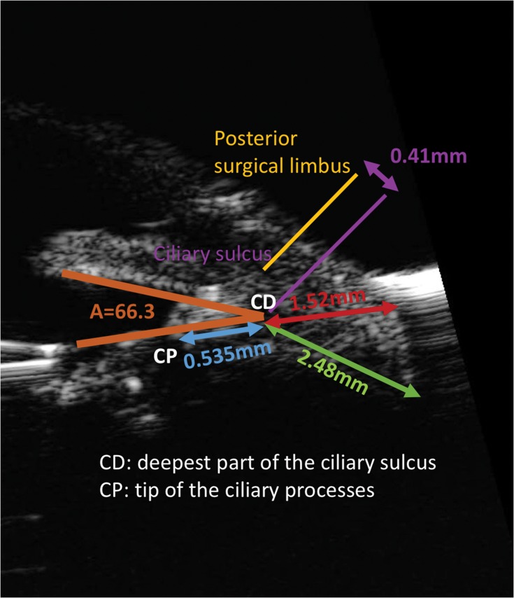
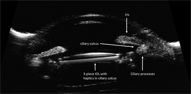
Recent findings: The development of IOLs designed specifically for placement in the ciliary sulcus continues to be an area of interest for the ophthalmic industry. Currently the one-piece PMMA (polymethylmethacrylate) lens or a three-piece IOL are the best available options for IOL placement in the ciliary sulcus space. Single piece acrylic (SPA) IOLs are not designed for sulcus placement and there is growing evidence of chronic complications related to their use in the ciliary sulcus. Many of these eyes ultimately require surgical intervention, including lens exchange. Endoscopic imaging and ultrasound biomicroscopy (UBM) have enabled a better understanding of ciliary sulcus anatomy and measurements in the living eye.
Summary: When the capsular bag is compromised, IOL placement in the ciliary sulcus is a reasonable option. In these circumstances, appropriate choice of IOL, knowledge of the sulcus anatomy, and correct technique can improve results and reduce postoperative complications.
Keywords: capsule rupture; ciliary sulcus; complications; intraocular lens; surgical technique.
© 2019 Mehta and Aref.
Conflict of interest statement
The authors report no conflicts of interest in this work.
Figures
References
-
- Randleman JB, Ahmed IIK, Editors. Intraocular Lens Surgery. New York: Thieme medical publishers; 2016:10–18, 138–178.
-
- Kim T, DelMonte DW, Gupta PK, Chang DF, Editors. Curbside Consultation in Cataract Surgery. 2nd ed. NJ: Slack incorporated; 2014:153–167.
Publication types
LinkOut - more resources
Full Text Sources
