Review
. 2019 Dec 2:13:2341-2352.
doi: 10.2147/OPTH.S220845. eCollection 2019.

Central Serous Chorioretinopathy: Pathogenesis and Management

Affiliations
Review

Central Serous Chorioretinopathy: Pathogenesis and Management

Francesco Semeraro et al. Clin Ophthalmol. .

Abstract

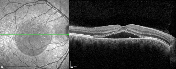
Central serous chorioretinopathy (CSC) is a common retina disease and has a relative high recurrence rate, etiology, and pathogenesis of which remains largely ambiguous. The effects on the retina are usually self-limited, although some people are left with permanent vision loss due to progressive and irreversible photoreceptor damage or retinal pigment epithelium atrophy. There have been a number of interventions used in CSC, including, but not limited to, laser treatment, photodynamic therapy (PDT), intravitreal injection of anti-vascular endothelial growth factor agents, and subthreshold lasers. It is not clear whether there is a clinically important benefit to treating acute CSC, which often resolves spontaneously as part of its natural history. Of the interventions studied to date, PDT and micropulse laser treatment appear the most promising. .

Keywords: acute central serous chorioretinopathy; central serous chorioretinopathy; chronic central serous chorioretinopathy.


Conflict of interest statement

Dr Andrea Pilotto reports personal fees from BioMarin, Chiesi, Nutricia, UCB, Zambon and Z-cube S.r.l.; grants from VItaflo Germany and Zambon Italy, Italian Ministry of Health, outside of the submitted work. The authors report no other conflicts of interest in this work.

Figures

Figure 1
Figure 1
A spectral domain optical coherence tomography image of an eye affected by acute central serous chorioretinopathy showing a convex-shaped blister of fluid separating the neurosensory retina from the retinal pigment epithelium and choroid.
Figure 2
Figure 2
A spectral domain optical coherence tomography image of an eye affected by chronic central serous chorioretinopathy showing a low level of subretinal fluid. At the level of serous detachment, the inner limit is represented by the ellipsoid zone, with elevated photoreceptor tips, and marked reflectivity.
Figure 3
Figure 3
Early and late fluorescein angiograms of a 29-year-old patient affected by chronic central serous chorio-retinopathy revealing an irregularity of the retinal pigment epithelium, resembling a “honeycomb” pattern, near the point of fluorescein leakage. Window defects and late-phase leakage from the expanding point in the superior macula are evident, toward the fovea in a “steam” configuration.

References

    1. Spaide RF, Campeas L, Haas A, et al. Central serous chorioretinopathy in younger and older adults. Ophthalmology. 1996;103(12):2070–2079. doi:10.1016/S0161-6420(96)30386-2 - DOI - PubMed
    1. Wang M, Munch IC, Hasler PW, Prünte C, Larsen M. Central serous chorioretinopathy. Acta Ophthalmol (Copenh). 2008;86(2):126–145. doi:10.1111/j.1600-0420.2007.00889.x - DOI - PubMed
    1. Liegl R, Ulbig MW. Central serous chorioretinopathy. Ophthalmologica. 2014;232(2):65–76. doi:10.1159/000360014 - DOI - PubMed
    1. N V B, Gürlü VP, Esgin H. Long-term macular function in eyes with central serous chorioretinopathy. Clin Experiment Ophthalmol. 2005;33(4):369–372. doi:10.1111/j.1442-9071.2005.01027.x - DOI - PubMed
    1. Mudvari SS, Goff MJ, Fu AD, et al. The natural history of pigment epithelial detachment associated with central serous chorioretinopathy. Retina. 2007;27(9):1168–1173. doi:10.1097/IAE.0b013e318156db8a - DOI - PubMed