. 2019 Nov 18:14:8989-9006.
doi: 10.2147/IJN.S209603. eCollection 2019.

MRI Detectable Polymer Microspheres Embedded With Magnetic Ferrite Nanoclusters For Embolization: In Vitro And In Vivo Evaluation

Affiliations

MRI Detectable Polymer Microspheres Embedded With Magnetic Ferrite Nanoclusters For Embolization: In Vitro And In Vivo Evaluation

Xiao-Ya Qin et al. Int J Nanomedicine. .

Abstract

Objective: The objective of this study was to develop magnetic embolic microspheres that could be visualized by clinical magnetic resonance imaging (MRI) scanners aiming to improve the efficiency and safety of embolotherapy.

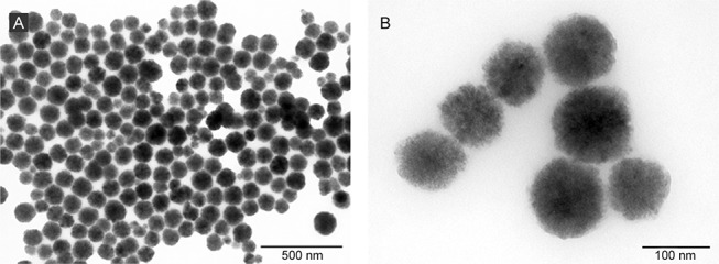
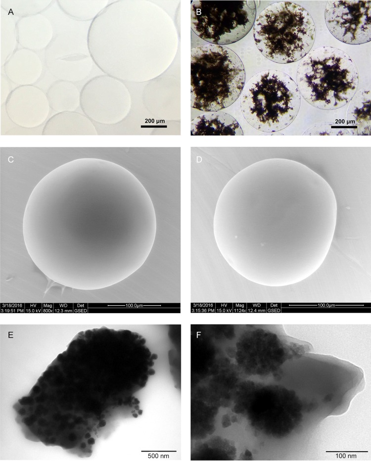
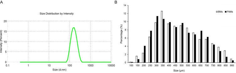
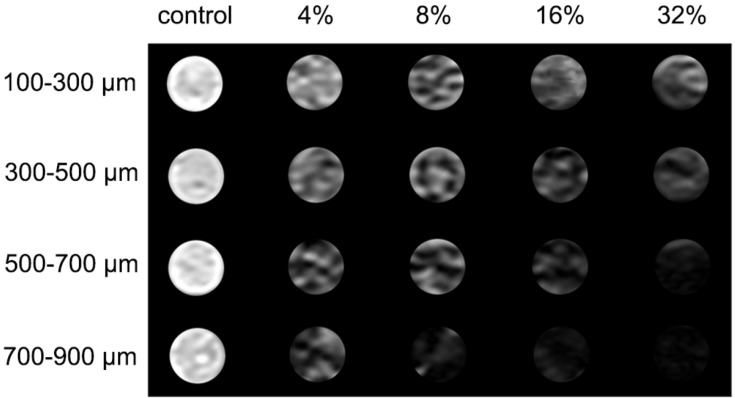
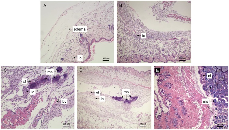
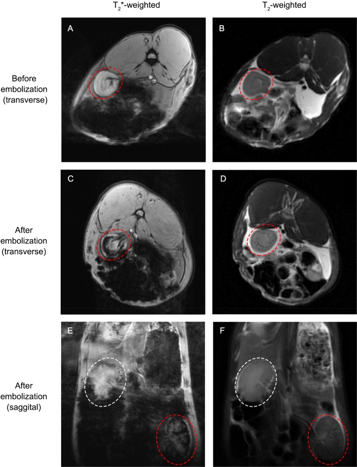
Methods and discussion: Magnetic ferrite nanoclusters (FNs) were synthesized with microwave-assisted solvothermal method, and their morphology, particle size, crystalline structure, magnetic properties as well as T2 relaxivity were characterized to confirm the feasibility of FNs as an MRI probe. Magnetic polymer microspheres (FNMs) were then produced by inverse suspension polymerization with FNs embedded inside. The physicochemical and mechanical properties (including morphology, particle size, infrared spectra, elasticity, etc.) of FNMs were investigated, and the magnetic properties and MRI detectable properties of FNMs were also assayed by vibrating sample magnetometer and MRI scanners. Favorable biocompatibility and long-term MRI detectability of FNMs were then studied in mice by subcutaneous injection. FNMs were further used to embolize rabbits' kidneys to evaluate the embolic property and detectability by MRI.

Conclusion: FNMs could serve as a promising MRI-visualized embolic material for embolotherapy in the future.

Keywords: embolization; magnetic ferrite nanoclusters; magnetic resonance imaging; microwave-assisted solvothermal method; polymerized microspheres.


Conflict of interest statement

The authors report no conflicts of interest in this work.

Figures

Figure 1
Figure 1
TEM images of FNs at a magnification of 10 k (A) and 40 k (B). Abbreviations: TEM, transmission electron microscopy; FNs, magnetic ferrite nanoclusters.
Figure 2
Figure 2
Morphology of microspheres. Notes: BMs under optical microscope (A); FNMs under optical microscope (B); BMs under ESEM (C); FNMs under ESEM (D); FNMs under TEM at a magnification of 8 k (E) and 50 k (F). Abbreviations: BMs, blank polymer microspheres; FNMs, magnetic polymer microspheres; ESEM, environmental scanning electron microscope; TEM, transmission electron microscopy.
Figure 3
Figure 3
DLS intensity distributions of FNs (A) and size distribution of the BMs (blank column) and FNMs (black column) (B). Abbreviations: DLS, dynamic light scattering; FNs, magnetic ferrite nanoclusters; BMs, blank polymer microspheres; FNMs, magnetic polymer microspheres.
Figure 4
Figure 4
FT-IR spectra of FNs (A), BMs (B) and FNMs (C). Abbreviations: FT-IR, Fourier transform infrared; FNs, magnetic ferrite nanoclusters; BMs, blank polymer microspheres; FNMs, magnetic polymer microspheres.
Figure 5
Figure 5
The XRD patterns of FNs (A) and FNMs (B). Abbreviations: XRD, X-ray diffraction; FNs, magnetic ferrite nanoclusters; FNMs, magnetic polymer microspheres.
Figure 6
Figure 6
The XPS spectra of FNs and FNMs. Notes: XPS survey spectrum (A) and Fe 2p XPS spectrum (B) of FNs; XPS survey spectrum (C) and Fe 2p XPS spectrum (D) of FNMs. Abbreviations: XPS, X-ray photoelectron spectroscopy; FNs, magnetic ferrite nanoclusters; FNMs, magnetic polymer microspheres.
Figure 7
Figure 7
The hysteresis loops of FNs (A) and FNMs (B) at room temperature. Notes: The inserts in (A) were photographs of the FNs dispersed in the water and responding to external magnetic field, respectively. The inserts in (B) were photographs of the FNMs dispersed in the water and responding to external magnetic field, respectively. Abbreviations: FNs, magnetic ferrite nanoclusters; FNMs, magnetic polymer microspheres.
Figure 8
Figure 8
Compression curves (A) and stress relaxation curves (B) of different microspheres. Abbreviations: BMs, blank polymer microspheres; FNMs, magnetic polymer microspheres.
Figure 9
Figure 9
T2 relaxivity measurement of FNs. Abbreviation: FNs, magnetic ferrite nanoclusters.
Figure 10
Figure 10
T2-weighted MR images of in vitro gel phantom from left to right column: BMs with the concentration of 32% (v/v) (as controls) and FNMs with concentrations of 4%, 8%, 16%, and 32% (v/v). Abbreviations: MR, magnetic resonance; BMs, blank polymer microspheres; FNMs, magnetic polymer microspheres.
Figure 11
Figure 11
The typical T2-weighted MR images of the same mouse before, immediately after, at 14 d and 28 d after subcutaneous injection of FNMs into the back (A) and the quantificational analysis of signal-to-noise changes at the corresponding time points after the injection into three mice (B). Note: The arrows denoted the dark signal area induced by FNMs. Abbreviations: MR, magnetic resonance; FNMs, magnetic polymer microspheres; △SNR, the change of signal-to-noise ratio.
Figure 12
Figure 12
Histological features of mice after subcutaneous injection of FNMs at 2 h (A), 2 d (B), 7 d (C), 14 d (D), and 28 d (E). Abbreviations: FNMs, magnetic polymer microspheres; ic, inflammatory cells; cf, collagen fibers; bv, blood vessels; ms, microspheres.
Figure 13
Figure 13
Arterial angiogram of a rabbit’s left kidney, before embolization (A) and immediately after embolization (B). Note: The peripheral blood vessels were occluded with FNMs (100–300 μm) after embolization. Abbreviation: FNMs, magnetic polymer microspheres.
Figure 14
Figure 14
MR images of embolized kidney with T2*- and T2-weighted sequences before and after embolization. Notes: The rabbit’s left embolized kidney was denoted by red elliptical rings while the right untreated kidney was denoted by white elliptical rings. Abbreviation: MR, magnetic resonance.

References

    1. Yamakado K, Nakatsuka A, Kobayashi S, et al. Radiofrequency ablation combined with renal arterial embolization for the treatment of unresectable renal cell carcinoma larger than 3.5 cm: initial experience. Cardiovasc Intervent Radiol. 2006;29(3):389–394. doi:10.1007/s00270-004-0090-9 - DOI - PubMed
    1. Goodwin SC, Wong GCH. Uterine artery embolization for uterine fibroids: a radiologist’s perspective. Clin Obstet Gynecol. 2001;44(2):412–424. doi:10.1097/00003081-200106000-00026 - DOI - PubMed
    1. Khan SU, Rahman KM, Siddiqui MR, et al. Endovascular embolization of life threatening intracranial arterio-venous malformation. Mymensingh Med J. 2010;19(3):438–441. - PubMed
    1. Murakami R, Ichikawa T, Kumazaki T, Kobayashi Y, Ogura J, Kurokawa A. Transcatheter arterial embolization for postpartum massive hemorrhage: a case report. Clin Imaging. 2000;24(6):368–370. - PubMed
    1. Sharma KV, Bascal Z, Kilpatrick H, et al. Long-term biocompatibility, imaging appearance and tissue effects associated with delivery of a novel radiopaque embolization bead for image-guided therapy. Biomaterials. 2016;103:293–304. doi:10.1016/j.biomaterials.2016.06.064 - DOI - PubMed