. 2019 Nov 28;17(4):1559325819891378.
doi: 10.1177/1559325819891378. eCollection 2019 Oct-Dec.

Assessment of Genomic Instability in Medical Workers Exposed to Chronic Low-Dose X-Rays in Northern China

Affiliations

Assessment of Genomic Instability in Medical Workers Exposed to Chronic Low-Dose X-Rays in Northern China

Lianying Fang et al. Dose Response. .

Abstract

The increasing use of ionizing radiation (IR) in medical diagnosis and treatment has caused considerable concern regarding the effects of occupational exposure on human health. Despite this concern, little information is available regarding possible effects and the mechanism behind chronic low-dose irradiation. The present study assessed potential genomic damage in workers occupationally exposed to low-dose X-rays. A variety of analyses were conducted, including assessing the level of DNA damage and chromosomal aberrations (CA) as well as cytokinesis-block micronucleus (CBMN) assay, gene expression profiling, and antioxidant level determination. Here, we report that the level of DNA damage, CA, and CBMN were all significantly increased. Moreover, the gene expression and antioxidant activities were changed in the peripheral blood of men exposed to low-dose X-rays. Collectively, our findings indicated a strong correlation between genomic instability and duration of low-dose IR exposure. Our data also revealed the DNA damage repair and antioxidative mechanisms which could result in the observed genomic instability in health-care workers exposed to chronic low-dose IR.

Keywords: DNA damage; antioxidants; biomarker; genomic instability; low-dose ionizing radiation.


Conflict of interest statement

Declaration of Conflicting Interests: The author(s) declared no potential conflicts of interest with respect to the research, authorship, and/or publication of this article.

Figures

Figure 1.
Figure 1.
DNA damage of lymphocytes in X-ray-exposed (exposure group) and nonexposed (reference group) workers as detected by a comet assay. (A) Representative comet image of lymphocytes from the reference group. (B) Representative comet image of lymphocytes from the exposure group. Tail DNA% (C), tail moment (D), and olive tail moment (E) values in lymphocytes of workers in the exposure group were all significantly greater than those in the reference group. The level of DNA damage was represented as the mean of 3 independent experiments and at least 200 cells were counted (*P < .05, **P < .01).
Figure 2.
Figure 2.
Cytokinesis-block micronucleus in the lymphocytes of the exposure and reference groups. (A) The rate of CBMN in lymphocytes of the exposure group was greater than that of the reference group. (B) Cytokinesis-block micronucleus frequency in lymphocytes of smoking workers was greater than that of nonsmoking workers in both the exposure and control groups. (C) Representative micronucleus image in a binucleated lymphocyte of workers exposed to low-dose IR. Experiments were performed independently in triplicate; more than 1000 cells were counted in each experiment (*P < .05, **P < .01). CBMN indicates cytokinesis-block micronucleus; IR, ionizing radiation.
Figure 3.
Figure 3.
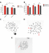
Chromosomal aberrations rate in the lymphocytes of exposure and reference workers. (A) Chromosomal aberrations rate in low-dose X-ray-exposed workers was greater than that of the reference group. Chromosomal aberrations rate of smoking participants was higher than that of nonsmoking participants in the exposure group. (B) Chromosomal aberrations rate in lymphocytes of workers exposed to low-dose X-ray radiation was higher than that in reference group for different age subgroups. (C) Representative normal chromosomal image obtained from the reference group. (D) and (E), Representative, abnormal chromosomal images of the low-dose, X-ray-exposed workers. A 2-way ANOVA was used to test the interactions between smoking and CA rate in lymphocytes (*P < .05, **P < .01). ANOVA indicates analysis of variance; dis, dicentric chromosomes; r, ring chromosomes.
Figure 4.
Figure 4.
Identification of genes responsive to low-dose X-ray exposure using gene profiling. Heat map showing the log (fold-changes) in DNA damage-related genes in exposure and control groups as determined by PCR array analysis. Fold changes were calculated versus the control group average. Color scale ranges from red to blue, which denotes up- or downregulated genes, respectively. Red and blue color indicate genes with a > 2-fold upregulation and genes with a > 2-fold downregulation, respectively. (To better interpret the color references in the figure legend, please refer to the online version of this article.)
Figure 5.
Figure 5.
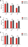
Malondialdehyde, SOD, and GSH levels in blood serum of the exposure and reference groups. (A) Malondialdehyde levels were significantly lower in the X-ray-exposed workers with more than 20 years of exposure. (B) Superoxide dismutase levels were significantly lower in X-ray-exposed workers with more than 20 years of exposure. (C) The relationship between the level of GSH and the X-ray exposure duration was not significant (*P < .05, **P < .01). GSH indicates glutathione; SOD, superoxide dismutase.

References

    1. Abosala A. A report on occupational ionizing radiation exposure by an orthopedic surgeon in a national health-care setting-clinical case perspective. J Orthop Case Rep. 2018;8(3):81–84. - PMC - PubMed
    1. Ciraj-Bjelac O, Rehani MM, Sim KH, Liew HB, Vano E, Kleiman NJ. Risk for radiation-induced cataract for staff in interventional cardiology: is there reason for concern? Catheter Cardiovasc Interv. 2010;76(6): 826–834. - PubMed
    1. Linet MS, Slovis TL, Miller DL, et al. Cancer risks associated with external radiation from diagnostic imaging procedures. CA Cancer J Clin. 2012;62(2):75–100. - PMC - PubMed
    1. Visweswaran S, Joseph S, S VH, O A, Jose MT, Perumal V. DNA damage and gene expression changes in patients exposed to low-dose X-radiation during neuro-interventional radiology procedures. Mutat Res. 2019;844:54–61. - PubMed
    1. Basheerudeen S, Kanagaraj K, Jose MT, et al. Entrance surface dose and induced DNA damage in blood lymphocytes of patients exposed to low-dose and low-dose-rate X-irradiation during diagnostic and therapeutic interventional radiology procedures. Mutat Res. 2017;818:1–6. - PubMed

LinkOut - more resources