

Diagnostic performance of diffusion-weighted magnetic resonance imaging in assessing lymph node metastasis of esophageal cancer compared with PET
- PMID: 31820208
- PMCID: PMC7316698
- DOI: 10.1007/s10388-019-00704-w
Diagnostic performance of diffusion-weighted magnetic resonance imaging in assessing lymph node metastasis of esophageal cancer compared with PET
Abstract
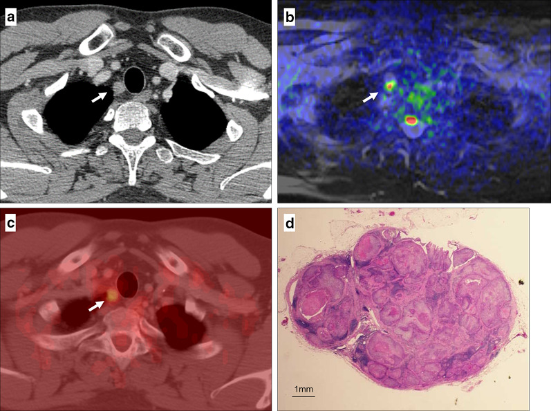
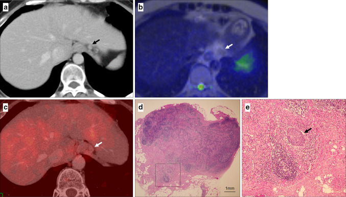
Background: Although diffusion-weighted magnetic resonance imaging (DWI) for detecting lymph node (LN) metastasis is reported to be a successful modality for primary malignant tumors, there are few studies relating to esophageal cancer. This study aimed to clarify the diagnostic performance of DWI for assessing LN metastasis compared with positron emission tomography (PET) in patients with esophageal squamous cell cancer (eSCC).
Methods: Seventy-six patients with histologically proven eSCC who underwent curative esophagectomy without neoadjuvant treatment were reviewed retrospectively. Harvested LNs were divided into 1229 node stations with 94 metastases. Diagnostic abilities and prognostic significance were compared.
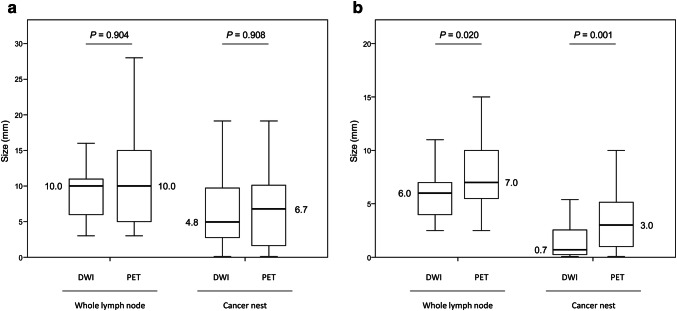
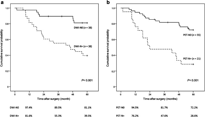
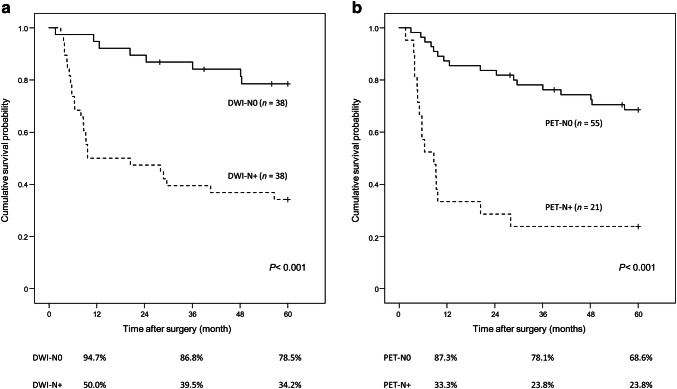
Results: In a station-by-station evaluation, the sensitivity was higher in DWI than PET (67% vs. 32%, P < 0.001). DWI showed more than 80% sensitivity for middle- and large-sized cancer nests and large area of cancer nests. The DWI-N0 group had a better 5-year relapse-free survival rate than the DWI-N+ group (78.5% vs. 34.2%, P < 0.001), as did the PET-N0 group. DWI-N status was an independent prognostic factor (hazard ratio [HR], 2.642; P = 0.048), as was PET-N status (HR 2.481; P = 0.033).
Conclusions: DWI, which depends on cancer cell volume followed by elevated intranodal density, is a non-invasive modality and showed higher sensitivity than PET. It has clinical impact in predicting postoperative survival for patients with eSCC alongside its diagnostic ability and has significant performance in clinical practice.
Keywords: Diffusion-weighted MRI; Esophageal cancer; Lymph node; PET.
Conflict of interest statement
The authors have no conflicts of interest and received no financial support for this study.
Figures
Similar articles
-
Association between prognosis and lymph node status using 18F-fluorodeoxyglucose-positron emission tomography in esophageal squamous cell carcinoma treated with esophagectomy post-neoadjuvant chemotherapy.World J Surg. 2024 Mar;48(3):650-661. doi: 10.1002/wjs.12067. Epub 2024 Jan 23. World J Surg. 2024. PMID: 38686781
-
Intravoxel incoherent motion diffusion-weighted imaging in evaluating preoperative staging of esophageal squamous cell carcinoma : Evaluation of preoperative stage of primary tumour and prediction of lymph node metastases from esophageal cancer using IVIM: a prospective study.Cancer Imaging. 2024 Aug 29;24(1):116. doi: 10.1186/s40644-024-00765-w. Cancer Imaging. 2024. PMID: 39210470 Free PMC article.
-
Clinical Significance of 18F-Fluorodeoxyglucose-Positron Emission Tomography-Positive Lymph Nodes to Outcomes of Trimodal Therapy for Esophageal Squamous Cell Carcinoma.Ann Surg Oncol. 2019 Jun;26(6):1869-1878. doi: 10.1245/s10434-019-07158-5. Epub 2019 Jan 23. Ann Surg Oncol. 2019. PMID: 30675704
-
A Comprehensive Comparison of CT, MRI, Positron Emission Tomography or Positron Emission Tomography/CT, and Diffusion Weighted Imaging-MRI for Detecting the Lymph Nodes Metastases in Patients with Cervical Cancer: A Meta-Analysis Based on 67 Studies.Gynecol Obstet Invest. 2017;82(3):209-222. doi: 10.1159/000456006. Epub 2017 Feb 10. Gynecol Obstet Invest. 2017. PMID: 28183074 Review.
-
Frequency and distribution pattern of lymph node metastasis after neoadjuvant chemoimmunotherapy for locally advanced esophageal squamous cell carcinoma.J Cancer Res Clin Oncol. 2024 Oct 24;150(10):476. doi: 10.1007/s00432-024-05967-0. J Cancer Res Clin Oncol. 2024. PMID: 39448397 Free PMC article. Review.
Cited by
-
Low anti-CFL1 antibody with high anti-ACTB antibody is a poor prognostic factor in esophageal squamous cell carcinoma.Esophagus. 2022 Oct;19(4):617-625. doi: 10.1007/s10388-022-00939-0. Epub 2022 Jul 3. Esophagus. 2022. PMID: 35780443
-
Progress of magnetic resonance imaging radiomics in preoperative lymph node diagnosis of esophageal cancer.World J Radiol. 2023 Jul 28;15(7):216-225. doi: 10.4329/wjr.v15.i7.216. World J Radiol. 2023. PMID: 37545645 Free PMC article. Review.
-
Development of a prediction model for the risk of recurrent laryngeal nerve lymph node metastasis in thoracolaparoscopic esophagectomy with cervical anastomosis.Ann Transl Med. 2021 Jun;9(12):990. doi: 10.21037/atm-21-2374. Ann Transl Med. 2021. PMID: 34277790 Free PMC article.
-
Diffusion-Weighted Magnetic Resonance Imaging for the Diagnosis of Lymph Node Metastasis in Patients with Biliary Tract Cancer.Cancers (Basel). 2024 Sep 13;16(18):3143. doi: 10.3390/cancers16183143. Cancers (Basel). 2024. PMID: 39335116 Free PMC article.
-
Radiomics diagnostic performance for predicting lymph node metastasis in esophageal cancer: a systematic review and meta-analysis.BMC Med Imaging. 2024 Jun 12;24(1):144. doi: 10.1186/s12880-024-01278-5. BMC Med Imaging. 2024. PMID: 38867143 Free PMC article.
References
-
- Kuwano H, Nakajima M, Miyazaki T, et al. Distinctive clinicopathological characteristics in esophageal squamous cell carcinoma. Ann Thorac Cardiovasc Surg. 2003;9:6–13. - PubMed
-
- Ando N, Kato H, Igaki H, et al. Randomized trial comparing postoperative adjuvant chemotherapy with cisplatin and 5-fluorouracil versus preoperative chemotherapy for localized advanced squamous cell carcinoma of the thoracic esophagus (JCOG9907) Ann Surg Oncol. 2012;19:68–74. doi: 10.1245/s10434-011-2049-9. - DOI - PubMed
-
- Choi JY, Lee KH, Shim YM, et al. Improved detection of individual nodal involvement in squamous cell carcinoma of the esophagus by FDG PET. J Nucl Med. 2000;41:808–815. - PubMed
Publication types
MeSH terms
LinkOut - more resources
Full Text Sources
Medical