. 2020 Jun;14(2):207-221.
doi: 10.1007/s12079-019-00539-1. Epub 2019 Dec 9.

Lutein reverses hyperglycemia-mediated blockage of Nrf2 translocation by modulating the activation of intracellular protein kinases in retinal pigment epithelial (ARPE-19) cells

Affiliations

Lutein reverses hyperglycemia-mediated blockage of Nrf2 translocation by modulating the activation of intracellular protein kinases in retinal pigment epithelial (ARPE-19) cells

Arpitha Haranahalli Shivarudrappa et al. J Cell Commun Signal. 2020 Jun.

Abstract

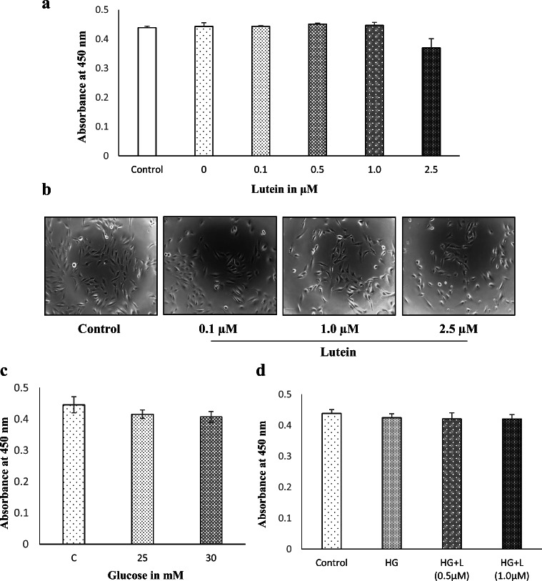
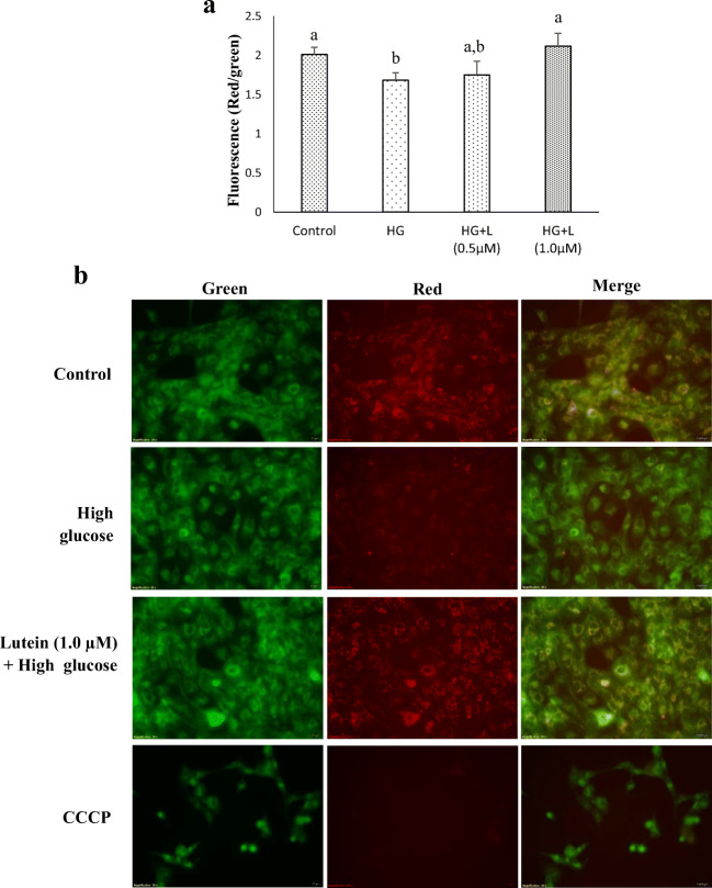
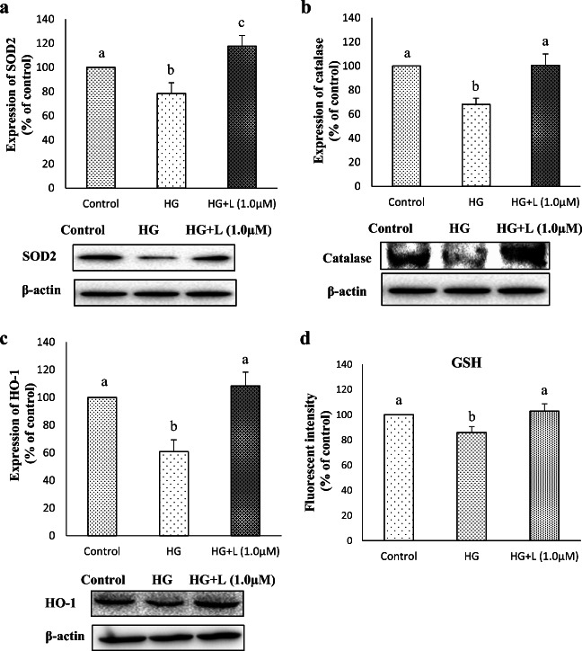
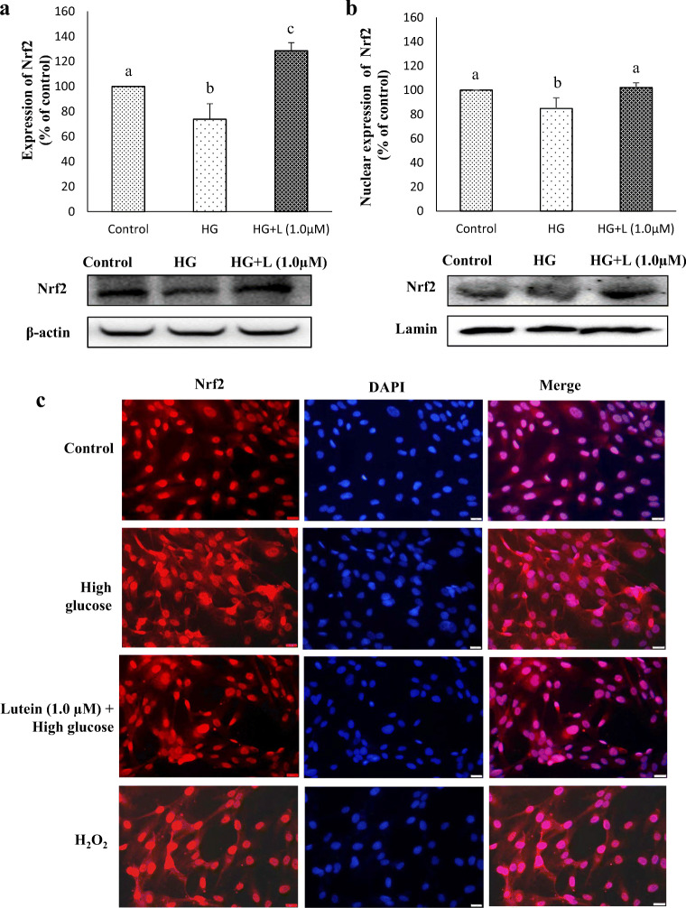
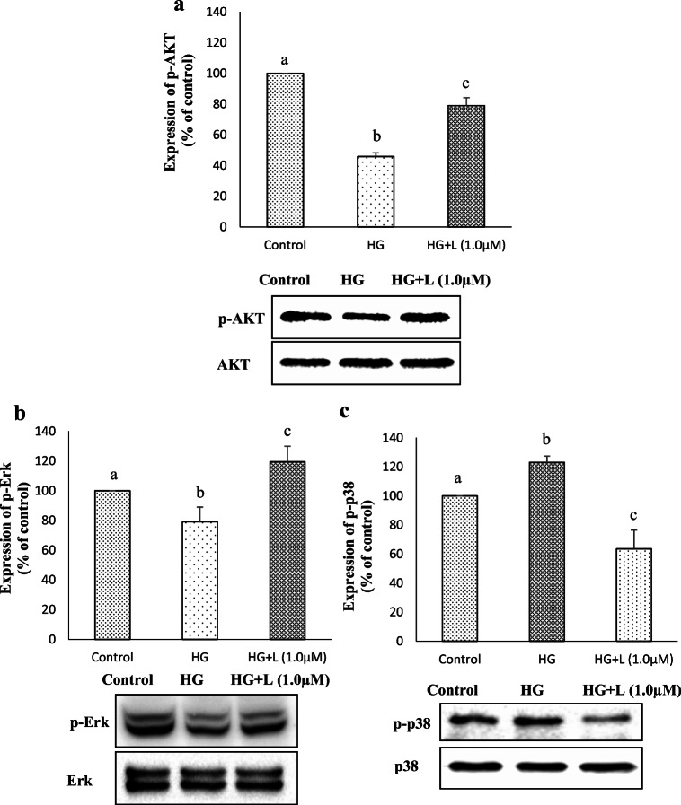
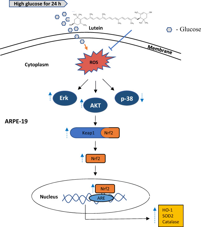
Diabetic retinopathy (DR) is a major cause of acquired blindness among working adults. The retinal pigment epithelium (RPE), constitutes an outer blood-retinal barrier, is vastly affected in diabetic humans and animals. Lower levels of lutein in the serum and retina of diabetic population, and beneficial effects of carotenoids supplementation in diabetic retinopathy patients created an interest to examine the protective effect of lutein on hyperglycemia-mediated changes in oxidative stress and antioxidant defense system in ARPE-19 cells. The WST-1 assay was performed to analyze the impact of glucose, and lutein on the viability of ARPE-19. The intracellular oxidative stress was measured by a DCF (dichlorofluorescein) assay, mitochondrial membrane potential (MMP) was monitored using a JC-10 MMP assay kit and GSH level was examined using GSH/GSSG ratio detection kit. The oxidative stress markers, protein carbonyl and malondialdehyde were spectrophotometrically measured using 2,4-dinitrophenylhydrazine and 2-thiobarbituric acid, respectively. The expression of endogenous antioxidant enzymes and regulatory proteins in ARPE-19 was quantified by western blotting. The localization of Nrf2 protein was examined by immunofluorescent staining. The results show that lutein (up to 1.0 μM) did not affect the viability of ARPE-19 grown in both normal and high-glucose conditions. Lutein treatment blocked high glucose-mediated elevation of intracellular ROS, protein carbonyl and malondialdehyde content in ARPE-19 cells. The decreased MMP and GSH levels observed in ARPE-19 grown under high-glucose condition were rescued by lutein treatment. Further, lutein protected high glucose-mediated down-regulation of a redox-sensitive transcription factor, Nrf2, and antioxidant enzymes, SOD2, HO-1, and catalase. This protective effect of lutein was linked with activated nuclear translocation of Nrf2, which was associated with increased activation of regulatory proteins such as Erk and AKT. Our study indicates that improving the concentration of lutein in the retina could protect RPE from diabetes-associated damage.

Keywords: ARPE-19; Hyperglycemia; Lutein; ROS; Redox signaling.


Conflict of interest statement

The authors declare no conflict of interest.

Figures

Fig. 1
Fig. 1
Isolation and identification of lutein. a Carotenoids profile of Chenopodium album.b HPLC chromatogram of purified lutein from the total carotenoid extract of C. album. c UV-VIS-spectrum of the respective peak of lutein
Fig. 2
Fig. 2
Effect of lutein and glucose on the viability and morphology of ARPE-19 cells. a The cell viability of ARPE-19 treated with lutein at the noted concentrations (0, 0.1, 0.5, 1.0 & 2.5 μM) for 24 h was analyzed by WST-1 assay. Values are mean ± SD (n = 3). b The cell morphology of ARPE-19 treated with lutein for 24 h was observed under a phase-contrast microscope, and the representative picture is presented. c The cell viability of ARPE-19 treated with glucose at the different concentrations (25 & 30 mM) for 24 h was analyzed by WST-1 assay. Values are mean ± SD (n = 3). d The effect of lutein (L-1 μM) on the viability of ARPE-19 cells treated with high-glucose (HG-25 mM) was analyzed by WST-1 assay. Values are mean ± SD (n = 3)
Fig. 3
Fig. 3
Effect of lutein on oxidative status of ARPE-19 cells upon glucose induction. a The intracellular ROS levels were measured as indicated in materials and methods section after pre-treating ARPE-19 with lutein (0.5 μM & 1 μM) for 3 h and with glucose (25 mM) for 24 h. Values are mean ± SD (n = 3); Bars with different letter indicate significant difference (p < 0.05) between the group. b The microscopic images of the intensity of DCF fluorescence of respective experimental group. c ARPE-19 cells were exposed to lutein (1 μM) for 3 h followed by glucose (25 mM) for 24 h. MDA content was assessed using Thiobarbituric Acid-Reactive Substances (TBARS) assay as described in materials and methods. Values are mean ± SD (n = 3); Bars with different letter indicate significant difference (p < 0.05) between the group. d The carbonyl content in ARPE-19 cells treated with lutein (1 μM) for 3 h followed by glucose (25 mM) for 24 h was measured by spectrophotometric DNPH assay as detailed in materials and methods. Values are mean ± SD (n = 3); Bars with different letter indicate significant difference (p < 0.05) between the group
Fig. 4
Fig. 4
Effect of lutein on mitochondrial membrane potential (MMP) in ARPE-19 cells. The MMP was analyzed in ARPE-19 cells treated with lutein (0.5 μM & 1 μM) for 3 h followed by glucose (25 mM) for 24 h using a JC-10 assay kit as indicated in materials and methods. a The quantitative analysis of MMP in the respective treatment group. Values are mean ± SD (n = 3); Bars with different letter indicate significant difference (p < 0.05) between the group. b The fluorescence images of MMP in the respective treatment group
Fig. 5
Fig. 5
Effect of lutein on the expression of redox markers in ARPE-19 cells upon glucose induction. Protein expression of SOD2 (a), catalase (b) and HO-1 (c), and GSH levels (d) in cellular extract of ARPE-19 pre-treated with lutein (1 μM) for 3 h followed by glucose (25 mM) for 24 h was detected by western blotting as described in materials and methods. Values are mean ± SD (n = 3); Bars with different letter indicate significant difference (p < 0.05) between the group. d GSH levels were assessed using GSH assay kit after pre-treatment with lutein (1 μM) followed by glucose (25 mM) treatment. Values are mean ± SD (n = 3); Bars with different letter indicate significant difference (p < 0.05) between the group
Fig. 6
Fig. 6
Effect of lutein on the expression and translocation of Nrf2. a Protein expression of Nrf2 in the cellular extract of ARPE-19 cells pre-treated with lutein (1 μM) for 3 h followed by glucose (25 mM) for 24 h was detected by western blotting as described in materials and methods. Values are mean ± SD (n = 3); Bars with different letter indicate significant difference (p < 0.05) between the group. b Nuclear translocation of Nrf2 in ARPE-19 cells was analyzed by western blotting using the nuclear extract as depicted in materials and methods. Values are mean ± SD (n = 3); Bars with different letter indicate significant difference (p < 0.05) between the group. c Nuclear translocation was confirmed by immunofluorescent assay
Fig. 7
Fig. 7
Effect of lutein on the regulatory markers of Nrf2. Relative activation of AKT (a), Erk (b) and p-38 (c) was analyzed by western blotting as described in materials and methods. Values are mean ± SD (n = 3); Bars with different letter indicate significant difference (p < 0.05) between the group
Fig. 8
Fig. 8
A putative model for therapeutic targets of lutein on hyperglycemia-mediated oxidative damage in ARPE-19. Lutein activates Nrf2 translocation through activating its upstream regulators, PI3K-AKT and Erk1/2, and thereby upregulates protective antioxidant enzymes in hyperglycemic ARPE-19 cells

References

    1. Bai Y, Cui W, Xin Y, Miao X, Barati MT, Zhang C, Chen Q, Tan Y, Cui T, Zheng Y, Cai L. Prevention by sulforaphane of diabetic cardiomyopathy is associated with up-regulation of Nrf2 expression and transcription activation. J Mol Cell Cardiol. 2013;57:82–95. doi: 10.1016/j.yjmcc.2013.01.008. - DOI - PubMed
    1. Ben-Dor A, Steiner M, Gheber L, Danilenko M, Dubi N, Linnewiel K, Zick A, Sharoni Y, Levy J. Carotenoids activate the antioxidant response element transcription system. Mol Cancer Ther. 2005;4:177–186. - PubMed
    1. Bernstein PS, Law WC, Rando RR. Isomerization of all-trans-retinoids to 11-cis-retinoids in vitro. Proc Natl Acad Sci U S A. 1987;84:1849–1853. doi: 10.1073/pnas.84.7.1849. - DOI - PMC - PubMed
    1. Bone RA, Landrum JT, Guerra LH, Ruiz CA. Lutein and zeaxanthin dietary supplements raise macular pigment density and serum concentrations of these carotenoids in humans. J Nutr. 2003;133:992–998. doi: 10.1093/jn/133.4.992. - DOI - PubMed
    1. Boulton M, Dayhaw-Barker P. The role of the retinal pigment epithelium: topographical variation and ageing changes. Eye. 2001;15:384–389. doi: 10.1038/eye.2001.141. - DOI - PubMed