

Management of suprapatellar synovial plica, a common cause of anterior knee pain: a clinical review
- PMID: 31821281
- PMCID: PMC7233704
- DOI: 10.23750/abm.v90i11-S.8781
Management of suprapatellar synovial plica, a common cause of anterior knee pain: a clinical review
Abstract
Background and aim of the work: Suprapatellar synovial plica is caused by a congenital thickening of the synovial membrane and is generally asymptomatic. In the literature, suprapatellar plicae are described as one of the causes of anterior knee pain however, their real role in determining symptoms is controversial. The aim of the current paper is to describe the anatomy, classifications, pathophysiology, symptoms and management of suprapatellar plica syndrome, as well as the differential diagnosis from other causes of anterior knee pain.
Method: Via a search within the MEDLINE/PubMed database, a current review was conducted, and the results summarized.
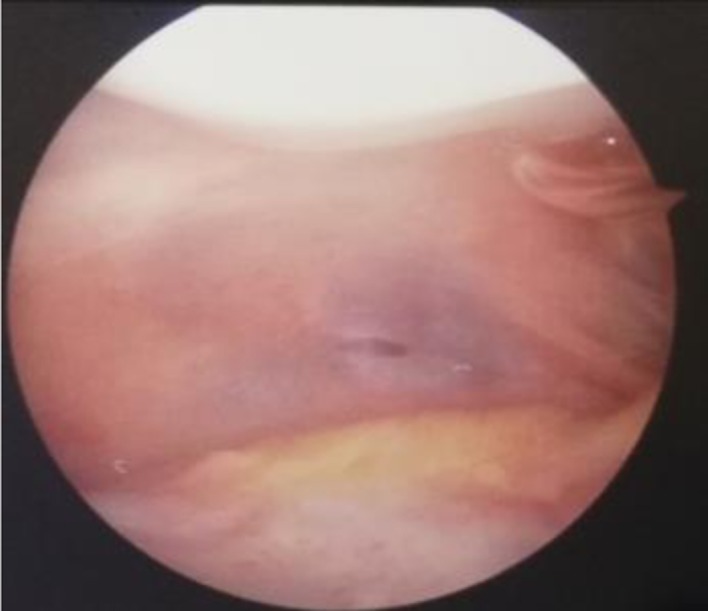
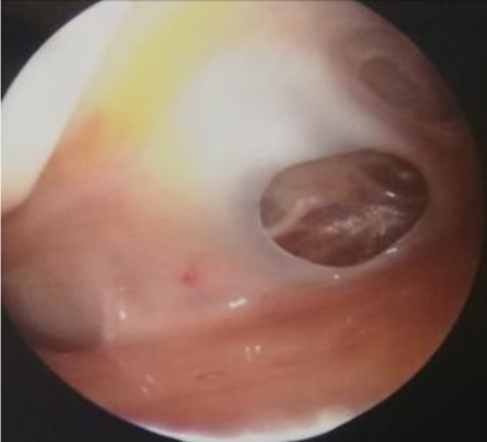
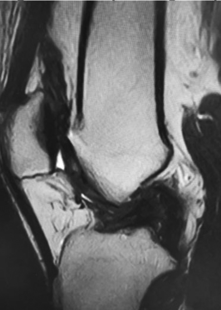
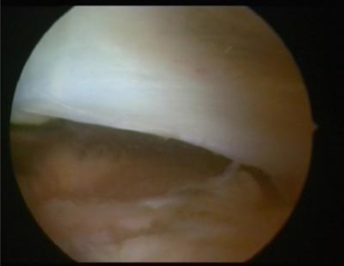
Results: Due to idiopathic, traumatic or inflammatory conditions, plicae can become pathological, causing anterior knee pain with possible knee clicking, swelling, giving way and locking after prolonged flexion of the knee. The diagnosis should be formulated based on an accurate medical history and clinical examination, followed by an appropriate imaging study. However, arthroscopy remains the "golden standard" for detecting all synovial plica.
Conclusions: In patients with anterior knee pain, where doubt is present in the imaging investigation for intraarticular or periarticular lesions, pathological suprapatellar synovial plica must be suspected. The treatment should initially be conservative, but in cases where symptoms persist, patients should undergo arthroscopy to confirm diagnosis and to determine a suitable treatment. In the presence of pathological plica associated with cartilage damage of the femoral condyle or patella at the time of diagnostic arthroscopy, plicae excision leads to favourable results in a high number of cases.
Conflict of interest statement
Each author declares that he or she has no commercial associations (e.g. consultancies, stock ownership, equity interest, patent/licensing arrangement etc.) that might pose a conflict of interest in connection with the submitted article
Figures
References
-
- Patel D. Arthroscopy of the plicae: synovial folds and their significance. Am J Sports Med. 1978;6:217–225. - PubMed
-
- Schindler OS. ‘The Sneaky Plica’ revisited: morphology, pathophysiology and treatment of synovial plicae of the knee. Knee Surg Sports Traumatol Arthrosc. 2014;22(2):247–262. - PubMed
-
- Zmerly H, Akkawi I, Citarella R, El Ghoch M. Clinical Management of Medial Patellar Plica Syndrome: Expert Point of View from Diagnosis to Treatment. Curr Rheumatol Rev. 2018 - PubMed
-
- Patel D. Plica as a cause of anterior knee pain. Orthop Clin North Am. 1986;17:273–7. - PubMed
Publication types
MeSH terms
LinkOut - more resources
Full Text Sources
Research Materials
