

Traumatic extensor tendons injuries of the foot in childhood: a case report
- PMID: 31821310
- PMCID: PMC7233706
- DOI: 10.23750/abm.v90i12-S.8933
Traumatic extensor tendons injuries of the foot in childhood: a case report
Abstract
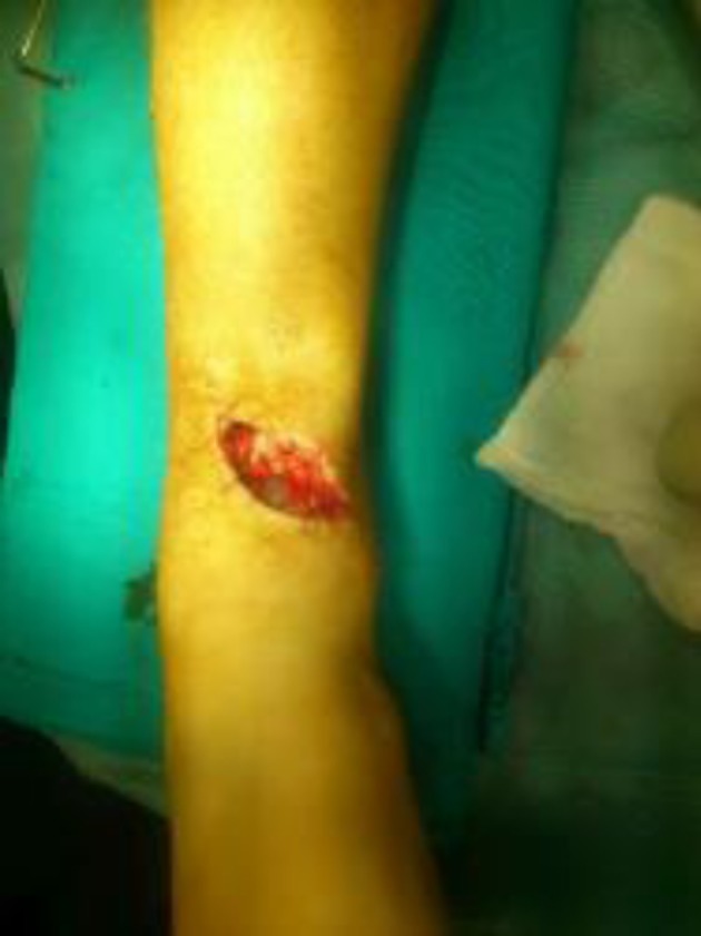
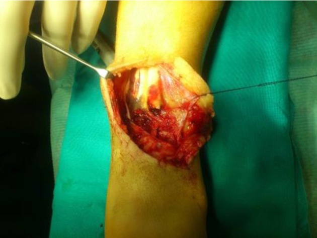
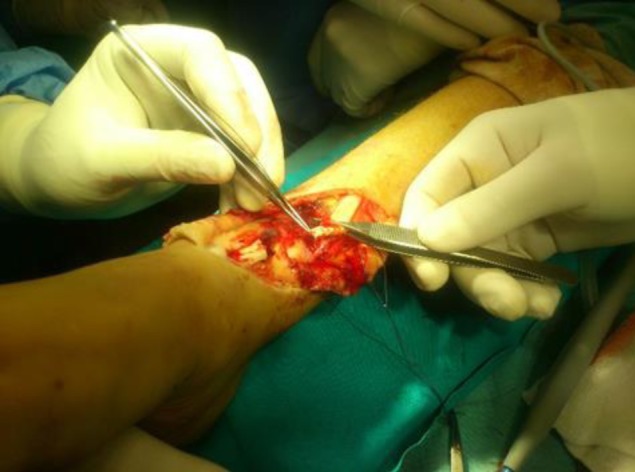
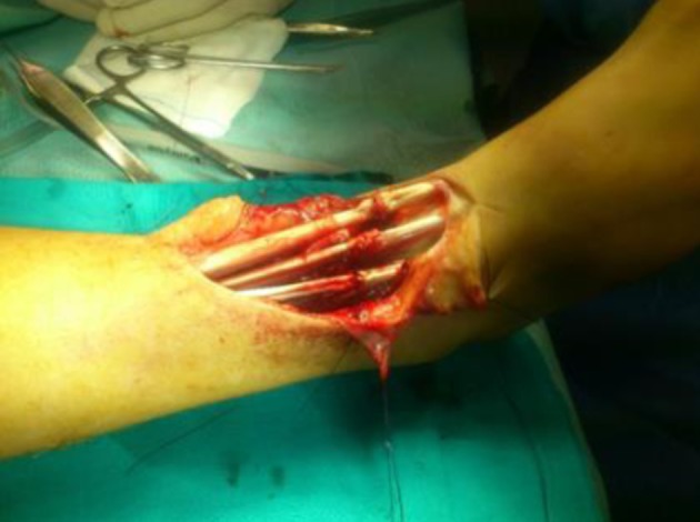
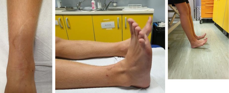
Background and aim of the work: Extensor tendon injuries of the foot in children represent a rare foot injury. We report a case of a 9 year-old male who suffered of a traumatic wound laceration in the distal third of the right leg with a glass the day before in another country, getting a combined injury of tibialis anterior (TA), extensor hallucis longus (EHL) and extensor digitorum longus (EDL).
Methods: After an initial clinical and radiological evaluation, antibiotic prophylaxis was immediately started. Surgery was necessary for the repair of the lesions and after rehabilitation the patient recovered a good function with a complete return to a normal life.
Results: 5 years follow-up clinical examination revealed a complete and painless range of movement comparable to the other foot. The patient regained active dorsiflexion without functional limitations, deformity or contracture.
Conclusions: Early exploration is important to allow full definition of the extent of injury and early surgical repair of tendons is recommended to avoid future disability.
Conflict of interest statement
Each author declares that he or she has no commercial associations (e.g. consultancies, stock ownership, equity interest, patent/licensing arrangement etc.) that might pose a conflict of interest in connection with the submitted article
Figures
References
-
- Sammarco VJ, Sammarco GJ. Injuries to the Tibialis Anterior, Peroneal Tendons, and Long Flexors of the Toes. Baxter’s the Foot and Ankle in Sport. 2008;2nd ed:121–46.
-
- Gwynne-Jones D, Garneti N, Wyatt M. Closed tibialis anterior tendon rupture: a case series. Foot & Ankle International. 2009;30:758–62. - PubMed
-
- Lee MH, Chung CB, Cho JH, Mohana-Borges AV, Pretterklieber ML, Trudell DJ, et al. Tibialis anterior tendon and extensor retinaculum: imaging in cadavers and patients with tendon tear. American Journal of Roentgenology. 2006;187:161–68. - PubMed
-
- Wong JC, Daniel JN, Raikin SM. Repair of acute extensor hallucis longus tendon injuries: a retrospective review. Foot & Ankle Specialist. 2013;7:45–51. - PubMed
-
- Pedrazzini A, Ceccarelli F, Martelli A, Marenghi L, Petraglia F, Romiti D, et al. Return to run after partial amputation of the ankle: clinical assessment and instrumental evaluation. Acta Biomedica. 2013;84:237–43. - PubMed
Publication types
MeSH terms
LinkOut - more resources
Full Text Sources
Medical
Research Materials
