

Persistence of Burkholderia thailandensis E264 in lung tissue after a single binge alcohol episode
- PMID: 31821337
- PMCID: PMC6903738
- DOI: 10.1371/journal.pone.0218147
Persistence of Burkholderia thailandensis E264 in lung tissue after a single binge alcohol episode
Abstract
Background: Binge drinking, an increasingly common form of alcohol use disorder, is associated with substantial morbidity and mortality; yet, its effects on the immune system's ability to defend against infectious agents are poorly understood. Burkholderia pseudomallei, the causative agent of melioidosis can occur in healthy humans, yet binge alcohol intoxication is increasingly being recognized as a major risk factor. Although our previous studies demonstrated that binge alcohol exposure increased B. pseudomallei near-neighbor virulence in vivo and increased paracellular diffusion and intracellular invasion, no experimental studies have examined the extent to which bacterial and alcohol dosage play a role in disease progression. In addition, the temporal effects of a single binge alcohol dose prior to infection has not been examined in vivo.
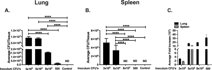
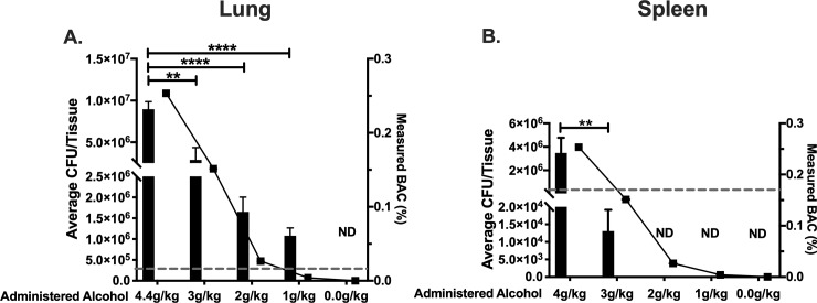
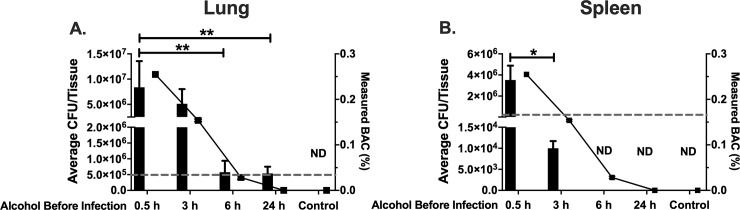
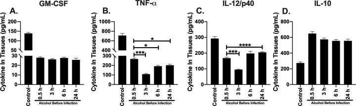
Principal findings: In this study, we used B. thailandensis E264 a close genetic relative of B. pseudomallei, as useful BSL-2 model system. Eight-week-old female C57BL/6 mice were utilized in three distinct animal models to address the effects of 1) bacterial dosage, 2) alcohol dosage, and 3) the temporal effects, of a single binge alcohol episode. Alcohol was administered comparable to human binge drinking (≤ 4.4 g/kg) or PBS intraperitoneally before a non-lethal intranasal infection. Bacterial colonization of lung and spleen was increased in mice administered alcohol even after bacterial dose was decreased 10-fold. Lung and not spleen tissue were colonized even after alcohol dosage was decreased 20 times below the U.S legal limit. Temporally, a single binge alcohol episode affected lung bacterial colonization for more than 24 h after alcohol was no longer detected in the blood. Pulmonary and splenic cytokine expression (TNF-α, GM-CSF) remained suppressed, while IL-12/p40 increased in mice administered alcohol 6 or 24 h prior to infection. Increased lung and not intestinal bacterial invasion was observed in human and murine non-phagocytic epithelial cells exposed to 0.2% v/v alcohol in vitro.
Conclusions: Our results indicate that the effects of a single binge alcohol episode are tissue specific. A single binge alcohol intoxication event increases bacterial colonization in mouse lung tissue even after very low BACs and decreases the dose required to colonize the lungs with less virulent B. thailandensis. Additionally, the temporal effects of binge alcohol alters lung and spleen cytokine expression for at least 24 h after alcohol is detected in the blood. Delayed recovery in lung and not spleen tissue may provide a means for B. pseudomallei and near-neighbors to successfully colonize lung tissue through increased intracellular invasion of non-phagocytic cells in patients with hazardous alcohol intake.
Conflict of interest statement
The authors have declared that no competing interests exist.
Figures
Similar articles
-
A mouse model of binge alcohol consumption and Burkholderia infection.PLoS One. 2018 Nov 28;13(11):e0208061. doi: 10.1371/journal.pone.0208061. eCollection 2018. PLoS One. 2018. PMID: 30485380 Free PMC article.
-
Effects of binge alcohol exposure on Burkholderia thailandensis-alveolar macrophage interaction.Alcohol. 2017 Nov;64:55-63. doi: 10.1016/j.alcohol.2017.04.004. Epub 2017 Aug 19. Alcohol. 2017. PMID: 28965656
-
Impact of Binge Alcohol Intoxication on the Humoral Immune Response during Burkholderia spp. Infections.Microorganisms. 2019 May 9;7(5):125. doi: 10.3390/microorganisms7050125. Microorganisms. 2019. PMID: 31075819 Free PMC article.
-
Inflammation patterns induced by different Burkholderia species in mice.Cell Microbiol. 2008 Jan;10(1):81-7. doi: 10.1111/j.1462-5822.2007.01016.x. Epub 2007 Jul 20. Cell Microbiol. 2008. PMID: 17645551
-
Alcohol Intoxication and Cognition: Implications on Mechanisms and Therapeutic Strategies.Front Neurosci. 2020 Feb 12;14:102. doi: 10.3389/fnins.2020.00102. eCollection 2020. Front Neurosci. 2020. PMID: 32116535 Free PMC article. Review.
Cited by
-
Thinking Outside the Bug: Targeting Outer Membrane Proteins for Burkholderia Vaccines.Cells. 2021 Feb 25;10(3):495. doi: 10.3390/cells10030495. Cells. 2021. PMID: 33668922 Free PMC article. Review.
-
Drivers of melioidosis endemicity: epidemiological transition, zoonosis, and climate change.Curr Opin Infect Dis. 2022 Jun 1;35(3):196-204. doi: 10.1097/QCO.0000000000000827. Curr Opin Infect Dis. 2022. PMID: 35665713 Free PMC article. Review.
-
Exploring patient characteristics and respiratory impacts of pulmonary melioidosis: A 5-year experience from endemic region of Thailand.PLoS Negl Trop Dis. 2025 Jun 25;19(6):e0013222. doi: 10.1371/journal.pntd.0013222. eCollection 2025 Jun. PLoS Negl Trop Dis. 2025. PMID: 40561162 Free PMC article.
-
Hyaladherins May be Implicated in Alcohol-Induced Susceptibility to Bacterial Pneumonia.Front Immunol. 2022 May 12;13:865522. doi: 10.3389/fimmu.2022.865522. eCollection 2022. Front Immunol. 2022. PMID: 35634317 Free PMC article. Review.
-
Dosage scaling of alcohol in binge exposure models in mice: An empirical assessment of the relationship between dose, alcohol exposure, and peak blood concentrations in humans and mice.Alcohol. 2020 Dec;89:9-17. doi: 10.1016/j.alcohol.2020.03.011. Epub 2020 Apr 4. Alcohol. 2020. PMID: 32259574 Free PMC article.
References
-
- Rush B. (1808). An inquiry into the effects of ardent spirits upon the human body and mind: With an account of the means of preventing, and of the remedies for curing them (4th ed.). Philadelphia: Printed for Thomas Dobson; Archibald Bartram, printer.
Publication types
MeSH terms
Substances
Supplementary concepts
Associated data
LinkOut - more resources
Full Text Sources
