. 2019 Dec 20;8(12):bio043950.
doi: 10.1242/bio.043950.

The interconnection between cytokeratin and cell membrane-bound β-catenin in Sertoli cells derived from juvenile Xenopus tropicalis testes

Affiliations

The interconnection between cytokeratin and cell membrane-bound β-catenin in Sertoli cells derived from juvenile Xenopus tropicalis testes

Thi Minh Xuan Nguyen et al. Biol Open. .

Abstract

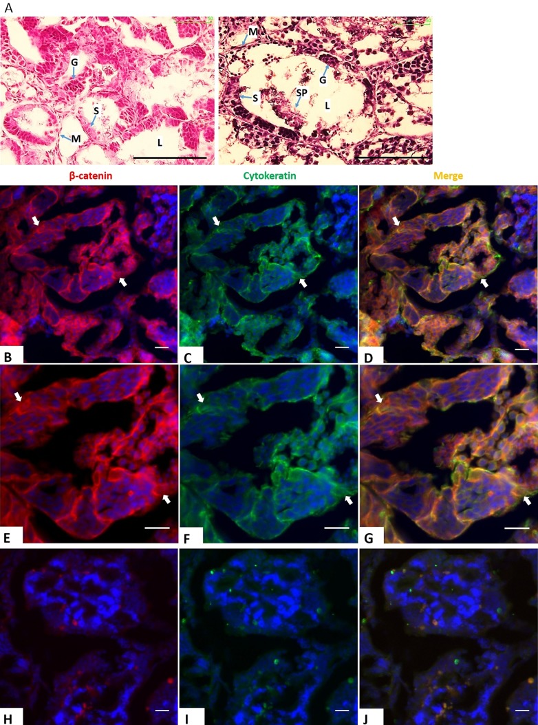
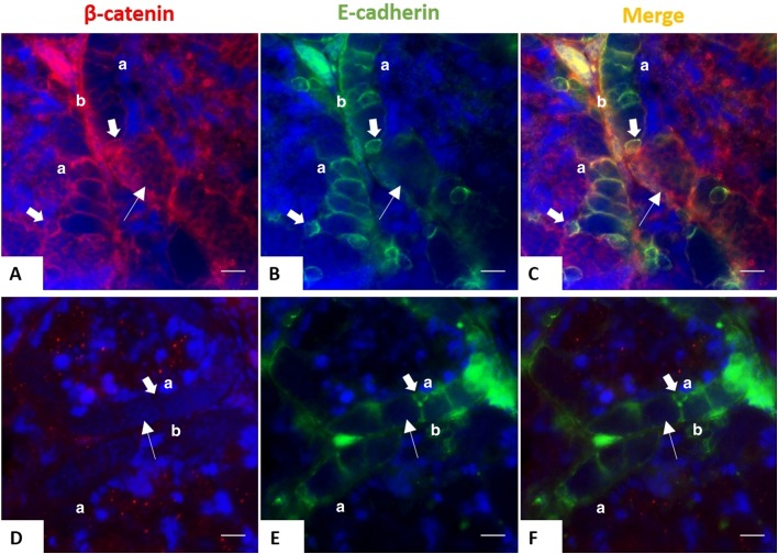
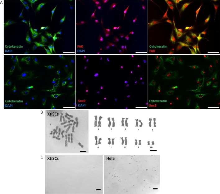
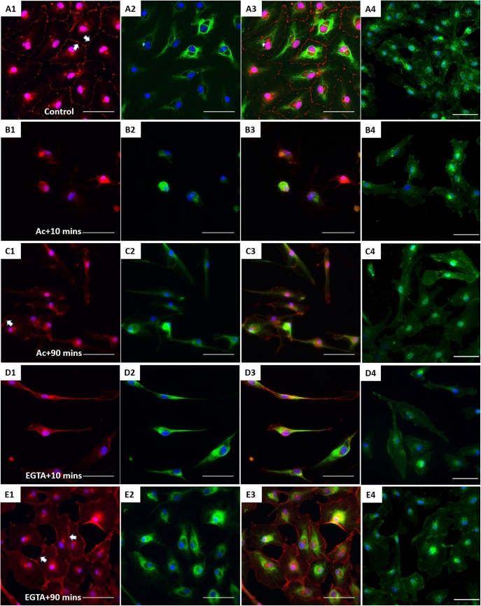
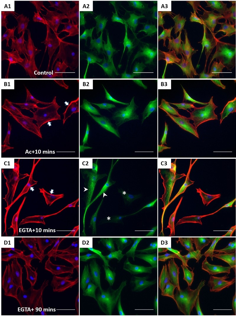
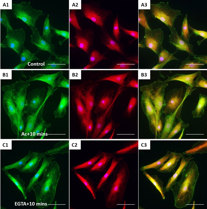
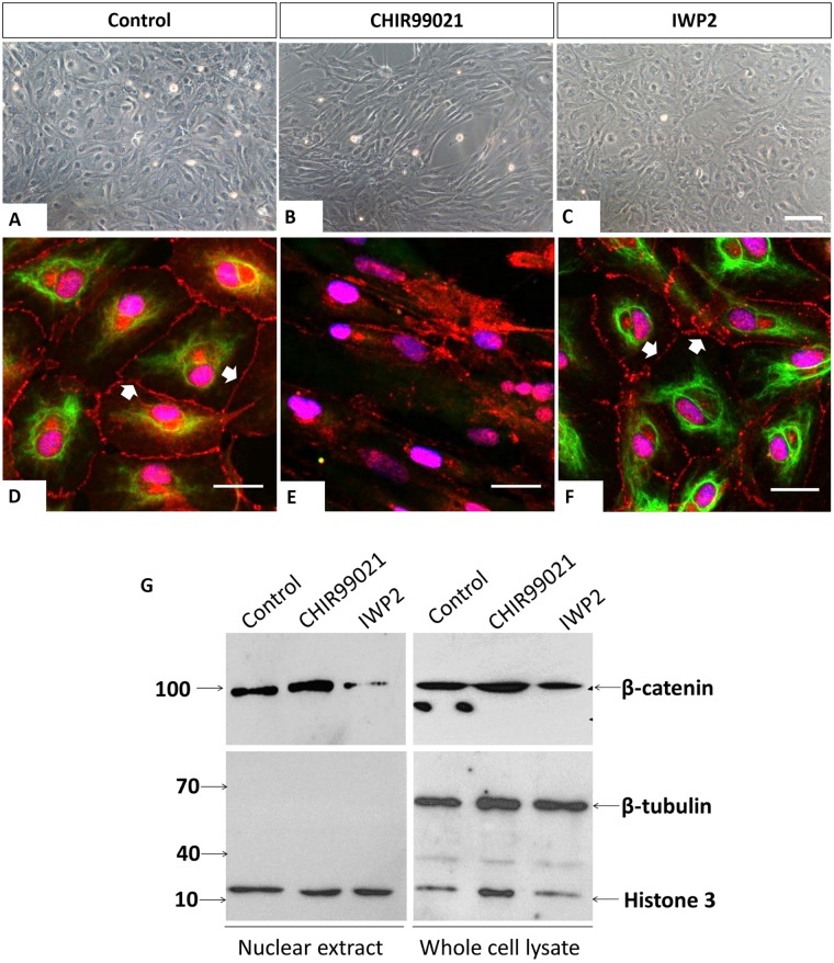
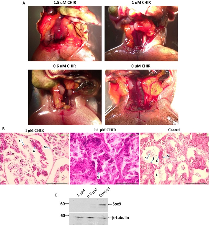
Sertoli cells (SCs) play a central role in the determination of male sex during embryogenesis and spermatogenesis in adulthood. Failure in SC development is responsible for male sterility and testicular cancer. Before the onset of puberty, SCs are immature and differ considerably from mature cells in post-pubertal individuals regarding their morphology and biochemical activity. The major intermediate filament (IF) in mature SCs is vimentin, anchoring germ cells to the seminiferous epithelium. The collapse of vimentin has resulted in the disintegration of seminiferous epithelium and subsequent germ cell apoptosis. However, another IF, cytokeratin (CK) is observed only transiently in immature SCs in many species. Nevertheless, its function in SC differentiation is poorly understood. We examined the interconnection between CK and cell junctions using membrane β-catenin as a marker during testicular development in the Xenopus tropicalis model. Immunohistochemistry on juvenile (5 months old) testes revealed co-expression of CK, membrane β-catenin and E-cadherin. Adult (3-year-old males) samples confirmed only E-cadherin expression; CK and β-catenin were lost. To study the interconnection between CK and β-catenin-based cell junctions, the culture of immature SCs (here called XtiSCs) was employed. Suppression of CK by acrylamide in XtiSCs led to breakdown of membrane-bound β-catenin but not F-actin and β-tubulin or cell-adhesion proteins (focal adhesion kinase and integrin β1). In contrast to the obvious dependence of membrane β-catenin on CK stability, the detachment of β-catenin from the plasma membrane via uncoupling of cadherins by Ca2+ chelator EGTA had no effect on CK integrity. Interestingly, CHIR99021, a GSK3 inhibitor, also suppressed the CK network, resulting in the inhibition of XtiSCs cell-to-cell contacts and testicular development in juvenile frogs. This study suggests a novel role of CK in the retention of β-catenin-based junctions in immature SCs, and thus provides structural support for seminiferous tubule formation and germ cell development.

Keywords: Adherens junctions; Cytokeratin; Immature Sertoli cells; Testicles; Xenopus tropicalis.


Conflict of interest statement

Competing interestsThe authors declare no competing or financial interests.

Figures

Fig. 1.
Fig. 1.
Co-expression of CK and β-catenin during testicular development. (A) H&E staining of 5-month-old (left) and 3-year-old (right) X. tropicalis testes. Scale bars: 100 μm. L, lumen; S, Sertoli cells; G, germ cells; SP, spermatid; M, mesenchyme. (B–J) Double staining of testicular sections of juvenile (B–G) and adult frogs (H–J) with CK (mouse, green) and β-catenin (rabbit, red) antibodies; E–G show higher magnification of young testes' staining. Nuclei were stained with DAPI (blue). Scale bars: 20 μm. Both proteins were found in the immature testes, but not in the adulthood. Arrows indicate SCs.
Fig. 2.
Fig. 2.
Expression of E-cadherin and β-catenin in SCs during testicular development. Double staining on testicular sections of juvenile (5-month-old) (A–C) and adult (3-year-old) frogs (D–F) with E-cadherin (mouse, green) and β-catenin (rabbit, red) antibodies. Nuclei were stained with DAPI (blue). Scale bars: 20 μm. Both proteins surrounded the SCs from juvenile testes, but only E-cadherin is expressed in adulthood. Thick arrows indicate SCs. Thinner arrows show germ cells. a, apical; b, basal.
Fig. 3.
Fig. 3.
Immunofluorescent, cytogenetic and transformation characteristics of isolated XtiSCs. (A) XtiSCs expressed SC proteins, including focal adhesion kinase (FAK, red), Sox9 (red) and CK (green), an immature SC marker. Nuclei were stained with DAPI. Chromosome analysis (B) and soft agar assay (C) showed XtiSCs as non-transformed cells. Scale bars: 50 μm (A), 10 μm (B), 400 μm (C).
Fig. 4.
Fig. 4.
The effect of CK network on the β-catenin-based cell junctions. XtiSCs were treated with vehicles (Control, A) or 10 mM acrylamide (Ac; B,C) or 2 mM EGTA (EGTA; D,E). After treatment, cells were washed and changed to the fresh medium and then collected at the indicated time points: 10 min (Ac+10 min or EGTA+10 min, B,D) or 90 min (Ac+90 min or EGTA+90 min, C,E) for immunofluorescent staining with antibodies against β-catenin (red, A1–E1), CK (green, A2–E2) and merge (A3–E3). (A4–E4) Fluorescent images of WGA-stained cells showing the cell shape and cytoplasmic membrane. Arrows show membrane β-catenin. Nuclei were stained with DAPI (blue). Scale bars: 50 μm.
Fig. 5.
Fig. 5.
The effect of acrylamide and EGTA on F-actin and tubulin. After treatment, XtiSCs were collected at the indicated time points for immunofluorescent staining with antibodies against F-actin (red, A1–D1), β-tubulin (green, A2–D2) and merge (A3–D3). Arrows show thick membrane F-actin; the aggregates of β-tubulin are marked by arrowheads and asterisks indicate the cells without β-tubulin. Nuclei were stained with DAPI (blue). Scale bars: 50 μm.
Fig. 6.
Fig. 6.
The effect of acrylamide and EGTA on cell adhesion proteins. Immunofluorescent images of control XtiSCs (A) and XtiSCs 10 min after washing out the acrylamide (B) or EGTA (C) staining with antibodies against integrin β1 (CD29, green, A1–C1), focal adhesion kinase (FAK, red, A2–C2) and merge (A3–C3). Nuclei were stained with DAPI (blue). Scale bars: 50 μm.
Fig. 7.
Fig. 7.
CK regulates plasma membrane β-catenin. (A–C) Phase-contrast images of XtiSCs after treatment with CHIR99021 and IWP2 for 3 days show the morphological changes in CHIR99021-treated cells. XtiSCs were collected for immunofluorescence (D–F) or immunoblotting analysis (G). Immunostaining of CHIR99021-treated XtiSCs against CK (green) and β-catenin (red) reveals the disruption of the CK network and cell-to-cell contact altogether with the disappearance of membrane β-catenin. Nuclei were stained with DAPI (blue). Scale bars: 20 μm. Arrows indicate membrane-bound β-catenin. The downregulation of nuclear β-catenin in media supplemented with IWP2 was confirmed by immunoblotting (G). Histone H3 is a marker of nuclei. The absence of β-tubulin shows the purity of the nuclear lysate.
Fig. 8.
Fig. 8.
In vivo treatment of CHIR99021 led to failure in testicular development. (A) 2.5 months after CHIR99021 was injected into the dorsal sac, no testes were observed in 1.5 μM-injected frogs. Arrows indicate testes. (B) H&E staining of testicular sections from 1, 0.6 and 0 μM CHIR-injected X. tropicalis frogs. The most serious damage, the detachment of germinal epithelium from tubules, was observed in the 1 μM group. Testes with 0.6 μM CHIR had unorganized seminiferous tubules. Scale bars: 100 μm. L, lumen; S, Sertoli cells; G, germ cells; SP, spermatid; M, mesenchyme. (C) Immunoblotting of testicular extract from 1, 0.6 and 0 μM CHIR-injected X. tropicalis with antibody against Sox9 (SC marker). β-tubulin is as a loading control.

References

    1. Ameen N. A., Figueroa Y. and Salas P. J. (2001). Anomalous apical plasma membrane phenotype in CK8-deficient mice indicates a novel role for intermediate filaments in the polarization of simple epithelia. J. Cell Sci. 114, 563-575. - PubMed
    1. Appert A., Fridmacher V., Locquet O. and Magre S. (1998). Patterns of keratins 8, 18 and 19 during gonadal differentiation in the mouse: sex- and time-dependent expression of keratin 19. Differentiation 63, 273 10.1046/j.1432-0436.1998.6350273.x - DOI - PubMed
    1. Barrionuevo F., Burgos M. and Jiménez R. (2011). Origin and function of embryonic Sertoli cells. Biomol. Concepts 2, 537-547. 10.1515/BMC.2011.044 - DOI - PubMed
    1. Bhattacharjee R., Goswami S., Dudiki T., Popkie A. P., Phiel C. J., Kline D. and Vijayaraghavan S. (2015). Targeted disruption of glycogen synthase kinase 3A (GSK3A) in mice affects sperm motility resulting in male infertility. Biol. Reprod. 92, 65 10.1095/biolreprod.114.124495 - DOI - PMC - PubMed
    1. Bilic J., Huang Y. L., Davidson G., Zimmermann T., Cruciat C. M., Bienz M. and Niehrs C. (2007). Wnt induces LRP6 signalosomes and promotes dishevelled-dependent LRP6 phosphorylation. Science 316, 1619-1622. 10.1126/science.1137065. - DOI - PubMed