. 2020 Mar;75(3):227-236.
doi: 10.1136/thoraxjnl-2019-213571. Epub 2019 Dec 10.

GLUT1-dependent glycolysis regulates exacerbation of fibrosis via AIM2 inflammasome activation

Affiliations

GLUT1-dependent glycolysis regulates exacerbation of fibrosis via AIM2 inflammasome activation

Soo Jung Cho et al. Thorax. 2020 Mar.

Abstract

Background: Idiopathic pulmonary fibrosis (IPF) is a rapidly progressive, fatal lung disease that affects older adults. One of the detrimental natural histories of IPF is acute exacerbation of IPF (AE-IPF), of which bacterial infection is reported to play an important role. However, the mechanism by which bacterial infection modulates the fibrotic response remains unclear.

Objectives: Altered glucose metabolism has been implicated in the pathogenesis of fibrotic lung diseases. We have previously demonstrated that glucose transporter 1 (GLUT1)-dependent glycolysis regulates fibrogenesis in a murine fibrosis model. To expand on these findings, we hypothesised that GLUT1-dependent glycolysis regulates acute exacerbation of lung fibrogenesis during bacterial infection via AIM2 inflammasome activation.

Results: In our current study, using a murine model of Streptococcus pneumoniae (S. pneumoniae) infection, we investigated the potential role of GLUT1 on mediating fibrotic responses to an acute exacerbation during bleomycin-induced fibrosis. The results of our current study illustrate that GLUT1 deficiency ameliorates S. pneumoniae-mediated exacerbation of lung fibrosis (wild type (WT)/phosphate buffered saline (PBS), n=3; WT/S. pneumoniae, n=3; WT/Bleomycin, n=5 ; WT/Bleomycin+S. pneumoniae, n=7; LysM-Cre-Glut1fl/f /PBS, n=3; LysM-Cre-Glut1fl/fl /S. pneumoniae, n=3; LysM-Cre-Glut1fl/fl /Bleomycin, n=6; LysM-Cre-Glut1fl/fl /Bleomycin+S. pneumoniae, n=9, p=0.041). Further, the AIM2 inflammasome, a multiprotein complex essential for sensing cytosolic bacterial DNA as a danger signal, is an important regulator of this GLUT1-mediated fibrosis and genetic deficiency of AIM2 reduced bleomycin-induced fibrosis after S. pneumoniae infection (WT/PBS, n=6; WT/Bleomycin+S. pneumoniae, n=15; Aim2-/-/PBS, n=6, Aim2-/-/Bleomycin+S. pneumoniae, n=11, p=0.034). GLUT1 deficiency reduced expression and function of the AIM2 inflammasome, and AIM2-deficient mice showed substantial reduction of lung fibrosis after S. pneumoniae infection.

Conclusion: Our results demonstrate that GLUT1-dependent glycolysis promotes exacerbation of lung fibrogenesis during S. pneumoniae infection via AIM2 inflammasome activation.

Keywords: idiopathic pulmonary fibrosis; interstitial fibrosis; pneumonia.


Conflict of interest statement

Competing interests: None declared.

Figures

Figure 1
Figure 1
Streptococcal pneumoniae infection exacerbates bleomycin-induced lung fibrosis. (A) Experimental layout of primary bleomycin-induced lung injury (day 0 instillation of bleomycin 0.01 mg/mouse followed by day 14 instillation of PBS) and S. pneumoniae infection (day 0 instillation of bleomycin 0.01 mg/mouse followed by day 14 instillation of 1×10⁵ CFU of S. pneumoniae). (B) Representative lung sections of bleomycin-treated mice subsequently infected with S. pneumoniae. Stained with Masson trichrome staining. Scale bars 200 μm. (C) Total lung collagen was quantified by Sircol assay (PBS/PBS, n=3; PBS/S. pneumoniae, n=3; bleomycin/PBS, n=14, bleomycin/S. pneumoniae, n=14). Fold change is relative to control lungs. Data are mean±95% Cl. *p<0.05 by analysis of variance. (D) Immunoblot analysis for collagen type 1 in bleomycin-treated lung tissue lysates in response to S. pneumoniae infection. β-actin served as the standard. Results are representative of three or more independent experiments.
Figure 2
Figure 2
GLUT1-dependent glycolysis is increased in bleomycin-treated mice after Streptococcal pneumoniae infection. (A) 18F-FDG-PET scan of lungs from primary bleomycin-induced lung injury (day 0 instillation of bleomycin followed by day 14 instillation of PBS) and S. pneumoniae infection 6 (day 0 instillation of bleomycin followed by day 14 instillation of 1×10⁶ CFU of S. pneumoniae). 18F-FDG uptake is more in bleomycin-treated lung in response to S.pneumoniae infection compared with controls. (B) lmmunoblot analysis for GLUT1 in bleomycin-treated lung tissue lysates in response to S. pneumoniae infection. β-actin served as the standard. (C) Immunohistochemical staining of GLUT1 (red) in bleomycin-treated lung reveals enhanced signal in fibrotic foci and inflammatory cells (solid arrow). Scale bars 200 µm. Results are representative of three or more independent experiments. GLUT1, glucose transporter 1; 18F-FDG-PET, 18F-FDG-positron emission tomography.
Figure 3
Figure 3
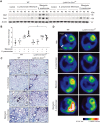
GLUT1-dependent glycolysis regulates exacerbation of lung fibrosis after Streptococcal pneumoniae infection. (A) Immunoblot analysis for Glut1 and collagen type 1 in bleomycin-treated WT and LysM-Cre-Glutimmicefl/fl after S. pneumoniae infection. β-actin served as the standard. (B) Total lung collagen was quantified by Sircol assay (WT/PBS, n = 3; WT/S. pneumoniae, n = 3, WT/Bleomycin n = 5, WT/Bleomycin+S . pneumoniae, n = 6; LysM-Cre-Glutlfl/fl/PBS, n = 3; LysM-Cre-Glut1fl/fl /S. pneumoniae, n = 3; LysM-Cre-Giutr1 fl/ flBleomycin, n=6; LysM-Cre-G/ut1fl/fl/Bleomycin+S . pneumoniae, n=6). Fold change is relative to PBS-treated WT lungs. Data are mean±95% Cl. *p<0.05 by ANOVA. (C) Representative lung sections of WT and LysM-Cre-Glut 1 fl/fl mice after bleomycin treatment followed by S. pneumoniae infection. Stained with Masson trichrome staining. Scale bars 200 µm. (D) 18F-FDG-PET scan showed that FDG uptake is less in LysM-Cre-Glut 1 fl/fl mice after bleomycin treatment followed by S. pneumoniae infection. Results are representative of two or more independent experiments.18F-FDG-PET, 18F-FDG-positron emission tomography; WT, wild type.
Figure 4
Figure 4
AIM2 inflammasome expression and activation is augmented in bleomycin-treated lung after Streptococcal pneumoniae infection. (A) Total cell count in bronchoalveolar lavage (BAL) in bleomycin-treated mice 3 days after S. pneumoniae infection. Data are mean±95% Cl. *p<0.05. (B) Immunohistochemical staining of AIM2 (red) in bleomycin-treated lung reveals enhanced signal in inflammatory cells (solid arrow). Scale bars 200 µm. (C) lmmunoblot analysis for AIM2, activated caspase-1, cleaved IL-1β (black arrows) in bleomycin-treated lung after S. pneumoniae infection.(β-actin served as the standard). Amounts of IL-1β (D) and IL-18 (E), as determined by ELISA in lung tissues (50 µg) after S. pneumoniae infection for 3 days (PBS/PBS, n=3; PBS/S. pneumoniae, n=3; Bleomycin/PBS, n=8, Bleomycin/S. pneumoniae, n=8). Data are mean±95% Cl. *p<0.05. Results are representative of two or more independent experiments. BAL, bronchoalveolar lavage; IL, interleukin.
Figure 5
Figure 5
GLUT1-dependent glycolysis regulates AIM2 inflammasome activation in vitro. (A) Extracellular acidification rate (ECAR) was measured in BMDMs from Nlrp3−/ mice (BMDMs, 1×10⁵ cells/well) transfected with non-target siRNA (control siRNA) or siRNA for Glut1 24 hours prior to LPS and poly(dA:dT) stimulation, a potent AIM2 inflammasome activator. Data are mean±SEM. (B) lmmunoblot analysis for activated caspase-1, cleaved IL-1β (black arrows) in BMDMs transfected with control siRNA or siRNA for Glut1 24 hours prior to LPS and poly(dA:dT) stimulation. β-actin served as the standard. (C) Quantification of IL-1β levels from BMDMs from Nlrp3−/ mice transfected with control siRNA or siRNA for Glut1 24 hours prior to LPS and poly(dA:dT) stimulation. Representative immunofluorescence images (n=5 individual images per group) of ASC speck formation (white arrows) images (D) and quantification (E) in BMDMs from Nlrp3−/ mice transfected with control siRNA or siRNA for Glut1 that were stimulated without or with LPS and poly(dA:dT). Scale bars, 20 µm. Throughout, data are mean±95% Cl. *p<0.05, **p<0.01 by analysis of variance. Results are representative of three or more independent experiments. BMDMs, bone marrow–derived macrophages; IL, interleukin; GLUT1, glucose transporter 1.
Figure 6
Figure 6
GLUT1-dependent glycolysis regulates AIM2 inflammasome activation in murine fibrosis exacerbation model. (A) Total BAL cell count in bleomycin-treated WT and LysM-Cre-Giut1fl/fl mice 3 days after Streptococcal pneumoniae infection. (B) lmmunoblot analysis for AIM2, activated caspase-1, cleaved IL-1β (black arrows) in bleomycin-treated lung after S. pneumoniae infection. β-actin served as the standard. Amounts of IL-1β (C), IL-18 (D), as determined by ELISA in lung tissues (50 µg) after S. pneumoniae infection for 3 days. (WT/PBS, n=3; WT/Bleomycin+S. pneumoniae, n=7; LysM-Cre-Giut1fl/fl///PBS, n=8, LysM-Cre-Giut1fl/fl/Bieomycin+S. pneumoniae, n=1 0). Throughout, data are mean±95% Cl. *p<0.05 by analysis of variance. Results are representative of three or more independent experiments. BAL, bronchoalveolar lavage; IL, interleukin; GLUT1, glucose transporter 1; WT, wild type.
Figure 7
Figure 7
Deficiency of AIM2 ameliorates fibrosis exacerbation in bleomycin-treated lung after Streptococcal pneumoniae infection. (A) Representative lung sections of WT and AIM2−/ mice after bleomycin treatment followed by S. pneumoniae infection. Stained with Masson trichrome staining. Scale bars 200 µm. (B) Total lung collagen was quantified by Sircol assay (WT/PBS, n=6; WT/Bleomycin+S. pneumoniae, n=15; AIM2−/ /PBS, n=6, AIM2/Bleomycin+S. pneumoniae, n=11). (C) Immunoblot analysis for collagen type 1 in bleomycin-treated WT and Aim2−/ mice after S. pneumoniae infection. β-actin served as the standard. (D) Immunoblot analysis for AIM2, activated caspase-1, cleaved IL-1β (black arrows) in bleomycin-treated WT and AIM2−/ mice after S. pneumoniae infection. β-actin served as the standard. Amounts of IL-β-β (E), IL-18 (F), as determined by ELISA in lung tissues (50 µg) after S. pneumoniae infection for 3 days (WT/PBS, n=3; WT/Bleomycin+S. pneumoniae, n=10; Aim2−/ /PBS, n=3, Aim2−/ /Bleomycin+S. pneumoniae, n=5). Throughout, data are mean±95% Cl. *p<0.05 by analysis of variance. Results are representative of three or more independent experiments. (G) Schematic of the proposed relationships between GLUT1-dependent glycolysis and AIM2 inflammasome activation-mediated fibrosis. IL, interleukin; GLUT1, glucose transporter 1; WT, wild type.

Comment in

References

    1. Raghu G, Collard HR, Egan JJ, et al. . An official ATS/ERS/JRS/ALAT statement: idiopathic pulmonary fibrosis: evidence-based guidelines for diagnosis and management. Am J Respir Crit Care Med 2011;183:788–824.10.1164/rccm.2009-040GL - DOI - PMC - PubMed
    1. Kim HJ, Perlman D, Tomic R. Natural history of idiopathic pulmonary fibrosis. Respir Med 2015;109:661–70.10.1016/j.rmed.2015.02.002 - DOI - PubMed
    1. Lederer DJ, Martinez FJ, Fibrosis IP. Idiopathic pulmonary fibrosis. N Engl J Med Overseas Ed 2018;378:1811–23.10.1056/NEJMra1705751 - DOI - PubMed
    1. Martinez FJ, Collard HR, Pardo A, et al. . Idiopathic pulmonary fibrosis. Nat Rev Dis Primers 2017;310.1038/nrdp.2017.74 - DOI - PubMed
    1. Molyneaux PL, Maher TM. The role of infection in the pathogenesis of idiopathic pulmonary fibrosis. Eur Respir Rev 2013;22:376–81.10.1183/09059180.00000713 - DOI - PMC - PubMed

Publication types

MeSH terms