. 2019 Dec 26;116(52):26614-26624.
doi: 10.1073/pnas.1915372116. Epub 2019 Dec 10.

Reactivation of Epstein-Barr virus by a dual-responsive fluorescent EBNA1-targeting agent with Zn2+-chelating function

Affiliations

Reactivation of Epstein-Barr virus by a dual-responsive fluorescent EBNA1-targeting agent with Zn2+-chelating function

Lijun Jiang et al. Proc Natl Acad Sci U S A. .

Erratum in

Abstract

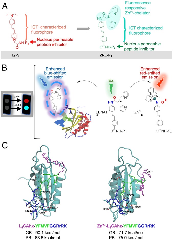
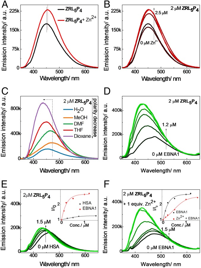
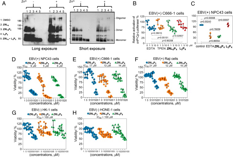
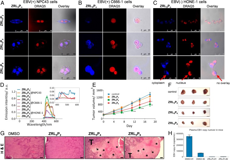
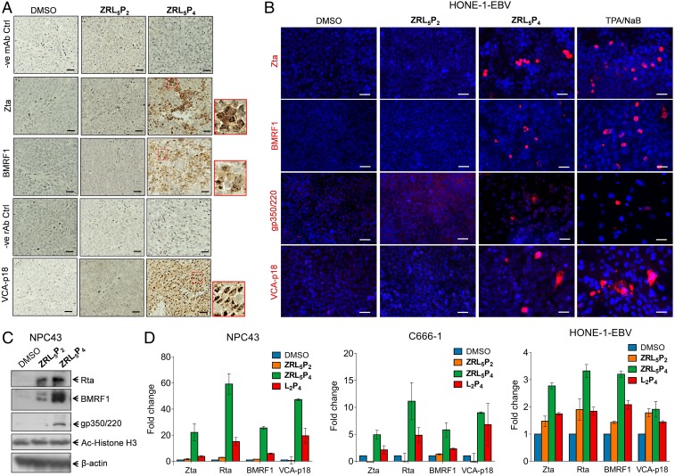
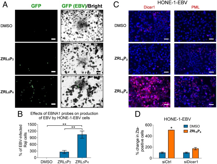
Epstein-Barr nuclear antigen 1 (EBNA1) plays a vital role in the maintenance of the viral genome and is the only viral protein expressed in nearly all forms of Epstein-Barr virus (EBV) latency and EBV-associated diseases, including numerous cancer types. To our knowledge, no specific agent against EBV genes or proteins has been established to target EBV lytic reactivation. Here we report an EBNA1- and Zn2+-responsive probe (ZRL5P4) which alone could reactivate the EBV lytic cycle through specific disruption of EBNA1. We have utilized the Zn2+ chelator to further interfere with the higher order of EBNA1 self-association. The bioprobe ZRL5P4 can respond independently to its interactions with Zn2+ and EBNA1 with different fluorescence changes. It can selectively enter the nuclei of EBV-positive cells and disrupt the oligomerization and oriP-enhanced transactivation of EBNA1. ZRL5P4 can also specifically enhance Dicer1 and PML expression, molecular events which had been reported to occur after the depletion of EBNA1 expression. Importantly, we found that treatment with ZRL5P4 alone could reactivate EBV lytic induction by expressing the early and late EBV lytic genes/proteins. Lytic induction is likely mediated by disruption of EBNA1 oligomerization and the subsequent change of Dicer1 expression. Our probe ZRL5P4 is an EBV protein-specific agent that potently reactivates EBV from latency, leading to the shrinkage of EBV-positive tumors, and our study also suggests the association of EBNA1 oligomerization with the maintenance of EBV latency.

Keywords: EBNA1-targeting agent; EBV-specific lytic inducer; dual-responsive fluorescent EBV probe.


Conflict of interest statement

Competing interest statement: K.-L.W., N.K.M., and L.J. are listed as inventors on a filed US nonprovisional patent titled “Zinc-binder based EBNA1-specific compounds” with the application number 16/249,987.

Figures

Fig. 1.
Fig. 1.
Chemical, fluorescent, and EBNA1-binding characteristics of the Zn2+ chelator EBNA1 probe ZRL5P4. (A) Chemical structures of L2P4 and ZRL5P4. (B) Schematic illustration of the emission response of ZRL5P4, binding to Zn2+ and EBNA1. (C) Representative conformations of ZRL5P4 and Zn2+-ZRL5P4 in the MD simulations. The calculated generalized Born (GB) and Poisson–Boltzmann (PB) values represent the binding free energy between EBNA1 and ZRL5P4 or Zn2+-ZRL5P4.
Fig. 2.
Fig. 2.
Dual-responsive emission of ZRL5P4 (2 µM; excitation 337 nm) toward Zn2+ and EBNA1. (A) Fluorescence spectral changes of ZRL5P4 in the presence of Zn2+ in aqueous solution (CH3CN/0.05 M Hepes [pH 7.4], 50:50). (B) Fluorescence spectral changes of ZRL5P4 upon gradual addition of Zn2+ in aqueous solution (CH3CN/0.05 M Hepes [pH 7.4], 50:50). (C) Solvation study of ZRL5P4 with a decrease of solvent polarity. Fluorescence spectral changes of ZRL5P4 in PBS upon addition of (D) EBNA1 and (E) HSA and upon addition of EBNA1 in the presence of (F) 1 equivalent Zn2+. (E, Inset) The selectivity of ZRL5P4 toward EBNA1 over HSA. (F, Inset) Interaction of ZRL5P4 with EBNA1 in the presence and absence of Zn2+. a.u., arbitrary units.
Fig. 3.
Fig. 3.
ZRL5P4 inhibits EBNA1 oligomerization and transactivation and cell viability. (A) Full- length EBNA1 self-association in the absence/presence of Zn2+ by ZRL5/ZRL5P4/L2P4/ZRL5 + L2P4; 0.1% DMSO serves as the solvent control. Long and short exposure images of the same blot are shown for comparison of various forms of EBNA1 self-association. Analysis of the effects of EBNA1 probes (ZRL5P4, L2P4) on the oriP-enhanced transactivation in EBV-positive (B) C666-1 and (C) NPC43 cells. The transactivation activities were detected by the oriP-Cp-luciferase reporter. EDTA and TPEN are the known chelators of Zn2+. (DH) Cytotoxic activities of the EBNA1 probes ZRL5P2, ZRL5P4, and ZRL5P6 in the EBV-positive and -negative cell lines. Cytotoxicity of EBV-positive (D) NPC43 cells, (E) C666-1 cells, and (F) Raji cells (concentrations 1, 3, 5, 10, 15, and 20 µM) and EBV-negative (G) HK-1 cells and (H) HONE-1 cells (concentrations 1, 5, 10, 20, 50, and 100 µM) were measured by the MTT assay. Cells were treated with different probes and then incubated for 5 d to test their cytotoxicity (half of the medium was replaced every 4 d with fresh medium containing the appropriate concentration of the probes). Data are expressed as the means ± SD.
Fig. 4.
Fig. 4.
Nuclear localization of the EBNA1 probes and their in vivo antitumor activities. (AC) Two-photon fluorescence imaging of ZRL5P2, ZRL5P4, and ZRL5P6 in living EBV-positive (A) NPC43 cells and (B) C666-1 cells and EBV-negative (C) HONE-1 cells. ZRL5Pn, signal emitted from the respective EBNA1 probe. DRAQ5 is a fluorescent dye used to label the cell nuclei of the living cells as indicated. (D) In vitro emission spectra (from confocal microscopy) of ZRL5P2, ZRL5P4, and ZRL5P6 in the nucleus of EBV-positive NPC43 and C666-1 and EBV-negative HONE-1 cells. Emission intensity was much greater for ZRL5P4 and ZRL5P6 in EBV-positive cells. (E) In vivo antitumor activity of ZRL5P2, ZRL5P4, and ZRL5P4. Mice transplanted with C666-1–derived tumors were treated twice weekly with 4 µg per injection of the probes for 18 d. Throughout the treatment period, tumor volumes were measured. At the experimental endpoint, tumors were excised. Data are expressed as the means ± SEM. *P < 0.05; **P < 0.01; ***P < 0.001 vs. control (0.1% DMSO). (Scale bars, 10 mm.) (F) Representative photographs of tumors. (G) Representative H&E staining images of tumor sections derived from the above in vivo animal study. Cell necrosis (acellular areas indicated by asterisk) was observed in the tumor nodules treated with ZRL5P4 and ZRL5P6. T, adjacent area with tumor cells. Magnification, 400×. (Scale bars, 20 µm.) (H) Response of plasma EBV DNA levels in mice transplanted with C666-1 cells after the treatment of ZRL5P4. The circulating EBV DNA level of each mouse (DMSO #1, #2; ZRL5P4 #1, #2) is shown.
Fig. 5.
Fig. 5.
EBV lytic induction analysis of EBV-positive tumors and cells in response to the EBNA1 probes. (A) IHC analysis of lytic proteins Zta, BMRF1, and VCA-p18 in the transplanted C666-1–derived tumor tissues as described in Fig. 4 EG. Representative results are shown. Nuclear and cytoplasmic staining of Zta, BMRF1, and VCA-p18 are observed in response to ZRL5P4. Insets (in the red boxes) are the enlarged images to indicate the cellular localization of the 3 proteins. Negative control staining images are included for the mouse antibodies (mAb) against Zta and BMRF1 and for the rat antibody (rAb) against VCA-p18. Magnification, 400×. (Scale bars, 20 µm.) (B) Representative images of immunofluorescent analysis of EBV lytic proteins, Zta, BMRF1, gp350/220, and VCA-p18 in HONE-1-EBV cells in response to 10 µM ZRL5P2 or ZRL5P4. TPA/NaB (20 ng/mL TPA, 3 mM NaB) serves as a positive control of lytic induction. The presence of these 4 lytic proteins is indicated by red signals. The nuclei were stained with DAPI. (Scale bars, 50 µm.) (C) Western blot analysis of Rta, BMRF1, and gp350/220 EBV lytic proteins in NPC43 cells cultured with or without 10 µM ZRL5P2 and ZRL5P4 for 7 d. Acetylated histone H3 (Ac-Histone H3) was also detected. β-actin serves as the loading control. (D) Gene expression analysis of EBV lytic genes, Zta, Rta, BMRF1, and VCA-p18 in NPC43, C666-1, and HONE-1-EBV cells cultured with or without 10 µM ZRL5P2, ZRL5P4, and L2P4, for 7 (NPC43 cells), 3 (C666-1 cells), or 5 (HONE-1-EBV) d. The gene expression was analyzed by qRT-PCR. The fold change of relative gene expression after each treatment was compared with the solvent control (DMSO). Data are expressed as the means ± CI.
Fig. 6.
Fig. 6.
Production of infectious EBV particle in response to ZRL5P4. The HONE-1-EBV cell line, which expresses GFP to indicate the presence of the EBV genome, was used. This cell line was treated with 10 µM ZRL5P2 or ZRL5P4 for 4 d, and the viral particles released in the culture medium were detected by the Raji cell assay. The culture medium was added to Raji cells for 3 d, and GFP expression reflects the reinfection by the HONE-1–released EBV particles. (A) Representative results are shown. The GFP signal was detected by ultraviolet light exposure, and the cell morphology was captured by phase-contrast light microscopy and the bright-field image was merged with the GFP image. Magnification, 400×. (Scale bars, 100 µm.) (B) Relative average viral titer in response to ZRL5P2 or ZRL5P4 was compared with the solvent control (DMSO). The Raji cell assay was performed in triplicate for each treatment. **P < 0.01, statistically significant difference. Data are expressed as the means ± SD. (C) Representative images of immunofluorescent analysis of Dicer1 and PML in HONE-1-EBV cells in response to 10 µM ZRL5P2 or ZRL5P4. The nuclei were counterstained with DAPI and indicated in blue. (Scale bars, 50 µm.) (D) Comparison of the number of Zta-positive cells after treatment with ZRL5P4 in the presence versus the absence of Dicer1. HONE-1-EBV and NPC43 were included. Gene silencing of Dicer1 was achieved by siRNA transfection. Zta was detected by immunofluorescent analysis. Control siRNA (siCTL) was used as a negative control. Relative percentage of Zta-positive was compared against the DMSO solvent control in the siRNA control cells. *P < 0.05.

References

    1. Cohen J. I., Epstein-Barr virus infection. N. Engl. J. Med. 343, 481–492 (2000). - PubMed
    1. Novalić Z., Rossen T. M. v., Greijer A. E., Middeldorp J. M., Agents and approaches for lytic induction therapy of Epstein-Barr virus associated malignancies. Med. Chem. 6, 449–466 (2016).
    1. Frappier L., Contributions of Epstein-Barr nuclear antigen 1 (EBNA1) to cell immortalization and survival. Viruses 4, 1537–1547 (2012). - PMC - PubMed
    1. Aras S., Singh G., Johnston K., Foster T., Aiyar A., Zinc coordination is required for and regulates transcription activation by Epstein-Barr nuclear antigen 1. PLoS Pathog. 5, e1000469 (2009). - PMC - PubMed
    1. Westhoff Smith D., Sugden B., Potential cellular functions of Epstein-Barr nuclear antigen 1 (EBNA1) of Epstein-Barr virus. Viruses 5, 226–240 (2013). - PMC - PubMed

LinkOut - more resources