. 2019 Dec 10;10(1):5634.
doi: 10.1038/s41467-019-13627-y.

Metabolic balancing by miR-276 shapes the mosquito reproductive cycle and Plasmodium falciparum development

Affiliations

Metabolic balancing by miR-276 shapes the mosquito reproductive cycle and Plasmodium falciparum development

Lena Lampe et al. Nat Commun. .

Abstract

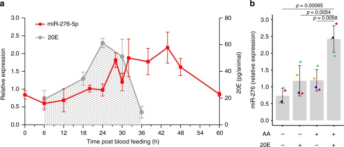
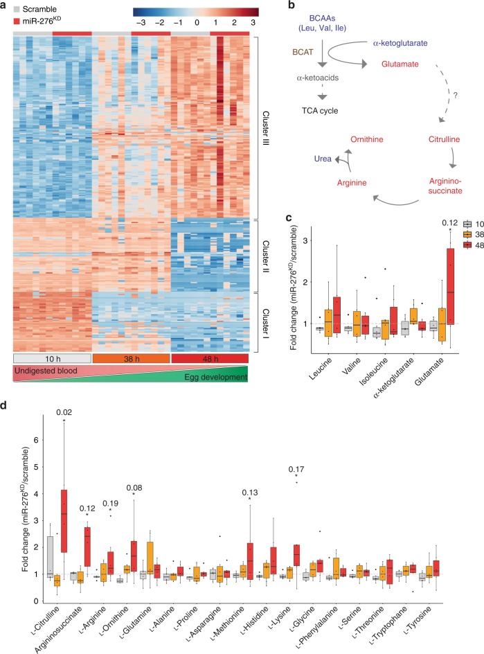
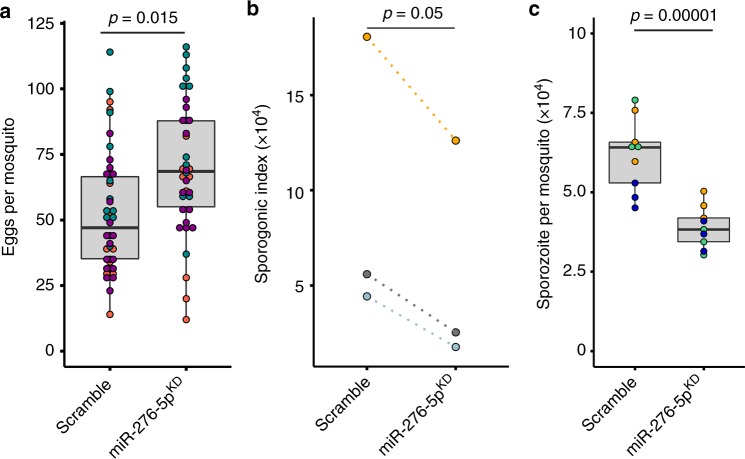
The blood-feeding behavior of Anopheles females delivers essential nutrients for egg development and drives parasite transmission between humans. Plasmodium growth is adapted to the vector reproductive cycle, but how changes in the reproductive cycle impact parasite development remains unclear. Here, we show that the bloodmeal-induced miR-276-5p fine-tunes the expression of branched-chain amino acid transferase to terminate the reproductive cycle. Silencing of miR-276 prolongs high rates of amino acid (AA) catabolism and increases female fertility, suggesting that timely termination of AA catabolism restricts mosquito investment into reproduction. Prolongation of AA catabolism in P. falciparum-infected females also compromises the development of the transmissible sporozoite forms. Our results suggest that Plasmodium sporogony exploits the surplus mosquito resources available after reproductive investment and demonstrate the crucial role of the mosquito AA metabolism in within-vector parasite proliferation and malaria transmission.


Conflict of interest statement

The authors declare no competing interest.

Figures

Fig. 1
Fig. 1. Amino acid (AA) and steroid hormone (20E) signalling regulate miR-276 expression.
a miR-276 (red) expression in the female fat body (n = 10, N = 6) and 20-hydroxyecdysone (20E) titres (gray) in the mosquito females (n = 9, N = 3) at different time points after blood feeding. The plots show mean ± SEM of independent experiments. b miR-276 expression in ex vivo fat body cultures of 3–4-day-old females (n = 3, N = 4) after incubation with medium with or without amino acids (AA) and 20E. miRNA expression levels were normalized using the ribosomal protein RPS7 gene. Boxplots show the median with first and third quartile, whiskers depict the min and max of independent experiments, colored dots show mean of each experiment. The statistical significance was tested by one-way ANOVA followed by Tukey’s post hoc test and the obtained p-values are shown above the horizontal lines (n = number of mosquitoes pooled for each independent experiment; N = number of independent experiments).
Fig. 2
Fig. 2. miR-276 post-transcriptionally represses the branched-chain amino acid transferase in the mosquito fat body.
a Expression of predicted miRNA targets at 38 h post blood feeding in the fat body of mosquitoes (n = 5, N = 6) injected with anti-miR-276 (miR-276, red) or scrambled antagomir (scramble, gray). Expression of the ATP synthase (ATPase V), branched chain amino acid transferase (BCAT), glycerol-3-phosphate dehydrogenase (GPD), methylmalonate-semialdehyde dehydrogenase (MMSDH), 3-methylcrotonyl-CoA carboxylase (MCC), NADH dehydrogenase (NADH (I)), branched-chain alpha-keto acid dehydrogenase (BCKDH) and mitochondrial ornithine receptor (MOR). Expression levels were normalized using the ribosomal protein RPS7 gene. Boxplots show the median with first and third quartile, whiskers depict the min and max. Statistical significance was tested by two-way ANOVA followed by Tukey’s post hoc test and significant differences are shown by the p-value above the horizontal line. b Sequence of the predicted miR-276 binding site in the BCAT 3′UTR. Red color indicates complementary nucleotides in the 3′UTR binding site and miR-276. c Dual luciferase reporter assay in vitro. A construct containing three copies of miR-276 binding sites served as positive control. Lines depict medians and colored dots show means of independent experiments (N = 3). Statistical significance of differences was tested by one-way ANOVA followed by Tukey’s post hoc test and significant differences are indicated by the p-values above the horizontal lines. d BCAT expression in the fat body during the reproductive cycle of mosquitoes injected with anti-miR-276 (miR-276KD, red) or scrambled antagomir (scramble, black). Expression levels were normalized using the ribosomal protein RPS7 gene. Means ± SEM (vertical lines) were plotted. Statistically significant differences between miR-276KD and control mosquitoes were determined by two-way ANOVA followed by Tukey’s post hoc test (N = 3) and significant differences are shown by the p-value (*) (n = number of mosquitoes pooled for each independent experiment; N = number of independent experiments).
Fig. 3
Fig. 3. miR-276 fine-tunes amino acid (AA) metabolism in the late phase of the mosquito reproductive cycle.
a Heatmap of annotated metabolite levels that vary across the reproductive cycle. Metabolite levels were measured by GC-MS and LC-MS from whole females (n = 10, N = 6) at 10, 38, and 48 h post P. falciparum-infected blood meal in control (scrambled, gray) and miR-276-depleted (miR-276KD, red) mosquitoes. Bottom bars indicate time points after blood feeding (10 h, gray; 38 h, orange; 48 h, red), and gradients below show the progression of blood digestion (red) and ovary development (green). Samples are plotted on the x-axis and metabolites on the y-axis. For heatmap visualization, data were log2 transformed and pareto scaled. b Overview of the classical enzymatic activity of the branched chain aminotransferase (BCAT) in animals. The amino group of BCAAs is transferred to α-ketoglutarate by BCAT resulting in glutamate. Glutamate serves as a nitrogen sink, which is passed on to the urea cycle in vertebrates. As mosquitoes lack the carbamoyl phosphate synthase, the link between glutamate and citrulline of the urea cycle is unknown. Within the classical urea cycle, citrulline is converted to argininosuccinate and subsequently arginine, which is metabolized to ornithine ultimately releasing urea. Metabolite changes upon miR-276 inhibition (48 hpb) are shown in red, unchanged metabolites are in blue, and undetected metabolites are in gray. c Fold-change differences in branched chain amino acids (leucine, valine, isoleucine), α-ketoglutarate and glutamate detected in miR-276KD as compared to scramble controls. d Fold change differences in metabolites of AA metabolism at 10 h (gray), 38 h (orange), 48 h (red) post blood feeding in whole females. Statistical significance of differences in metabolite levels of miR-276KD and scramble controls was tested by t-test (n = number of mosquitoes pooled for each independent experiment; N = number of independent experiments).
Fig. 4
Fig. 4. Effects of miR-276 silencing on mosquito fertility and P. falciparum sporogonic development.
a Egg laying rates (fertility) of individual females injected with miR-276 (miR-276KD) or control antagomir (scramble) after a blood meal. Boxplots show the median with first and third quartile, whiskers depict min and max values. Each dot represents one mosquito and dot colors indicate independent experiments (N = 3, Supplementary Table 3). b Females injected with miR-276 (miR-276KD) or control (scramble) antagomir were infected with P. falciparum. At day 11 post infection, oocyst number and size were measured. The median of P. falciparum oocysts per midgut and mean of P. falciparum oocyst size per experiment were multiplied to generate a sporogonic index (N = 3, Supplementary Table 3). c The numbers of the salivary gland sporozoites per mosquito were quantified on day 14 post infection. Boxplots show the median with first and third quartile, whiskers depict min and max values. Colors show replicates of each independent experiment. Statistically significant differences are indicated by p-values above the horizontal lines deduced by one-way ANOVA and Tukey’s post-hoc test (N = 3, Supplementary Table 3), N = number of independent experiments.
Fig. 5
Fig. 5. Summary of miR-276 function and model of mosquito reproductive investment and P. falciparum development interaction.
a Amino acids (AA) and 20-hydroxyecdysone (20E) induce expression of miR-276 after blood feeding and, thereby, initiate the termination of the fat body AA catabolism by inhibiting the branched chain amino acid transferase (BCAT). This catabolism to anabolism switch in the fat body restricts mosquito investment into reproduction. P. falciparum sporogonic stages parasitize spare mosquito resources for their own development. b Mosquito nutritional status and the extent of the reproductive investment shape the within-vector environment which shapes P. falciparum sporogonic development.

References

    1. Hou Y, et al. Temporal coordination of carbohydrate metabolism during mosquito reproduction. PLoS Genet. 2015;11:e1005309. doi: 10.1371/journal.pgen.1005309. - DOI - PMC - PubMed
    1. Wang Xueli, Hou Yuan, Saha Tusar T., Pei Gaofeng, Raikhel Alexander S., Zou Zhen. Hormone and receptor interplay in the regulation of mosquito lipid metabolism. Proceedings of the National Academy of Sciences. 2017;114(13):E2709–E2718. doi: 10.1073/pnas.1619326114. - DOI - PMC - PubMed
    1. Ziegler R, Ibrahim MM. Formation of lipid reserves in fat body and eggs of the yellow fever mosquito, Aedes aegypti. J. Insect Physiol. 2001;47:623–627. doi: 10.1016/S0022-1910(00)00158-X. - DOI - PubMed
    1. Briegel H, Hefti M, DiMarco E. Lipid metabolism during sequential gonotrophic cycles in large and small female Aedes aegypti. J. Insect Physiol. 2002;48:547–554. doi: 10.1016/S0022-1910(02)00072-0. - DOI - PubMed
    1. Carpenter VK, et al. SLC7 amino acid transporters of the yellow fever mosquito Aedes aegypti and their role in fat body TOR signaling and reproduction. J. insect Physiol. 2012;58:513–522. doi: 10.1016/j.jinsphys.2012.01.005. - DOI - PMC - PubMed