. 2019 Dec 10;9(1):18692.
doi: 10.1038/s41598-019-54887-4.

The human-specific paralogs SRGAP2B and SRGAP2C differentially modulate SRGAP2A-dependent synaptic development

Affiliations

The human-specific paralogs SRGAP2B and SRGAP2C differentially modulate SRGAP2A-dependent synaptic development

Ewoud R E Schmidt et al. Sci Rep. .

Abstract

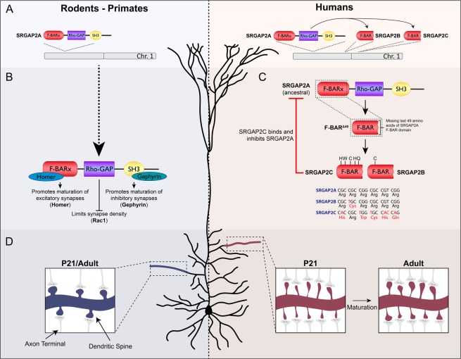
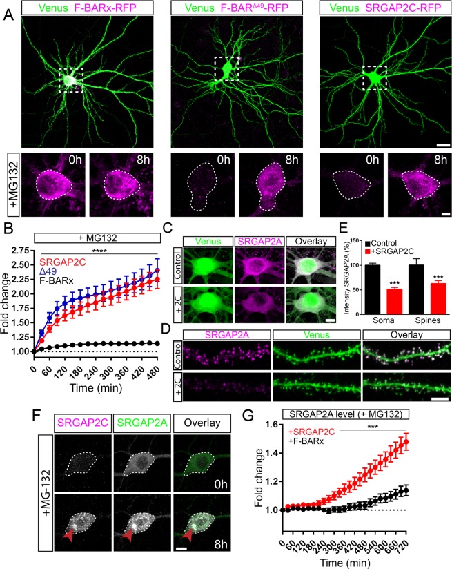
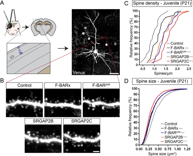
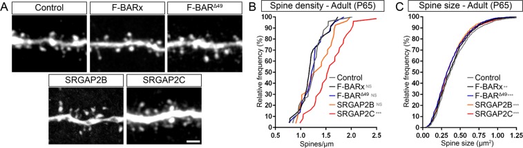
Human-specific gene duplications (HSGDs) have recently emerged as key modifiers of brain development and evolution. However, the molecular mechanisms underlying the function of HSGDs remain often poorly understood. In humans, a truncated duplication of SRGAP2A led to the emergence of two human-specific paralogs: SRGAP2B and SRGAP2C. The ancestral copy SRGAP2A limits synaptic density and promotes maturation of both excitatory (E) and inhibitory (I) synapses received by cortical pyramidal neurons (PNs). SRGAP2C binds to and inhibits all known functions of SRGAP2A leading to an increase in E and I synapse density and protracted synapse maturation, traits characterizing human cortical neurons. Here, we demonstrate how the evolutionary changes that led to the emergence of SRGAP2 HSGDs generated proteins that, in neurons, are intrinsically unstable and, upon hetero-dimerization with SRGAP2A, reduce SRGAP2A levels in a proteasome-dependent manner. Moreover, we show that, despite only a few non-synonymous mutations specifically targeting arginine residues, SRGAP2C is unique compared to SRGAP2B in its ability to induce long-lasting changes in synaptic density throughout adulthood. These mutations led to the ability of SRGAP2C to inhibit SRGAP2A function and thereby contribute to the emergence of human-specific features of synaptic development during evolution.


Conflict of interest statement

The authors declare no competing interests.

Figures

Figure 1
Figure 1
SRGAP2A and the emergence of the human-specific paralogs SRGAP2B and SRGAP2C. (A) The ancestral copy SRGAP2A, present in rodents, primates, and humans, was duplicated in the human lineage to form the truncated copies SRGAP2B and SRGAP2C. (B) SRGAP2A contains three distinct protein domains. Through binding of Homer1 to an EVH1 site in the F-BAR domain, SRGAP2A promotes maturation of excitatory synapses, while binding of Gephyrin to the SH3 domain promotes inhibitory synapse maturation. The Rho-GAP domain is involved in limiting synapse density through Rac1. (C) Duplication of SRGAP2A in humans generated a truncated protein, F-BARΔ49, containing the F-BARx domain of SRGAP2A that lacks the last C-terminal 49 amino acids. Subsequent nonsynonymous base pair mutations converted arginines to non-polar residues which led to the formation of SRGAP2B and SRGAP2C. Binding of SRGAP2C to SRGAP2A inhibits all functions of SRGAP2A. (D) In rodents, synaptic development of cortical pyramidal neurons is complete at P21. At this stage, both synapse size and synaptic density reach levels of those observed in adulthood. In contrast, expression of SRGAP2C in mouse cortical pyramidal neurons binds and inhibits SRGAP2A resulting in protracted synaptic maturation and increased synaptic density.
Figure 2
Figure 2
Proteasome degradation of SRGAP2 proteins. (A) Expression of RFP-tagged F-BARx, F-BARΔ49, or SRGAP2C in mouse cortical pyramidal neurons cultured for 18–21 days in vitro (DIV). Low levels of expression were observed for F-BARΔ49 and SRGAP2C, while addition of the proteasome inhibitor MG-132 resulted in a strong increase after 8 h of treatment. F-BARx is highly expressed throughout the experiment. Scale bar top panels: 25 µm, bottom panels: 5 µm. (B) Quantification of fluorescence intensity after MG-132 treatment. nF-BARx = 31 neurons, nF-BARΔ49 = 50 neurons, nSRGAP2C = 47 neurons; multiple t tests with Holm-Sidak correction for multiple comparisons (α = 0.05); ****p < 0.0001; mean ± SEM. (C–E) Co-expression of SRGAP2A and SRGAP2C in cultured mouse cortical neurons. SRGAP2A levels are reduced when co-expressed with SRGAP2C in both soma and dendritic spines. Scale bars: 5 µm. (E) Quantification of SRGAP2A fluorescence intensity. Soma: nControl = 110, nSRGAP2C = 68; Spines: nControl = 969, nSRGAP2C = 1259; Mann-Whitney test; ***p < 0.001; mean ± SEM. (F) Treatment of cultured mouse cortical neurons expressing both SRGAP2A-GFP and SRGAP2C-RFP with MG-132. Levels for both SRGAP2A and SRGAP2C increased upon treatment, and co-localization of SRGAP2A and SRGAP2C protein clusters were observed (red arrow). Scale bar: 10 µm. (G) Quantification of SRGAP2A fluorescent intensity when co-expressed with either F-BARx or SRGAP2C in the presence of MG-132. nF-BAR = 60 neurons, nSRGAP2C = 59 neurons; multiple t tests with Holm-Sidak correction for multiple comparisons (α = 0.05); ***p < 0.001; mean ± SEM.
Figure 3
Figure 3
In vivo analysis of spine density and spine size at juvenile stage. (A) Schematic illustrating in utero electroporation approach targeting layer 2/3 cortical pyramidal neurons and subsequent quantification of dendritic spine density and head size. Scale bar: 50 µm. (B) Representative images showing apical oblique dendrites of neurons at P21 co-expressing either Venus and tdTomato (Control), or mRFP-tagged F-BARx, F-BARΔ49, SRGAP2B, or SRGAP2C. Scale bar: 2 µm. (C,D) Quantification of spine density (C) and spine head size (D). Quantifications are shown as frequency distributions. Spine density (segments): nControl = 26, nF-BARx = 25, nF-BARΔ49 = 34, nSRGAP2B = 25, nSRGAP2C = 25; Spine size (number of spines): nControl = 1693, nF-BARx = 2054, nF-BARΔ49 = 3267, nSRGAP2B = 2900, nSRGAP2C = 2448; one-way ANOVA with Holm-Sidak’s post hoc multiple comparison test; *p < 0.05, ****p < 0.0001 compared to control.
Figure 4
Figure 4
In vivo analysis of spine density and spine size at the adult stage. (A) Representative images showing apical oblique dendrites of adult neurons co-expressing either Venus (shown) and tdTomato (Control), or mRFP-tagged F-BARx, F-BARΔ49, SRGAP2B, or SRGAP2C (not shown). Scale bar: 2 µm. (B,C) Quantification of spine density (B) and spine head size (C). Quantifications are shown as frequency distributions. Spine density (segments): nControl = 25, nF-BARx = 24, nF-BARΔ49 = 22, nSRGAP2B = 27, nSRGAP2C = 25; Spine size (number of spines): nControl = 1872, nF-BARx  = 2049, nF-BARΔ49 = 1843, nSRGAP2B = 2295, nSRGAP2C = 2630; one-way ANOVA with Holm-Sidak’s post hoc multiple comparison test; NS: p > 0.05, *p < 0.05, ***p < 0.001, ****p < 0.0001 compared to control.

References

    1. Sousa AMM, Meyer KA, Santpere G, Gulden FO, Sestan N. Evolution of the Human Nervous System Function, Structure, and Development. Cell. 2017;170:226–247. doi: 10.1016/j.cell.2017.06.036. - DOI - PMC - PubMed
    1. Suzuki IK, et al. Human-Specific NOTCH2NL Genes Expand Cortical Neurogenesis through Delta/Notch Regulation. Cell. 2018;173:1370–1384.e16. doi: 10.1016/j.cell.2018.03.067. - DOI - PMC - PubMed
    1. Fiddes IT, et al. Human-Specific NOTCH2NL Genes Affect Notch Signaling and Cortical Neurogenesis. Cell. 2018;173:1356–1369.e22. doi: 10.1016/j.cell.2018.03.051. - DOI - PMC - PubMed
    1. Florio M, et al. Human-specific gene ARHGAP11B promotes basal progenitor amplification and neocortex expansion. Science. 2015;347:1465–1470. doi: 10.1126/science.aaa1975. - DOI - PubMed
    1. Florio M, et al. Evolution and cell-type specificity of human-specific genes preferentially expressed in progenitors of fetal neocortex. Elife. 2018;7:1–37. doi: 10.7554/eLife.32332. - DOI - PMC - PubMed

Publication types

Substances

LinkOut - more resources