. 2019 Dec 10;9(1):18756.
doi: 10.1038/s41598-019-55128-4.

Enteric neuronal cell therapy reverses architectural changes in a novel diphtheria toxin-mediated model of colonic aganglionosis

Affiliations

Enteric neuronal cell therapy reverses architectural changes in a novel diphtheria toxin-mediated model of colonic aganglionosis

Sukhada Bhave et al. Sci Rep. .

Abstract

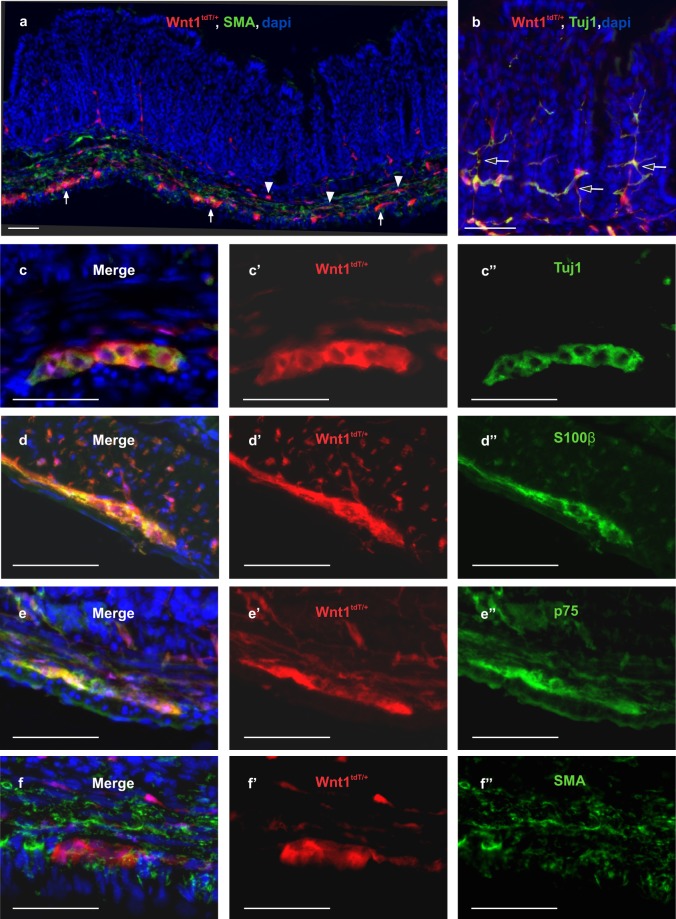
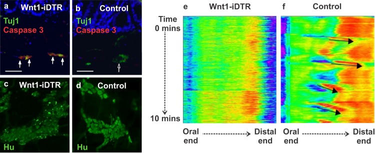
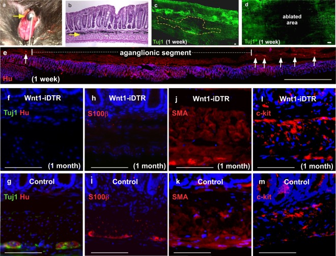
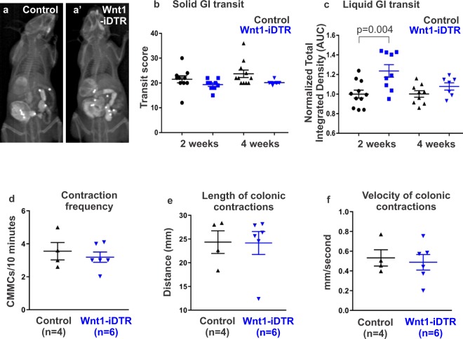
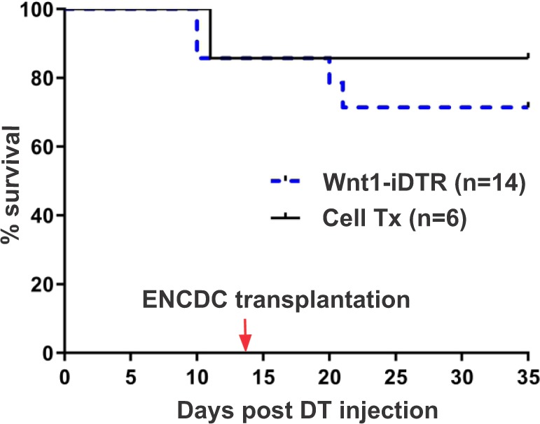
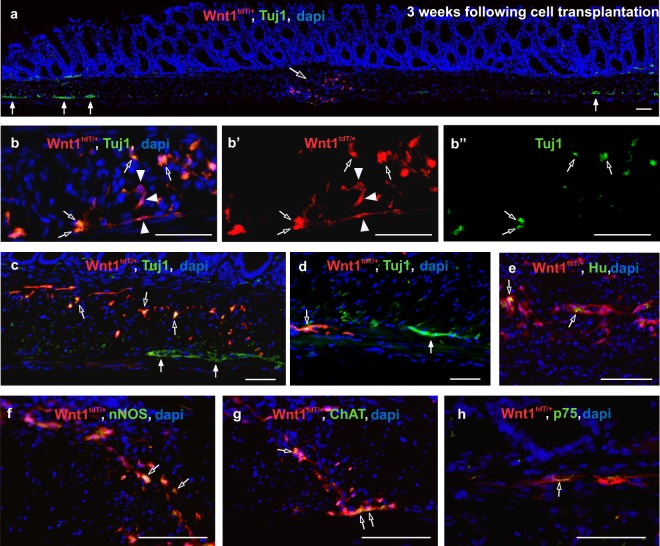
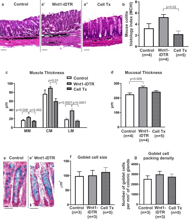
Hirschsprung disease (HSCR) is characterized by absence of the enteric nervous system (ENS) in the distal bowel. Despite removal of the aganglionic segment, gastrointestinal (GI) problems persist. Cell therapy offers potential treatment but use of genetic models is limited by their poor survival. We have developed a novel model of aganglionosis in which enteric neural crest-derived cells (ENCDCs) express diphtheria toxin (DT) receptor. Local DT injection into the colon wall results in focal, specific, and sustained ENS ablation without altering GI transit or colonic contractility, allowing improved survival over other aganglionosis models. Focal ENS ablation leads to increased smooth muscle and mucosal thickness, and localized inflammation. Transplantation of ENCDCs into this region leads to engraftment, migration, and differentiation of enteric neurons and glial cells, with restoration of normal architecture of the colonic epithelium and muscle, reduction in inflammation, and improved survival.


Conflict of interest statement

The authors declare no competing interests.

Figures

Figure 1
Figure 1
Characterization of colonic ENS in Wnt1tdT/+ mice. Colon of Wnt1tdT/+ mice expresses tdT in the submucosal (a, arrowheads) and myenteric plexi (a, arrows). tdT + fibers are found projecting to the mucosa (b, open arrows). Cross sections of Wnt1tdT/+ colon show tdT colocalizing with Tuj1 (c-c”), S100β (d-d”), and p75 (e-e”), but not with SMA (f-f”). Scale bar 50 µm (a–e).
Figure 2
Figure 2
Systemic administration of DT to Wnt1-iDTR mice leads to enteric neuronal loss and colonic dysmotility. Intraperitoneal delivery of DT causes enteric neuronal apoptosis in the colon of Wnt1-iDTR mice (a, arrows) but not iDTR-negative controls (b). This is confirmed by wholemount immunostaining showing disrupted Hu-expression in Wnt1-iDTR mice (c,d). Spatiotemporal mapping of colonic contractility shows absence of CMMCs in DT-treated Wnt1-iDTR mice (e), as compared to normal contractile activity in DT-treated control mice (f, arrows). Scale bar 50 µm (a,b).
Figure 3
Figure 3
Intracolonic injection of DT results in focal aganglionosis. DT was injected into the mid-colon via laparotomy (a). Injection site is marked by India ink (a,b arrows). Immunofluorescence one week later shows focal loss of myenteric neurons (ablated area marked by a dotted line; c) by wholemount staining for Tuj1 (c,d) and Hu staining of a longitudinal section of mid-colon (e, arrows mark endogenous neurons at margins of DT ablation). One month after DT injection, focal ablation of Tuj1 + and Hu + enteric neurons (f,g) and S100β + enteric glia (h,i) was observed, with no obvious loss of SMA + smooth muscle (j,k) or c-kit + interstitial cells of Cajal (l,m). Scale bar 100 µm (c,d), 1 mm (e), and 100 µm (f–m).
Figure 4
Figure 4
DT-induced focal aganglionosis does not affect total GI transit and colonic motility. A radiologic-based transit assay (a,a’) was used to measure solid and liquid transit following DT injection. No difference was observed in solid transit at 2 and 4 weeks after DT injection (b). Liquid transit showed a delay at 2 weeks, but not at 4 weeks (c). Ex vivo analysis by spatiotemporal mapping of the DT-injected colon from Wnt1-iDTR mice showed no changes in CMMC frequency (d), length (e), or velocity (f), as compared to controls.
Figure 5
Figure 5
ENCDC transplantation improves survival following ENS ablation in Wnt1-iDTR mice. Kaplan-Meier survival curves show that ENCDC transplantation results in an improved five-week survival rate in mice where ENS ablation was induced by DT injection.
Figure 6
Figure 6
Transplanted ENCDCs survive, engraft, and migrate in the aganglionic colon. Two weeks after DT injection, tdT + neurospheres were transplanted into the colon wall and analysis was performed at 3 weeks after cell transplantation. Transplanted tdT + neurospheres (a, open arrow) survive in the aganglionic segment of the colon (a, closed arrows mark endogenous ganglia). Transplanted cells consist of Tuj1 + neurons (b-b”, open arrows) and Tuj1-negative neural crest-derived cells (b-b’, arrowheads). Transplanted tdT + cells (c, open arrows) are found near endogenous ganglia (c, closed arrows) and project fibers within the muscle layers (d, open arrow; closed arrow marks endogenous myenteric plexus. Wnt1-tdT ENCDCs express Tuj1 (b,c,d), Hu (e), nNOS (f), ChAT (g), and p75 (h). Scale bar 50 µm (ah).
Figure 7
Figure 7
ENCDC transplantation reverses the effects of DT-induced aganglionosis on colonic architecture. Representative images are shown of H&E stained colon 4–5 weeks following DT injection into control (a), non-transplanted Wnt1-iDTR (a’), and ENCDC-transplanted Wnt1-iDTR (a”) mice. MCHI scores show low-grade inflammation in all three groups (b), with transplanted colon showing significantly less inflammation. DT-mediated ENS ablation leads to increased thickness of muscularis mucosa (MM) and longitudinal muscle (LM), with no significant change in circular muscle (CM), and these changes are reversed following cell transplantation (c). DT-mediated ENS ablation also increases mucosal thickness, which is partially normalized by ENCDC transplantation (d). Alcian blue staining (e,e’) shows no change in goblet cell size (f) or density (g) among the groups. Scale bar 100 µm (a-a”) and 50 µm (e,e’).

References

    1. Nagy N, Goldstein AM. Enteric nervous system development: A crest cell’s journey from neural tube to colon. Semin Cell Dev Biol. 2017;66:94–106. doi: 10.1016/j.semcdb.2017.01.006. - DOI - PMC - PubMed
    1. Rao M, Gershon MD. Neurogastroenterology: The dynamic cycle of life in the enteric nervous system. Nat Rev Gastroenterol Hepatol. 2017;14:453–454. doi: 10.1038/nrgastro.2017.85. - DOI - PubMed
    1. Burns Alan J., Goldstein Allan M., Newgreen Donald F., Stamp Lincon, Schäfer Karl-Herbert, Metzger Marco, Hotta Ryo, Young Heather M., Andrews Peter W., Thapar Nikhil, Belkind-Gerson Jaime, Bondurand Nadege, Bornstein Joel C., Chan Wood Yee, Cheah Kathryn, Gershon Michael D., Heuckeroth Robert O., Hofstra Robert M.W., Just Lothar, Kapur Raj P., King Sebastian K., McCann Conor J., Nagy Nandor, Ngan Elly, Obermayr Florian, Pachnis Vassilis, Pasricha Pankaj J., Sham Mai Har, Tam Paul, Vanden Berghe Pieter. White paper on guidelines concerning enteric nervous system stem cell therapy for enteric neuropathies. Developmental Biology. 2016;417(2):229–251. doi: 10.1016/j.ydbio.2016.04.001. - DOI - PMC - PubMed
    1. Heuckeroth RO. Hirschsprung disease - integrating basic science and clinical medicine to improve outcomes. Nat Rev Gastroenterol Hepatol. 2018;15:152–167. doi: 10.1038/nrgastro.2017.149. - DOI - PubMed
    1. Gershon MD, Ratcliffe EM. Developmental biology of the enteric nervous system: pathogenesis of Hirschsprung’s disease and other congenital dysmotilities. Semin Pediatr Surg. 2004;13:224–235. doi: 10.1053/j.sempedsurg.2004.10.019. - DOI - PMC - PubMed

Publication types

MeSH terms

Substances