Review
. 2020 Feb;16(2):75-86.
doi: 10.1038/s41584-019-0334-3. Epub 2019 Dec 10.

Asymptomatic hyperuricaemia: a silent activator of the innate immune system

Affiliations
Review

Asymptomatic hyperuricaemia: a silent activator of the innate immune system

Leo A B Joosten et al. Nat Rev Rheumatol. 2020 Feb.

Abstract

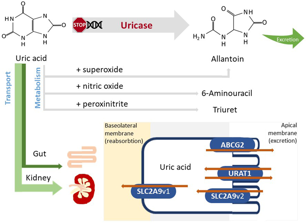
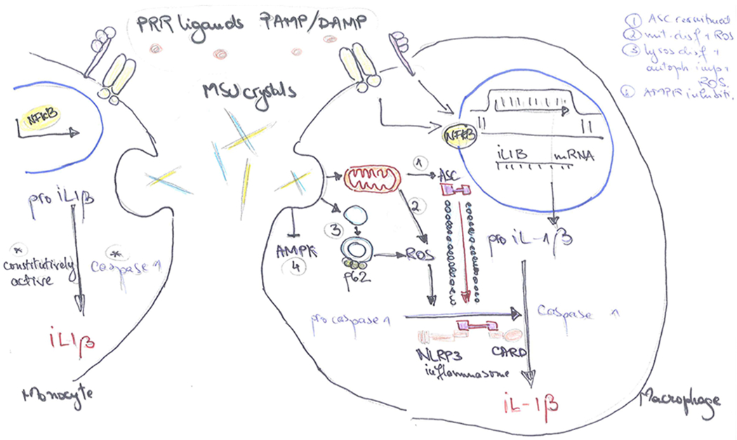
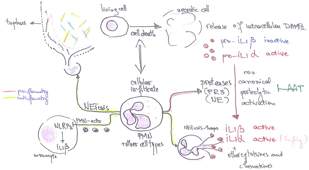
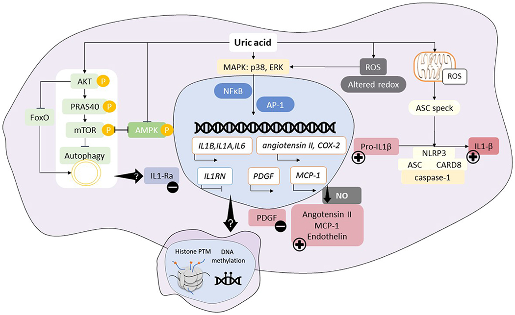
Asymptomatic hyperuricaemia affects ~20% of the general population in the USA, with variable rates in other countries. Historically, asymptomatic hyperuricaemia was considered a benign laboratory finding with little clinical importance in the absence of gout or kidney stones. Yet, increasing evidence suggests that asymptomatic hyperuricaemia can predict the development of hypertension, obesity, diabetes mellitus and chronic kidney disease and might contribute to disease by stimulating inflammation. Although urate has been classically viewed as an antioxidant with beneficial effects, new data suggest that both crystalline and soluble urate activate various pro-inflammatory pathways. This Review summarizes what is known about the role of urate in the inflammatory response. Further research is needed to define the role of asymptomatic hyperuricaemia in these pro-inflammatory pathways.


Conflict of interest statement

Competing interests statement

Dr Johnson is an inventor on several patents and patent applications related to the role of fructose and urate metabolism in hypertension, metabolic syndrome and kidney disease. He also has equity with XORT therapeutics which is developing novel xanthine oxidase inhibitors, and Colorado Research Partners LLC, which is developing inhibitors of fructose metabolism. Finally, he has received honoraria from Eli Lilly, Astra Zeneca and Horizon Pharmaceuticals. Dr. Bjornstad has received consulting fees or speaking honorarium or both from Horizon Pharma, Boehringer Ingelheim, Bayer, and Bristol-Myers Squibb. He also serves on a scientific advisory board for XORTX.

Figures

Figure 1.
Figure 1.. Regulation of serum urate.
Following sequential mutations in the uricase gene, humans and higher primates do not express functional uricase, needed to metabolize uric acid to allantoin. Serum urate can interact with reactive species generating substances or radicals such as allantoin, 6-aminouracil or triuret. However, most of the serum urate will be excreted via the kidney or, alternatively, via the gut. Both proximal tubule and intestinal cells express several urate transporters (collectively known as the transportasome) that are responsible for the excretion and/or reabsorption of urate. Several main urate transporters are illustrated: URAT1, GLUT9, OAT4 and OAT10 are involved in urate reabsorption at the level of the apical membrane (brush border) of proximal renal tubular cells; GLUT9 is responsible for urate transport out of the cell via the basolateral membrane into the blood; ABCG2 is a unidirectional transporter mediating the secretion of urate via the apical membrane; OAT1 and OAT3 localized on the basolateral membrane are involved in urate excretion. OAT, organic anion transporter; URAT1, urate transporter 1; GLUT9, glucose transporter 9; ABCG2, ATP-binding cassette super-family G member 2.
Figure 2.
Figure 2.. Inflammasome-dependent activation of IL-1β in response to MSU crystals.
IL-1β production is initiated by exogenous or endogenous stimuli (PAMPs or DAMPs) which use the PRR machinery to induce an inflammatory response. Engagement of PRR ligands to the receptors leads to the transduction of signal to the nucleus and transcription of the inactive pro-IL-1β precursor. In the intracellular space, pro-IL-1β is proteolytically activated by specific cleavage performed by caspase-1. Caspase-1 is, in turn, present in the cytosol in a precursor form (procaspase-1). MSU crystals act as inducers of the protein platform that cleaves procaspase-1 to caspase-1, thus mediating the activation and release of biologically active IL-1β. In the monocytes (left), caspase-1 is constitutively active, thus MSU crystals potentiate IL-1β maturation. MSU crystals also upregulate mTOR gene transcription, leading to enhanced gene transcription of IL-1β and other inflammatory cytokines, and drive monocyte cell death that could, indirectly, further stimulate the inflammasome. In the macrophages (right), caspase-1 is not constitutively active, MSU crystals drive changes inside the cell that result in the assembly of the inflammasome components and caspase-1 generation. MSU crystals can do this in multiple ways: (1) induce mitochondrial ASC recruitment to the site of inflammasome assembly in a microtubule dependent manner, (2) induce mitochondrial dysfunction and ROS generation, (3) lysosomal dysfunction with impairment of autophagy, increased levels of p62 and ROS generation, (4) AMPK inhibition in an NLRP3 dependent manner. PAMP, Pathogen Associated Molecular Patterns; DAMP, Damage Associated Molecular Patterns; PRR, pattern recognition receptor; ASC, Apoptosis-Associated Speck-Like Protein Containing CARD; ROS, reactive oxygen species; AMPK, AMP-activated protein kinase.
Figure 3.
Figure 3.. Inflammasome-independent regulation of inflammation in response to MSU crystals.
MSU crystal deposition leads to cell death and cellular infiltrate, mostly characterized by neutrophil recruitment at the site of crystal deposition, especially in the affected joints of patients with gout. Dying cells release intracellular molecules (DAMPs, alarmins) in the extracellular space, including precursors of the IL-1 cytokines. Pro-IL-1β is inactive while pro-IL-1α already has biological activity in its precursor form, therefore is able to mediate inflammatory responses in the surrounding tissue. Infiltrating cells, the majority of which are neutrophils, contain and release proteolytic enzymes (such as PR3 or NE, among others) that have the potential to cleave IL-1 precursors at alternative cleavage sites and activate IL-1β or enhance the bioactivity of IL-1α. This mechanism of protease induced activation of precursor cytokines can be limited by anti-proteases such as AAT. Conversely, neutrophils can undergo NETosis (neutrophil extracellular trap formation), which consists in the externalization of chromatin and binding of intercellular molecules. This phenomenon is considered to play an important role in the resolution of inflammation in gout: NETs can bind active cytokines or chemokines and facilitate their enzymatic digestion contributing to the limitation of inflammation; moreover, NETosis can contribute to the sequestration of MSU crystals and encapsulation of crystals within the tophus; neutrophil microvesicles (PMN-Ecto) can downregulate inflammatory pathways in paracrine manner, limiting the magnitude of the cytokine production during the acute phase by inhibiting NLRP3 or activating SOCS3. DAMP, Damage Associated Molecular Patterns; PR3, Proteinase 3; NE, Neutrophil Elastase, NLRP3, NACHT, LRR and PYD domains-containing protein 3; SOCS3, Supressor of Cytokine Signalling 3.
Figure 4.
Figure 4.. Intracellular pathways involved in proinflammatory effects of soluble uric acid.
Soluble uric acid has been shown to modulate cytokine production and inflammatory outcomes via several pathways. Uric acid enhances AKT phosphorylation, leading to PRAS40 phosphorylation, and resulting in autophagy inhibition; this is in line with downregulation of expression in genes involved in FOXO pathway. Uric acid was shown to inhibit AMPK phosphorylation, which is consistent with mTOR activation. Several transcription factors can be induced by uric acid signaling (NFκB or AP-1) following p38 and ERK MAPK activation, leading to enhanced transcription of innate cytokines in addition to the remarkable downregulation of natural IL-1 antagonist, IL-1Ra. In vascular endothelial cells, smooth muscle cells or adipocytes, enhanced transcription of vasoactive substances is known to lead to elevated angiotensin II, MCP-1, endothelin and low availability of nitric oxide which can mediate hypertension. In vascular cells, growth factors such as PDGF are upregulated, leading to smooth muscle cells proliferation and promotion of atherosclerosis. Soluble uric acid has also been shown to induce oxidative stress, alter the redox status of the cell which, on the one hand, activates signaling pathways for gene transcription and, on the other hand, lead to ASC speck formation and inflammasome activation, thus contributing to cleavage of pro-IL-1β. The transcriptional rewiring of uric acid stimulated cells is reminiscent of innate immune memory developed in response to endogenous sterile stimuli, leading to a scenario in which soluble uric acid acts as a silent stimulus that drives epigenetic reprogramming in circulating or tissue resident cells, thereby causing persistent inflammatory effects in response to continuous exposure to uric acid. We propose a model in which cells exposed to high uric acid levels are more prone to develop a strong proinflammatory response when exposed to specific triggers due to innate immune memory, epigenetic changes in exposed cells or persistence of exposure to high uric acid levels.

References

    1. Zhu Y, Pandya BJ & Choi HK Prevalence of gout and hyperuricemia in the US general population: the National Health and Nutrition Examination Survey 2007-2008. Arthritis and rheumatism 63, 3136–3141, doi: 10.1002/art.30520 (2011). - DOI - PubMed
    1. Neogi T & Mikuls TR To Treat or Not to Treat (to Target) in Gout. Ann Intern Med 166, 71–72, doi: 10.7326/M16-2401 (2017). - DOI - PMC - PubMed
    1. Jensen T et al. Fructose and sugar: A major mediator of non-alcoholic fatty liver disease. J Hepatol 68, 1063–1075, doi: 10.1016/j.jhep.2018.01.019 (2018). - DOI - PMC - PubMed
    1. Johnson RJ et al. Hyperuricemia, Acute and Chronic Kidney Disease, Hypertension, and Cardiovascular Disease: Report of a Scientific Workshop Organized by the National Kidney Foundation. Am J Kidney Dis, doi: 10.1053/j.ajkd.2017.12.009 (2018). - DOI - PMC - PubMed
    1. Grayson PC, Kim SY, LaValley M & Choi HK Hyperuricemia and incident hypertension: a systematic review and meta-analysis. Arthritis Care Res (Hoboken) 63, 102–110, doi: 10.1002/acr.20344 (2011). - DOI - PMC - PubMed

Publication types