. 2020 Feb;25(1):135-149.
doi: 10.1007/s00775-019-01747-1. Epub 2019 Dec 10.

Spectroscopic and biochemical insight into an electron-bifurcating [FeFe] hydrogenase

Affiliations

Spectroscopic and biochemical insight into an electron-bifurcating [FeFe] hydrogenase

Nipa Chongdar et al. J Biol Inorg Chem. 2020 Feb.

Abstract

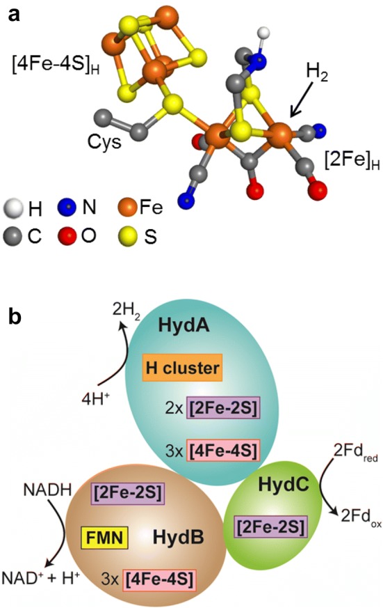
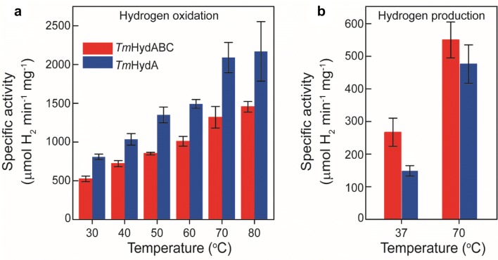
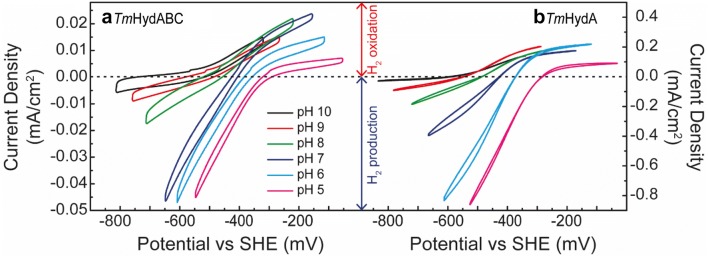
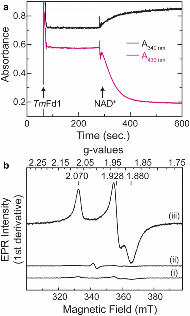
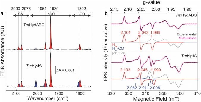
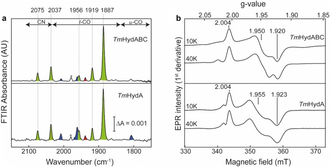
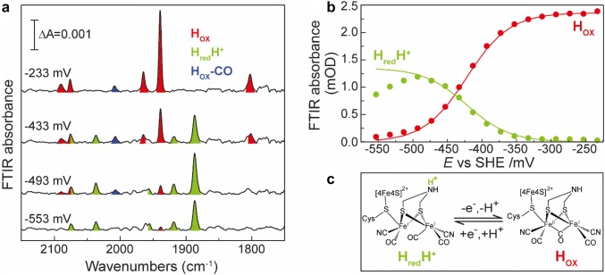
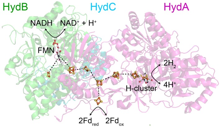
The heterotrimeric electron-bifurcating [FeFe] hydrogenase (HydABC) from Thermotoga maritima (Tm) couples the endergonic reduction of protons (H+) by dihydronicotinamide adenine dinucleotide (NADH) (∆G0 ≈ 18 kJ mol-1) to the exergonic reduction of H+ by reduced ferredoxin (Fdred) (∆G0 ≈ - 16 kJ mol-1). The specific mechanism by which HydABC functions is not understood. In the current study, we describe the biochemical and spectroscopic characterization of TmHydABC recombinantly produced in Escherichia coli and artificially maturated with a synthetic diiron cofactor. We found that TmHydABC catalyzed the hydrogen (H2)-dependent reduction of nicotinamide adenine dinucleotide (NAD+) in the presence of oxidized ferredoxin (Fdox) at a rate of ≈17 μmol NADH min-1 mg-1. Our data suggest that only one flavin is present in the enzyme and is not likely to be the site of electron bifurcation. FTIR and EPR spectroscopy, as well as FTIR spectroelectrochemistry, demonstrated that the active site for H2 conversion, the H-cluster, in TmHydABC behaves essentially the same as in prototypical [FeFe] hydrogenases, and is most likely also not the site of electron bifurcation. The implications of these results are discussed with respect to the current hypotheses on the electron bifurcation mechanism of [FeFe] hydrogenases. Overall, the results provide insight into the electron-bifurcating mechanism and present a well-defined system for further investigations of this fascinating class of [FeFe] hydrogenases.

Keywords: Electrochemistry; Electron bifurcation; Ferredoxin; Spectroscopy; [FeFe] hydrogenase.


Conflict of interest statement

The authors declare that they have no conflict of interest.

Figures

Fig. 1
Fig. 1
a Ball and stick representation of the structure of the H-cluster. The figure was created in Pymol using PDB file 4XDC. b Schematic diagram showing the three subunits of TmHydABC. The cofactors bound to the protein were predicted from amino acid sequence analysis [26]. The arrangement of subunits and the designated reactions occurring at each subunit are based on the proposition by Buckel and Thauer [23]
Fig. 2
Fig. 2
Activity assays of artificially maturated TmHydABC and TmHydA. a Temperature dependence of H2 oxidation of both proteins was measured in the range from 30 to 80 °C with benzyl viologen as the electron acceptor. b H2 production activity of TmHydABC and TmHydA was measured at 37 °C and 70 °C with reduced methyl viologen as electron donor
Fig. 3
Fig. 3
Cyclic voltammetry of TmHydABC (a) and TmHydA (b) adsorbed onto the surface of a pyrolytic graphite electrode. The applied potential was swept from open-circuit potential to 250 mV more positive and then to 250 mV more negative, to compare the catalytic currents at the same overpotential. The measurements were performed at 35 °C under 100% H2
Fig. 4
Fig. 4
Activity assay of TmHydABC using physiological partners. a Simultaneous reduction of TmFd1 (decrease in A430nm) and NAD+ (increase in A340nm) by TmHydABC at 70 °C under an atmosphere of 100% H2 was monitored by UV–Vis spectroscopy. To a 1 mL reaction mixture containing  ≈ 680 ng TmHydABC and 50 µM FMN in 200 mM potassium phosphate (pH 8) buffer, ≈35 µM TmFd1 was added (indicated by the first arrow). To the same reaction mixture, 0.5 mM NAD+ was added (indicated by the second arrow). b Reduction of TmFd1 monitored by EPR spectroscopy. In the absence of TmHydABC, H2 did not reduce TmFd1 (trace i). In the absence of NAD+, TmHydABC could not catalyze reduction of TmFd1 from H2 (trace ii). However, when NAD+ is present, TmHydABC could reduce TmFd1 using H2 as indicated by the appearance of the typical rhombic spectrum corresponding to the reduced [4Fe–4S] cluster. The conditions of the reactions used for preparing EPR samples were the same as for the UV–Vis measurements. The EPR spectra were measured at 10 K and at 0.2 mW power
Fig. 5
Fig. 5
FTIR and EPR spectra of artificially maturated TmHydABC and TmHydA under oxidizing conditions. a Top panel shows the FTIR spectrum of 400 µM TmHydABC oxidized with 20 mM NAD+ and the lower panel shows the FTIR spectrum of 400 µM TmHydA oxidized with 1 mM thionine. Both samples were in 0.1 M Tris–HCl pH 8 buffer, 0.15 M NaCl and the spectra were measured at room temperature. The peaks belonging to the Hox state are shaded in red and those belonging to the Hox–CO state are shaded in blue. b CW X-band EPR spectra of the oxidized TmHydABC and TmHydA samples at 20 K, 0.1 mW microwave power. The samples were prepared in the same way as for the FTIR measurements. The experimental spectra are overlaid with spectral simulations (dotted magenta lines) and the component spectra are shown underneath. The red trace (Component 1) corresponds to the Hox state, the blue trace (Component 2) corresponds to the Hox–CO state and the gray trace (Component 3) corresponds to one of the reduced F-clusters
Fig. 6
Fig. 6
FTIR and EPR spectra of TmHydABC and TmHydA after CO inhibition. a FTIR spectra of CO-inhibited (Hox–CO) artificially maturated TmHydABC and TmHydA. The ‘as-isolated’ protein samples (≈ 400 µM) were purged for 20 min with 100% CO, followed by incubation for an additional 60 min. Spectra were measured at room temperature. The peak marked with an asterisk belongs to an unidentified species. b CW X-band EPR spectra of CO-inhibited (Hox–CO) TmHydABC and TmHydA measured at 20 K and 0.1 mW microwave power. Approximately, 150 μM of each protein in 0.1 M Tris–HCl buffer pH 8, 0.15 M NaCl, 20% glycerol, were purged for 20 min with 100% CO, before measuring the spectra. The experimental spectra are overlaid with the spectral simulations (dotted magenta line) and the components are shown underneath
Fig. 7
Fig. 7
FTIR and EPR spectra of the reduced TmHydABC and TmHydA. a FTIR spectra of reduced TmHydABC or TmHydA samples are shown in the upper and lower panels, respectively. Approximately 400 µM of ‘as-isolated’ protein samples were incubated with 20 mM of sodium dithionite (NaDT) at room temperature for 5 min before measuring the spectra at room temperature. The peaks corresponding to the HredH+ state are shaded in green. The minor peaks shaded in blue and red belong to the Hox–CO and Hox states. The peak marked with an asterisk belongs to an unidentified species. b CW X-band EPR spectra of reduced protein samples (TmHydABC and TmHydA) were measured at 10 K (0.01 mW microwave power) and 40 K (1 mW microwave power). EPR sample composition: 150 μM, 10 mM of NaDT, 0.1 M Tris–HCl buffer pH 8, 0.15 M NaCl, 20% glycerol
Fig. 8
Fig. 8
Spectroelectrochemical FTIR of TmHydABC. a Selected FTIR spectra recorded at − 233, − 433, − 493 and − 553 mV are shown. The experiment was performed with ≈1 mM TmHydABC in 200 mM phosphate buffer (pH 8) and 200 mM KCl with redox mediators at 15 °C. b Changes in FTIR absorbance with changing electrode potential, during reductive titration, at peak position 1939 cm−1 (Hox) and 1887 cm−1 (HredH+) are shown by red and green circles, respectively. The solid red and green lines represent the Nernst-fit corresponding to the model shown in c
Fig. 9
Fig. 9
Proposed arrangement of subunits and cofactors in TmHydABC. The TmHydABC subunits are homologous to the Nqo1, Nqo2, and Nqo3 subunits of the structurally characterized complex I from Thermus thermophilus. Based on this homology, the arrangements of the conserved cofactors can be predicted. The figure shows the protein subunits Nqo1 (HydB, green), Nqo2 (HydC, blue), and Nqo3 (HydA, pink) in the cartoon representation (PDB: 4HEA [53],), with the cofactors from Nqo1 and Nqo2 from complex I, and the cofactors from the [FeFe] hydrogenase CpHydA (PDB: 4XDC [54]), overlaid. CpHydA was aligned to Nqo3 in Pymol giving almost perfect alignment of the homologous clusters. HydA contains an additional [2Fe–2S] cluster, for which CpHydA does not contain a homologous cluster, and HydB contains an additional two [4Fe–4S] clusters and one [2Fe–2S] cluster, for which complex I does not contain homologous clusters

References

    1. Lubitz W, Ogata H, Rüdiger O, Reijerse E. Chem Rev. 2014;114:4081–4148. doi: 10.1021/cr4005814. - DOI - PubMed
    1. Peters JW, Schut GJ, Boyd ES, Mulder DW, Shepard EM, Broderick JB, King PW, Adams MW. Biochim Biophys Acta. 2015;1853:1350–1369. doi: 10.1016/j.bbamcr.2014.11.021. - DOI - PubMed
    1. Glick BR, Martin WG, Martin SM. Can J Microbiol. 1980;26:1214–1223. doi: 10.1139/m80-203. - DOI - PubMed
    1. Peters JW, Lanzilotta WN, Lemon BJ, Seefeldt LC. Science. 1998;282:1853–1858. doi: 10.1126/science.282.5395.1853. - DOI - PubMed
    1. Nicolet Y, Piras C, Legrand P, Hatchikian CE, Fontecilla-Camps JC. Structure. 1999;7:13–23. doi: 10.1016/S0969-2126(99)80005-7. - DOI - PubMed

Publication types

LinkOut - more resources