Review
. 2019 Nov 22:7:337.
doi: 10.3389/fbioe.2019.00337. eCollection 2019.

Modern Trends for Peripheral Nerve Repair and Regeneration: Beyond the Hollow Nerve Guidance Conduit

Affiliations
Review

Modern Trends for Peripheral Nerve Repair and Regeneration: Beyond the Hollow Nerve Guidance Conduit

Cristiana R Carvalho et al. Front Bioeng Biotechnol. .

Abstract

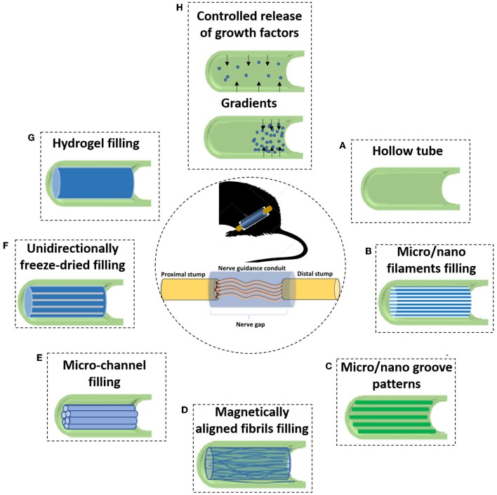
Peripheral nerve repair and regeneration remains among the greatest challenges in tissue engineering and regenerative medicine. Even though peripheral nerve injuries (PNIs) are capable of some degree of regeneration, frail recovery is seen even when the best microsurgical technique is applied. PNIs are known to be very incapacitating for the patient, due to the deprivation of motor and sensory abilities. Since there is no optimal solution for tackling this problem up to this day, the evolution in the field is constant, with innovative designs of advanced nerve guidance conduits (NGCs) being reported every day. As a basic concept, a NGC should act as a physical barrier from the external environment, concomitantly acting as physical guidance for the regenerative axons across the gap lesion. NGCs should also be able to retain the naturally released nerve growth factors secreted by the damaged nerve stumps, as well as reducing the invasion of scar tissue-forming fibroblasts to the injury site. Based on the neurobiological knowledge related to the events that succeed after a nerve injury, neuronal subsistence is subjected to the existence of an ideal environment of growth factors, hormones, cytokines, and extracellular matrix (ECM) factors. Therefore, it is known that multifunctional NGCs fabricated through combinatorial approaches are needed to improve the functional and clinical outcomes after PNIs. The present work overviews the current reports dealing with the several features that can be used to improve peripheral nerve regeneration (PNR), ranging from the simple use of hollow NGCs to tissue engineered intraluminal fillers, or to even more advanced strategies, comprising the molecular and gene therapies as well as cell-based therapies.

Keywords: biomaterials; luminal fillers; nerve guidance conduit; peripheral nerve; tissue engineering.


Figures

Figure 1
Figure 1
Basic anatomy of a peripheral nerve. A connective tissue layer, endoneurium, involves the individual axons. An arrangement of axons, designed fascicles, is surrounded by the perineurium, and groups of fascicles are separated by the epineurium. External to this layer is the blood supply derived from major arteries and the latter is involved by the mesoneurium. Reproduced with permission from Pedrosa et al. (2016).
Figure 2
Figure 2
Schematic representation of injury and regenerative process involved in peripheral nerves. (A) Represents the intact nerve, with myelin enwrapping healthy axons; (B) The moment where an injury occurs, instantaneous tissue damage happens at the injury site. After a few hours, macrophages gather at the lesion; (C) The normal Wallerian degeneration process starts roughly 1 day after the initial trauma and axons start to disintegrate; Growth factors are released by Schwann cells in the distal end. (D) Enrolment of Galectin-3 macrophages, which contribute to myelin degradation and removal of myelin debris. Growth factors are retrogradely transported to the proximal end, toward the cell body; and (E) The typical degradation of the distal nerve end happens with the participation of the Galectin-3 macrophages and Schwann cells. These cellular components scavenge deteriorated myelin and axonal matter. After the clearance of the debris, Schwann cells proliferate and align, forming the Bunger bands, for future guiding of the regenerating axons. Reproduced with the permission from Rotshenker (2011).
Figure 3
Figure 3
The classic TE model, where a triad of components interact with each other: scaffolds, cells and biological molecules. Overall, it includes the combination of living cells isolated from the patient donor tissue (nerve) and expanded in culture, with a natural, synthetic, or bioartificial matrix or scaffold. Together with the addition of biological stimuli such as growth factors, a 3D living construct that is structurally, mechanically, and functionally equal to a nerve tissue. The engineered construct can be implanted in the patient in order to restore the damaged tissue.
Figure 4
Figure 4
Five different phases of nerve regeneration inside a hollow NGC. The phase corresponds to the sequenced phases of Wallerian degeneration and resulting regeneration mechanism. Phase I corresponds to the fluid phase, where the conduit is filled with plasma exudate containing neurotrophic factors and ECM molecules. This phase takes place a few hours after injury. Phase II corresponds to the matrix formation, where fibrin cables are formed along the gap around 1 week after injury. Phase III is the cellular phase, where Schwann cells invade the gap, migrate and proliferate. They tend to align along the fibrin cable, forming the Bands of Bungner. Phase IV is axonal phase, which occurs around 2 weeks after injury. The re-growing immature axons use the biological cues provided by Schwann cells to reach their distal targets. Phase V corresponds to the myelin phase. At this time, around 3 weeks post-injury, Schwann cells shift to a myelinating phenotype and produce myelin which is wrapped around each axon, forming the mature myelinated axons.
Figure 5
Figure 5
Due to the incapacity of hollow NGCs to bridge larger nerve gaps, various filler materials and designs have been developed to enhance the performance of NGCs. (A) The initial strategy consisted of simple hollow NGCs; When considering luminal fillers, experiments suggested that the regenerating axons prefer aligned features rather than random orientation. Therefore, many of the approaches focus on obtaining an anisotropic topography. With this strategy in mind, many types of luminal matrices are considered (B) Micro- or nano- filaments resembling the structure of endoneurial tubes; (C) Micro/nano groove-patterns; (D) Magnetically aligned fibrils or cells; (E) Micro-channel filling; (F) Unidirectionally freeze-dried; (G) Another strategy consist in inserting permissive hydrogels as luminal fillers, being a soft support to regenerative axons; and (H) One branch of PNR research also focuses on the controlled delivery of growth factors. That can be achieved, for instance, using crescent gradients of growth factors from the proximal to the distal sites, acting as a biochemical cue and attracting regenerating neurons to reach the final target.
Figure 6
Figure 6
Anisotropic guiding cues have been successfully produced as NGCs luminal fillers. (AI) Transverse and longitudinal Micro-CT sections of the hollow conduit; (AII) Transverse and longitudinal Micro-CT sections of the oriented chitosan-gelatin cryogel as luminal filler; (BI) DRG explants seeded on the longitudinal sections of the directionally orientated collagen-chitosan filler, where neurites align in the matrix; (BII) 3D reconstruction of axonal regeneration and Schwann cell migration on the orientated collagen-chitosan filler; (CI) Schematic drawing of the peripheral nerve structure; (CII) SEM micrograph of the produced silk fibroin NGC fabricated incorporating microchannels, which looks like the depicted schematic; Scale bar 200 μm; (DI) DRG explants seeded in 0.25% volume of the anisogel, presenting isotropic structure, in which neurites do not orient; (DII) DRGs explants seeded in 1% anisogel in which neurites decide to orient; (E) Representative images of DRG explants neurites cultured on random patterns achieved with nanoimprinting lithography with metallic stampers made of three different spacings: (EI) on a flat surface; (EII) On a Blu-Ray disc spacing; (EIII) On a digital video disc spacing; and (EIV) On a compact disc spacing. Scale bar: 200 μm. Figures have been reprinted and adapted from: (A) (Singh et al., 2019), (B) (Huang L. et al., 2018), (C) (Dinis et al., 2015), (D) (Rose et al., 2017), and (E) (Huang L. et al., 2018).
Figure 7
Figure 7
Different strategies for incorporation and delivery of GFs from NGCs. One of the simplest approaches is based on simply blending the NTFs on the polymer, with or without further crosslinking of the polymer. Microspheres containing NTFs can also be blended in the polymer. At the surface, NTFs can be found just after an adsorption process or conjugated with other molecules for stronger entrapment or covalent links. When considering the delivery of NTFs from the lumen, several approaches can be followed, such as using engineered cells, nanofibers or hydrogels capable of loading and releasing the NTFs.
Figure 8
Figure 8
Proof of concept regarding the fabrication of a silk fibroin NGCs incorporating a gradient of GFs. (A) Schematic of a NGC incorporating two different concentrations of GFs in the wall of the conduit. In principle, the gradient of GFs increases from proximal to the distal, therefore attracting the growing axons to reach their distal target. (B) Stereomicrograph of a silk fibroin NGC presenting a gradient along the walls. The orange color represents the chosen Concentration 1, followed by the white color, representing Concentration 2. As it can be assessed, there is no separation in the conduit between the different concentrations, as the conduit is totally uniform. Scale bar: 1,000 μm.
Figure 9
Figure 9
Schematic on some known mechanisms of how miRNAs can intrinsically control and impact peripheral nerve injury and regeneration. After an injury, the myelin and axons degrade, and Schwann cells dedifferentiate. As these phenomena happen, the molecular regulators (e.g., miRNA-221, miRNA 222, and Let-7) can influence neurite outgrowth and modulate phenotypic changes in Schwann cells, as well as their myelinating capacity, among others.
Figure 10
Figure 10
Scheme representing the neuroplasticity that occurs throughout the CNS after PNIs. (A) Healthy peripheral nerve being subjected to an injury. Immediately after the trauma, the functionality of the nerve is affected, and the correct neurotransmission is interrupted. Neuroplasticity that occurs in the CNS following PNI is thought to occur through several mechanisms, with two of the most prominent theories being: (B) Unmasking of existing synaptic connections. In this process, there is the unmasking of neural paths which were not normally used for a specific purpose, and new neural paths are activated when the normally used system fails; and (C) Sprouting of new nerve terminals, where there is collateral sprouting from intact healthy cellular components to a denervated region, in an attempt to reestablish the neuronal connection.

References

    1. Abdallah B. M., Kassem M. (2008). Human mesenchymal stem cells: from basic biology to clinical applications. Gene Ther. 15, 109–116. 10.1038/sj.gt.3303067 - DOI - PubMed
    1. Aebischer P., Salessiotis A. N., Winn S. R. (1989). Basic fibroblast growth factor released from synthetic guidance channels facilitates peripheral nerve regeneration across long nerve gaps. J. Neurosci. Res. 23, 282–289. 10.1002/jnr.490230306 - DOI - PubMed
    1. Andreone B. J., Lacoste B., Gu C. (2015). Neuronal and vascular interactions. Annu. Rev. Neurosci. 38, 25–46. 10.1146/annurev-neuro-071714-033835 - DOI - PMC - PubMed
    1. Atala A. (2004). Tissue engineering and regenerative medicine: concepts for clinical application. Rejuvenation Res. 7, 15–31. 10.1089/154916804323105053 - DOI - PubMed
    1. Bartel D. P. (2009). MicroRNAs: target recognition and regulatory functions. Cell 136, 215–233. 10.1016/j.cell.2009.01.002 - DOI - PMC - PubMed