. 2019 Oct;6(4):045012.
doi: 10.1117/1.NPh.6.4.045012. Epub 2019 Dec 10.

Spatiotemporal dynamics of pial collateral blood flow following permanent middle cerebral artery occlusion in a rat model of sensory-based protection: a Doppler optical coherence tomography study

Affiliations

Spatiotemporal dynamics of pial collateral blood flow following permanent middle cerebral artery occlusion in a rat model of sensory-based protection: a Doppler optical coherence tomography study

Jiang Zhu et al. Neurophotonics. 2019 Oct.

Abstract

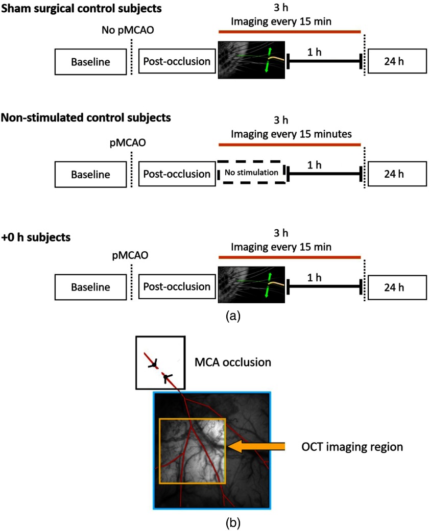
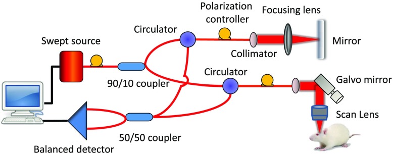
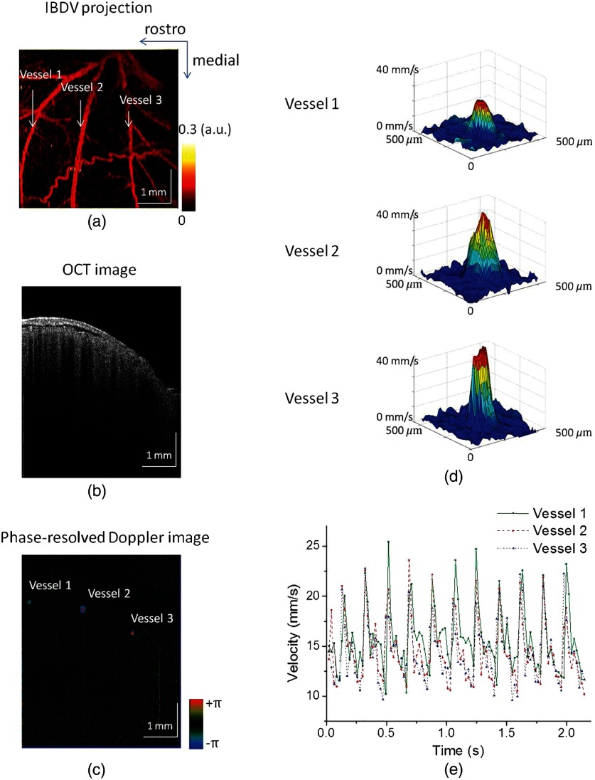
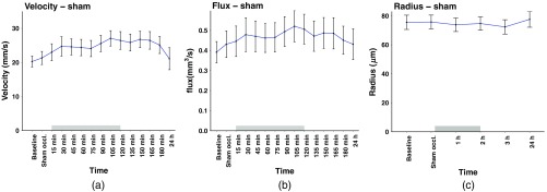
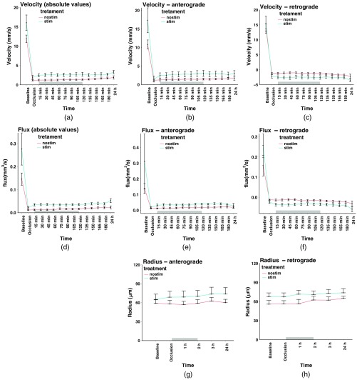
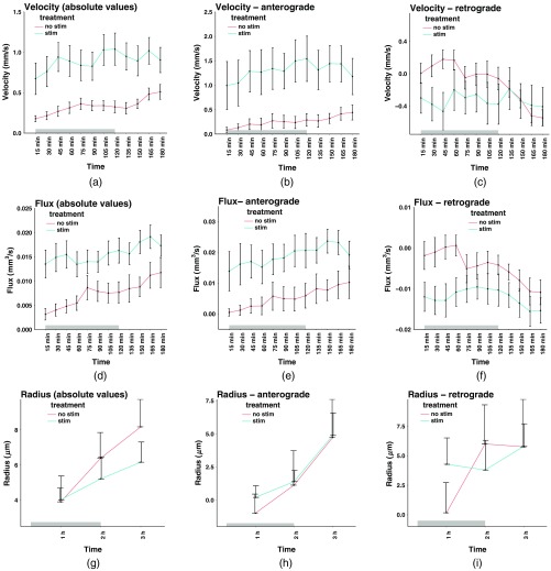
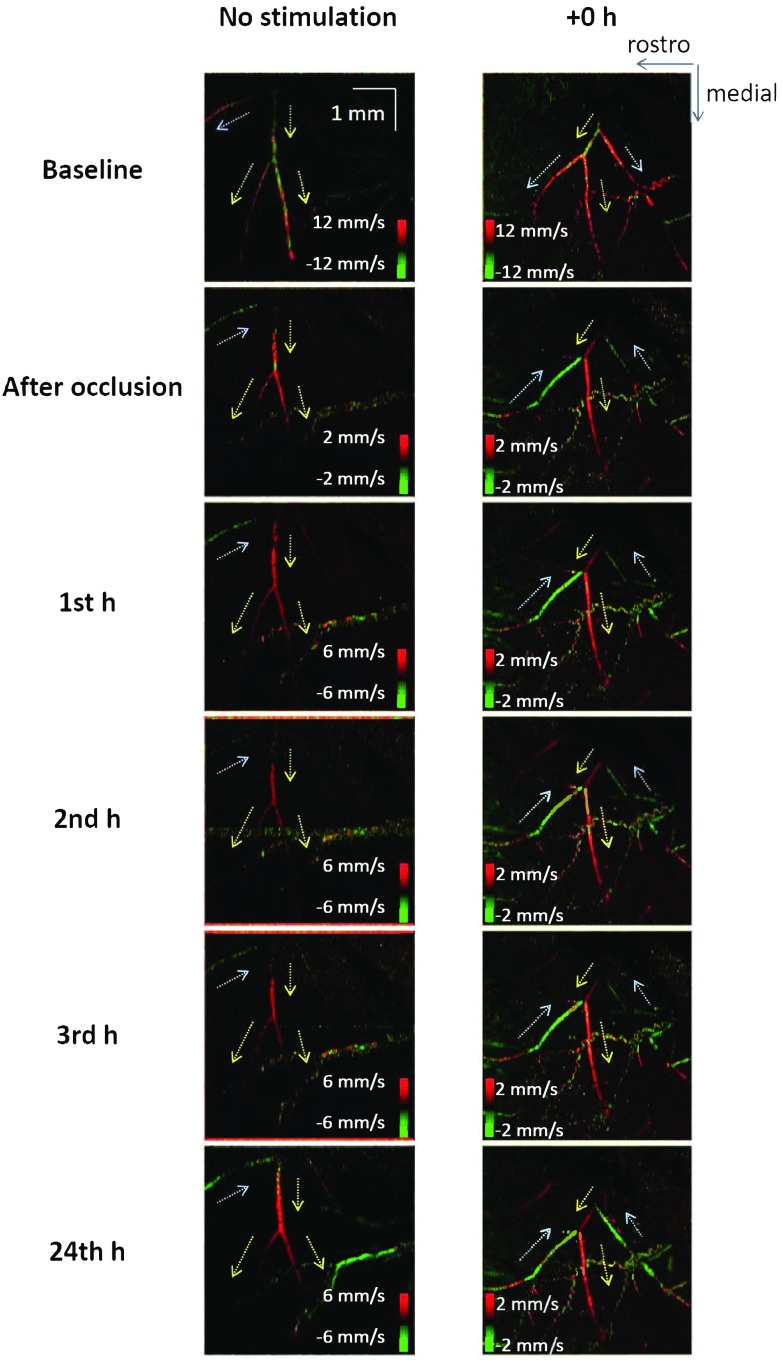
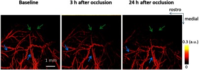
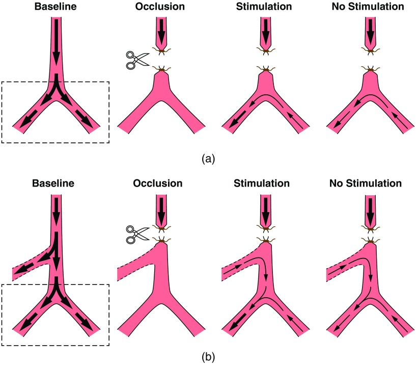
There is a growing recognition regarding the importance of pial collateral flow in the protection from impending ischemic stroke both in preclinical and clinical studies. Collateral flow is also a major player in sensory stimulation-based protection from impending ischemic stroke. Doppler optical coherence tomography has been employed to image spatiotemporal patterns of collateral flow within the dorsal branches of the middle cerebral artery (MCA) as it provides a powerful tool for quantitative in vivo flow parameters imaging (velocity, flux, direction of flow, and radius of imaged branches). It was employed prior to and following dorsal permanent MCA occlusion (pMCAo) in rat models of treatment by protective sensory stimulation, untreated controls, or sham surgery controls. Unexpectedly, following pMCAo in the majority of subjects, some MCA branches continued to show anterograde blood flow patterns over time despite severing of the MCA. Further, in the presence of protective sensory stimulation, the anterograde velocity and flux were stronger and lasted longer than in retrograde flow branches, even within different branches of single subjects, but stimulated retrograde branches showed stronger flow parameters at 24 h. Our study suggests that the spatiotemporal patterns of collateral-based dorsal MCA flow are dynamic and provide a detailed description on the differential effects of protective sensory stimulation.

Keywords: Doppler optical coherence tomography; cortex; leptomeningeal anastomoses; leptomeningeal collaterals; neurovascular.


Figures

Fig. 1
Fig. 1
(a) Experimental timelines of OCT imaging. Three experimental subjects were imaged, including sham surgical control subjects, pMCAo group without whisker stimulation (no-stimulation control subjects), and pMCAo group with whisker stimulation (+0  h subjects). (b) Doppler OCT imaging window that was used in the experiments; MCA imaged branches are highlighted in red. Note that the MCA occlusion site (the M1 section of the MCA) is always outside the DOCT imaging window.
Fig. 2
Fig. 2
Schematic of OCT system. The OCT system is based on a swept source with a wavelength of 1310 nm and A-line speed of 50 kHz.
Fig. 3
Fig. 3
Flow measurements using a flow phantom. (a) System setup for the flow measurements. (b) Comparison between theoretical flow velocities controlled by a syringe pump and measured phase shifts by Doppler OCT imaging.
Fig. 4
Fig. 4
Blood flow measurements in a sham surgical control subject. (a) IBDV projection from OCT imaging shows a microvascular network in the MCA region of a rat brain. A sampling A-line interval of 0.40 ms was used. (b) Cross-sectional OCT image. (c) Phase-resolved Doppler OCT cross-sectional scan for flow quantification. Three major MCA branches can be identified clearly under the dura mater. A sampling interval of 0.02 ms was used. (d) Distribution of flow velocities in three branches at one time. (e) Flow velocities in three branches over time. The changes of flow velocities were caused by the heartbeats.
Fig. 5
Fig. 5
Sham-surgery flow parameters and radii in the presence of sensory stimulation. In the presence of stimulation (denoted by a gray rectangle above the x-axis) as compared to presham-pMCAo there was (a) an increase in velocity as analyzed for the 15 to 180 min time points period, (b) increase in flux, but (c) no change in radii of the imaged branches.
Fig. 6
Fig. 6
Change in flow patterns in pMCAo groups (+0  h and no-stimulation) prior and following pMCAo and up to 24 h. Changes are shown for stimulated (green) versus nonstimulated (red) branches for velocity: (a) absolute values, (b) anterograde branches, and (c) retrograde branches; flux: (d) absolute values, (e) anterograde branches, and (f) retrograde branches. For the radii (sampled in a lower rate), absolute values were not relevant, and therefore, only (g) anterograde and (h) retrograde branches are shown. Stimulus duration is denoted by a gray rectangle above the x-axis. No stim group is denoted in red; stim group is denoted in green. Note that “retrograde” and “anterograde” labels have no meaning before pMCAo. See text for details.
Fig. 7
Fig. 7
Detailed analysis of difference in flow parameters and radii analyzed as differences from the occlusion point to 180 min in pMCAo groups (+0  h and no-stimulation) where colors denote stimulated (green) versus nonstimulated (red) branches. (a) Velocity (absolute values), (b) anterograde, and (c) retrograde. (d) Flux (absolute values), (e) anterograde, and (f) retrograde. (g) Radius (absolute values), (h) anterograde branches, and (i) retrograde branches. Stimulus duration is denoted by a gray rectangle above the x-axis. See text for details.
Fig. 8
Fig. 8
Example of maximum intensity projection using a phase-resolved Doppler method. Blue arrows denote retrograde flow and yellow arrows denote anterograde flow. Note reversal of flow direction following occlusion in each example. The green points in the red branch and the red points in the green branch represent phase wrapping due to larger velocities.
Fig. 9
Fig. 9
En face projections of IBDV images of vascular networks in the MCA region of a +0  h subject. A sampling A-line interval of 2.00 ms was used to highlight blood vessels with slower flow speed. After occlusion, there was a major reduction in blood flow speed in the far dorsal parts of major MCA branches as indicated by the green arrows, yet flow speed remained sustained in the more proximal parts of the same MCA branches as indicated by blue arrows.
Fig. 10
Fig. 10
Cartoon of the main the dorsal MCA branches and their blood flow patterns before and after pMCAo. (a) Intuitive explanation of our findings. Two main dorsal MCA branches are imaged by DOCT (imaged area is denoted by a rectangle on the left) with strong natural anterograde flow (wide, bold arrows). Following occlusion, retrograde flow in a branch that is connected to functional collateral supplies blood to the anterograde branch. In the presence of sensory stimulation, the anterograde flow becomes stronger than the retrograde flow as denoted by the width of the arrows. However, it is not clear how the retrograde and anterograde flows can show different flow patterns if the former is the only provider for the latter. Therefore, in (b) a different hypothesis is shown. In this case, there is a third MCA branch (denoted by dashed lines) outside the imaged area (denoted by the rectangle). Following pMCAo, retrograde flow in the unimaged branch supplies additional blood flow to the anterograde branch, explaining the discrepancy between retrograde and anterograde branches shown in (a). See text for details on another, complementary hypothesis about the increase in radius of anterograde vessels over time.

References

    1. Mozaffarian D., et al. , “Heart disease and stroke statistics-2016 update: a report from the American Heart Association,” Circulation 133(4), e38–e360 (2016).CIRCAZ10.1161/CIR.0000000000000350 - DOI - PubMed
    1. Bhatia R., et al. , “Low rates of acute recanalization with intravenous recombinant tissue plasminogen activator in ischemic stroke: real-world experience and a call for action,” Stroke 41(10), 2254–2258 (2010).SJCCA710.1161/STROKEAHA.110.592535 - DOI - PubMed
    1. Goyal M., et al. , “Randomized assessment of rapid endovascular treatment of ischemic stroke,” N. Engl. J. Med. 372(11), 1019–1030 (2015).NEJMAG10.1056/NEJMoa1414905 - DOI - PubMed
    1. Yoo A. J., Andersson T., “Thrombectomy in acute ischemic stroke: challenges to procedural success,” J. Stroke 19(2), 121–130 (2017).10.5853/jos.2017.00752 - DOI - PMC - PubMed
    1. Burnett M. G., et al. , “Electrical forepaw stimulation during reversible forebrain ischemia decreases infarct volume,” Stroke 37(5), 1327–1331 (2006).SJCCA710.1161/01.STR.0000217305.82123.d8 - DOI - PubMed