. 2020 Feb 1;318(2):C372-C379.
doi: 10.1152/ajpcell.00135.2019. Epub 2019 Dec 11.

Activation of the PKA signaling pathway stimulates oxalate transport by human intestinal Caco2-BBE cells

Affiliations

Activation of the PKA signaling pathway stimulates oxalate transport by human intestinal Caco2-BBE cells

Donna Arvans et al. Am J Physiol Cell Physiol. .

Abstract

Most kidney stones are composed of calcium oxalate, and small increases in urine oxalate enhance the stone risk. The mammalian intestine plays a crucial role in oxalate homeostasis, and we had recently reported that Oxalobacter-derived factors stimulate oxalate transport by human intestinal Caco2-BBE (C2) cells through PKA activation. We therefore evaluated whether intestinal oxalate transport is directly regulated by activation of the PKA signaling pathway. To this end, PKA was activated with forskolin and IBMX (F/I). F/I significantly stimulated (3.7-fold) [14C]oxalate transport by C2 cells [≥49% of which is mediated by the oxalate transporter SLC26A6 (A6)], an effect completely blocked by the PKA inhibitor H89, indicating that it is PKA dependent. PKA stimulation of intestinal oxalate transport is not cell line specific, since F/I similarly stimulated oxalate transport by the human intestinal T84 cells. F/I significantly increased (2.5-fold) A6 surface protein expression by use of immunocytochemistry. Assessing [14C]oxalate transport as a function of increasing [14C]oxalate concentration in the flux medium showed that the observed stimulation is due to a F/I-induced increase (1.8-fold) in Vmax and reduction (2-fold) in Km. siRNA knockdown studies showed that significant components of the observed stimulation are mediated by A6 and SLC26A2 (A2). Besides enhancing A6 surface protein expression, it is also possible that the observed stimulation is due to PKA-induced enhanced A6 and/or A2 transport activity in view of the reduced Km. We conclude that PKA activation positively regulates oxalate transport by intestinal epithelial cells and that PKA agonists might therapeutically impact hyperoxalemia, hyperoxaluria, and related kidney stones.

Keywords: PKA; SLC26A2; SLC26A6; T84 cells; intestinal oxalate transport.


Conflict of interest statement

H. Hassan is cofounder and Chief Scientific Officer for Oxalo Therapeutics. None of the other authors has any conflicts of interest, financial or otherwise, to disclose.

Figures

Fig. 1.
Fig. 1.
Effect of forskolin/IBMX (F/I) on [14C]oxalate uptake by Caco2-BBE (C2) cells. C2 cells were untreated (UT) or were treated apically with F/I (625/500 µM) for 30 min in NaCl buffer, and then [14C]oxalate uptake was measured as described in materials and methods. Values are means ± SE of 24 independent experiments, each done in triplicate. F/I significantly stimulated [14C]oxalate uptake by C2 cells (*P < 2.4E−11, by unpaired t test; n = 24).
Fig. 2.
Fig. 2.
Effect of different concentrations of forskolin/IBMX (F/I) on [14C]oxalate uptake by Caco2-BBE (C2) cells. C2 cells were untreated (UT) or were treated apically with different concentrations of F/I (5/500 = F5/I500, 25/500 = F25/I500, 125/500 = F125/I500, and 625/500 = F625/I500 µM) for 30 min in NaCl buffer, and then [14C]oxalate uptake was measured as described in materials and methods. Values are means ± SE of 3 independent experiments, each done in triplicate. F/I significantly stimulated [14C]oxalate uptake by C2 cells (*P < 0.05, < 0.01, < 0.001, and < 0.001 for UT vs. F5/I500, F25/I500, F125/I500, and F625/I500 µM, respectively; `P < 0.001 for F625/I500 vs. F5/I500, F25/500, and F125/I500; `P < 0.05 for F125/I500 vs. F5/I500, by ANOVA; n = 3).
Fig. 3.
Fig. 3.
Effect of different preincubation periods with forskolin/IBMX (F/I) on [14C]oxalate uptake by Caco2-BBE (C2) cells. To determine the time course over which the stimulatory effects of F/I occur, C2 cells were untreated (UT) or were treated apically with F/I (625/500 µM) for 5, 15, 30, and 60 min in NaCl buffer, and then [14C]oxalate uptake was measured as described in materials and methods. Values are means ± SE of 3–4 independent experiments, each done in triplicate. F/I significantly stimulated [14C]oxalate uptake by C2 cells over the 15-, 30-, and 60-min time points (*P < 0.05, < 0.001, and < 0.001 for UT vs. 15, 30, and 60 min, respectively; `P < 0.001 and < 0.01 for 30 min vs. 5 and 30 min, respectively; `P < 0.01 and < 0.05 for 60 min vs. 5 and 30 min, respectively; `P < 0.05 for 15 min vs. 5 min, by ANOVA; n = 3–4).
Fig. 4.
Fig. 4.
Effect of forskolin/IBMX (F/I) on [14C]oxalate uptake by T84 cells. T84 cells were untreated (UT) or were treated apically with F/I (625/500 µM) for 30 min in NaCl buffer, and then [14C]oxalate uptake was measured as described in materials and methods. Values are means ± SE of 6 independent experiments, each done in triplicate. F/I significantly stimulated [14C]oxalate uptake by T84 cells (*P < 0.02 for F/I vs. UT, by unpaired t test; n = 6).
Fig. 5.
Fig. 5.
Effect of PKA inhibitor H89 on the forskolin/IBMX (F/I)-induced stimulation of [14C]oxalate uptake by Caco2-BBE (C2) cells. C2 cells were untreated (UT) or were treated apically with F/I (625/500 µM) for 30 min in NaCl buffer, and then [14C]oxalate uptake was measured as described in materials and methods. C2 cells were also treated apically with PKA inhibitor H89 (20 µM) for 30 min followed by F/I (625/500 µM) for 30 min with continued presence of H89 (F/I+H89), or with H89 (20 µM) alone for 60 min, and then [14C]oxalate uptake was similarly measured. Values are means ± SE of 4 independent experiments, each done in triplicate. F/I significantly stimulated [14C]oxalate uptake by C2 cells, an effect completely blocked by H89 (*P < 0.001 for F/I vs. UT, F/I+H89, and H89, by ANOVA; n = 4).
Fig. 6.
Fig. 6.
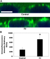
Effect of forskolin/IBMX (F/I) on SLC26A6 (A6) surface expression in Caco2-BBE (C2) cells assayed by immunocytochemistry. A: C2 cells were treated apically with vehicle (Control) or F/I (625/500 µM) for 30 min in NaCl buffer before immunofluorescence confocal microscopy on sections of paraformaldehyde-fixed C2 cells was performed, as described in materials and methods. Scale bar, 10 μm. F/I led to increased A6 surface protein expression. B: analysis of images in ImageJ. Peak immunofluorescence intensity was determined in the z direction from the average peak intensity of 5 different areas in each image. Values are means ± SE of 4 independent experiments. F/I significantly increased A6 surface protein expression (*P < 0.03, by paired t test; n = 4).
Fig. 7.
Fig. 7.
Effect of forskolin/IBMX (F/I) on kinetic characteristics of oxalate transport in Caco2-BBE (C2) cells. C2 cells were untreated (UT) or were treated apically with F/I (625/500 µM) for 30 min in NaCl buffer, and then [14C]oxalate uptake as a function of increasing [14C]oxalate concentration in the flux medium (0.3, 1, 3, 10, 30, 100, and 225 µM) was assessed. Values are means ± SE of 3–5 independent experiments, each done in triplicate. F/I significantly stimulated [14C]oxalate uptake by C2 cells at the 0.3, 1, 3, 10, 30, and 100 µM oxalate concentrations (*P < 0.02, < 0.007, < 0.00007, < 0.006, < 0.0008, and < 0.02 for F/I vs. UT at 0.3, 1, 3, 10, 30, and 100 µM [14C]oxalate concentrations, respectively, by unpaired t test; n = 3–5).
Fig. 8.
Fig. 8.
Effect of SLC26A6 (A6) knockdown on forskolin/IBMX (F/I)-induced stimulation of [14C]oxalate uptake by Caco2-BBE (C2) cells. Control (untransfected) and A6 siRNA (transfected with the siRNA targeting A6) C2 cells were untreated (UT) or were treated apically with forskolin/IBMX (F/I; 625/500 µM) in NaCl buffer, and then [14C]oxalate uptake was measured as described in materials and methods. Values are means ± SE of 10 independent experiments, each done in triplicate. Silencing A6 significantly reduced F/I-induced stimulation of oxalate transport by C2 cells [*P < 0.001 for F/I (Control) vs. UT (Control), UT (A6 siRNA), and F/I (A6 siRNA); #P < 0.001 for UT (Control) vs. UT (A6 siRNA), by Wilcoxon rank-sum test; n = 10].
Fig. 9.
Fig. 9.
Effect of SLC26A2 (A2) knockdown on forskolin/IBMX (F/I)-induced stimulation of [14C]oxalate uptake by Caco2-BBE (C2) cells. Control (untransfected) and A2 siRNA (transfected with the siRNA targeting A2) C2 cells were untreated (UT) or were treated apically with forskolin/IBMX (F/I; 625/500 µM) in NaCl buffer, and then [14C]oxalate uptake was measured as described in materials and methods. Values are means ± SE of 15 independent experiments, each done in triplicate. Silencing A2 significantly reduced F/I-induced stimulation of oxalate transport by C2 cells [*P < 0.001 for F/I (Control) vs. UT (Control), UT (A2 siRNA), and F/I (A2 siRNA); #P < 0.007 for UT (Control) vs. UT (A2 siRNA), by Wilcoxon rank-sum test; n = 15].

Comment in

References

    1. Alexander RT, Hemmelgarn BR, Wiebe N, Bello A, Morgan C, Samuel S, Klarenbach SW, Curhan GC, Tonelli M; Alberta Kidney Disease Network . Kidney stones and kidney function loss: a cohort study. BMJ 345: e5287, 2012. doi:10.1136/bmj.e5287. - DOI - PMC - PubMed
    1. Alper SL, Sharma AK. The SLC26 gene family of anion transporters and channels. Mol Aspects Med 34: 494–515, 2013. doi:10.1016/j.mam.2012.07.009. - DOI - PMC - PubMed
    1. Amin R, Asplin J, Jung D, Bashir M, Alshaikh A, Ratakonda S, Sharma S, Jeon S, Granja I, Matern D, Hassan H. Reduced active transcellular intestinal oxalate secretion contributes to the pathogenesis of obesity-associated hyperoxaluria. Kidney Int 93: 1098–1107, 2018. doi:10.1016/j.kint.2017.11.011. - DOI - PMC - PubMed
    1. Amin R, Sharma S, Ratakonda S, Hassan HA. Extracellular nucleotides inhibit oxalate transport by human intestinal Caco-2-BBe cells through PKC-δ activation. Am J Physiol Cell Physiol 305: C78–C89, 2013. doi:10.1152/ajpcell.00339.2012. - DOI - PMC - PubMed
    1. Arvans D, Jung YC, Antonopoulos D, Koval J, Granja I, Bashir M, Karrar E, Roy-Chowdhury J, Musch M, Asplin J, Chang E, Hassan H. Oxalobacter formigenes-derived bioactive factors stimulate oxalate transport by intestinal epithelial cells. J Am Soc Nephrol 28: 876–887, 2017. doi:10.1681/ASN.2016020132. - DOI - PMC - PubMed

Publication types

MeSH terms

Substances

LinkOut - more resources