Comparative Study
. 2020 Mar 1;318(3):C615-C626.
doi: 10.1152/ajpcell.00540.2019. Epub 2019 Dec 11.

Comparative profiling of skeletal muscle models reveals heterogeneity of transcriptome and metabolism

Affiliations
Comparative Study

Comparative profiling of skeletal muscle models reveals heterogeneity of transcriptome and metabolism

Ahmed M Abdelmoez et al. Am J Physiol Cell Physiol. .

Abstract

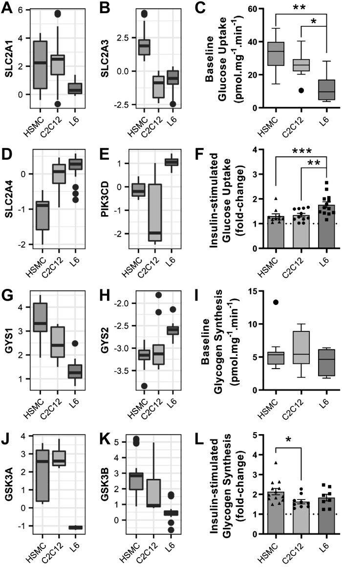
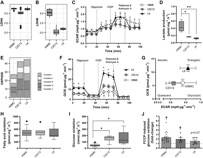
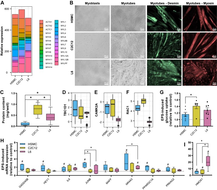
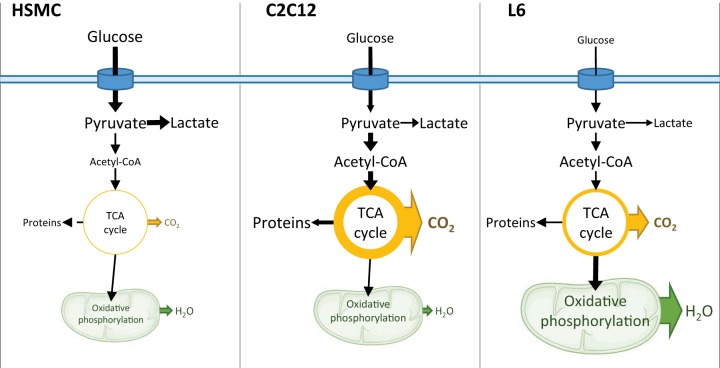
Rat L6, mouse C2C12, and primary human skeletal muscle cells (HSMCs) are commonly used to study biological processes in skeletal muscle, and experimental data on these models are abundant. However, consistently matched experimental data are scarce, and comparisons between the different cell types and adult tissue are problematic. We hypothesized that metabolic differences between these cellular models may be reflected at the mRNA level. Publicly available data sets were used to profile mRNA levels in myotubes and skeletal muscle tissues. L6, C2C12, and HSMC myotubes were assessed for proliferation, glucose uptake, glycogen synthesis, mitochondrial activity, and substrate oxidation, as well as the response to in vitro contraction. Transcriptomic profiling revealed that mRNA of genes coding for actin and myosin was enriched in C2C12, whereas L6 myotubes had the highest levels of genes encoding glucose transporters and the five complexes of the mitochondrial electron transport chain. Consistently, insulin-stimulated glucose uptake and oxidative capacity were greatest in L6 myotubes. Insulin-induced glycogen synthesis was highest in HSMCs, but C2C12 myotubes had higher baseline glucose oxidation. All models responded to electrical pulse stimulation-induced glucose uptake and gene expression but in a slightly different manner. Our analysis reveals a great degree of heterogeneity in the transcriptomic and metabolic profiles of L6, C2C12, or primary human myotubes. Based on these distinct signatures, we provide recommendations for the appropriate use of these models depending on scientific hypotheses and biological relevance.

Keywords: C2C12; metabolism; skeletal muscle; transcriptomics.


Conflict of interest statement

No conflicts of interest, financial or otherwise, are declared by the authors.

Figures

Fig. 1.
Fig. 1.
Transcriptomic profiling of rat L6, mouse C2C12, and human primary myotubes. Publicly available data sets for mouse, rat, and human skeletal muscle tissue and cell models were downloaded from the Gene Expression Omnibus (GEO) repository. A: raw data were processed, normalized, and annotated according to the official human gene ortholog nomenclature. B: principal component (PC) analysis demonstrated a clear segregation of all samples from each other. C: correlation matrix comparing all transcripts from one model to the other five. D: chord diagram presenting the overlap of the top 100 expressed genes in each model. The thickness of a band is proportional to the number of common genes between 2 models. RMA, robust multi-array.
Fig. 2.
Fig. 2.
Gene enrichment in L6, C2C12, and human primary myotubes. A: example of a gene (GRIN2B) exhibiting species differences. B: example of a gene (LDHD) differentially expressed in cells compared with skeletal muscle tissue. C: example of a gene (PLN) which expression is affected by both species and cell/tissue sample. D: gene ontology enrichment of genes differentially expressed in one model compared with the other two (false discovery rate < 0.05). Gene ratio is the number of significantly different genes divided by the total number of genes associated with a specific pathway. eIF2, eukaryotic translation initiation factor 2; HSMC, human skeletal muscle cells; p.adjust, adjusted P value.
Fig. 3.
Fig. 3.
Proliferation rates of L6, C2C12, and human primary myotubes. A: mRNA expression of the proliferation marker MKI67. B: total area covered by cells during 3 days of proliferation. Quantification of images was done with ImageJ, as described in methods. Mean ± SE, Exponential (Malthusian) growth fit; n totals are as follows: HSMC: n = 7 replicates from 7 independent donors, C2C12: n = 5 independent experiments, L6: n = 5 independent experiments, *P < 0.05 in human skeletal muscle cells (HSMC) compared with L6 and C2C12. C: doubling time calculated from the proliferation curves. Box-and-whisker Tukey plot; n totals are as follows: HSMC: n = 7 replicates from 7 independent donors, C2C12: n = 5 independent experiments, L6: n = 5 independent experiments, Kruskal-Wallis test with Dunn’s multiple comparison, *P < 0.05. D: BrdU incorporation into DNA measured as described in methods. Box-and-whisker Tukey plot; n totals are as follows: HSMC: n = 6 replicates from 3 independent donors, C2C12: n = 6 independent experiments, L6: n = 6 independent experiments, one-way ANOVA with Tukey’s multiple comparison, *P < 0.05. E–G: differentiation time course of mRNA expression of skeletal muscle markers creatine kinase (CKM), myogenin (MYOG), myogenic differentiation 1 (MYOD1), and myogenic factor 5 (MYF5). Mean ± SE, Dot plot; n totals are as follows: HSMC: n = 5 replicates from 5 independent donors, C2C12: n = 4 independent experiments, L6: n = 4 independent experiments, paired one-way ANOVA with Dunnett’s multiple testing, *P < 0.05 compared with undifferentiated cells. A.U., arbitrary units.
Fig. 4.
Fig. 4.
Insulin-induced glucose uptake and glycogen synthesis in L6, C2C12, and human primary myotubes. A and B: mRNA expression of glucose transporter 1 and 3 (SLC2A1 and SLC2A3). C: basal glucose uptake, as measured using 2-[1,2-3H]deoxy-d-glucose. Box-and-whisker Tukey plot; n totals are as follows: HSMC: n = 11 replicates from 11 independent donors, C2C12: n = 11 independent experiments, L6: n = 13 independent experiments, one-way ANOVA with Tukey’s multiple comparison. D and E: mRNA expression of glucose transporter 4 (SLC2A4) and the delta subunit of phosphatidylinositol 3-kinase (PIK3CD). F: insulin-stimulated glucose uptake, as measured using 2-[1,2-3H]deoxy-d-glucose. Means ± SE and individual data points: n totals are as follows: HSMC: n = 11 replicates from 11 independent donors, C2C12: n = 11 independent experiments, L6: n = 13 independent experiments, one-way ANOVA with Tukey’s multiple comparison. G and H: glycogen synthase (GS) mRNA expression [GS 1 isoform (GYS1) and GS 2 isoform (GYS2)]. I: basal glycogen synthesis, as measured by the incorporation of [14C]glucose into glycogen. Box-and-whisker Tukey plot; n totals are as follows: HSMC: n = 12 replicates from 12 independent donors, C2C12: n = 9 independent experiments, L6: n = 8 independent experiments, Kruskal-Wallis test with Dunn’s multiple comparison. J and K: glycogen synthase kinase 3 (GSK3) mRNA expression. L: insulin-stimulated glycogen synthesis as measured by the incorporation of [14C]glucose into glycogen. Means ± SE and individual data points; n totals are as follows: HSMC: n = 12 replicates from 12 independent donors, C2C12: n = 9 independent experiments, L6: n = 8 independent experiments, one-way ANOVA with Tukey’s multiple comparison, *P < 0.05, **P < 0.01, ***P < 0.001. HSMC, human skeletal muscle cells.
Fig. 5.
Fig. 5.
Oxygen consumption and substrate use in L6, C2C12, and human primary myotubes. A and B: mRNA expression of lactate dehydrogenase subunits (LDHA, LDHB). C: extracellular acidification rate (ECAR) during a Mito Stress Test measured with Seahorse technology. Means ± SE; n totals are as follows: HSMC: n = 13 replicates from 8 independent donors, C2C12: n = 7 independent experiments, L6: n = 8 independent experiments, two-way ANOVA with Tukey’s multiple comparison. D: lactate accumulation in the extracellular medium measured as described in methods. Box-and-whisker Tukey plot; n totals are as follows: HSMC: n = 5 replicates from 4 independent donors, C2C12: n = 5 independent experiments, L6: n = 5 independent experiments, one-way ANOVA with Tukey’s multiple comparison, *P < 0.05, **P < 0.01. E: mRNA expression of mitochondrial complexes. F: oxygen consumption rate (OCR) during a Mito Stress Test measured with Seahorse technology. Means ± SE; n totals are as follows: HSMC: n = 13 replicates from 8 independent donors, C2C12: n = 7 independent experiments, L6: n = 8 independent experiments, two-way ANOVA with Tukey’s multiple comparison, *P < 0.05 compared with both C2C12 and human skeletal muscle cells (HSMC), †P < 0.05 compared with C2C12 and L6. G: energy plot from the Mito Stress Test. H: unstimulated fatty acid oxidation, measured by the oxidation of [3H]palmitate into H2O. Box-and-whisker Tukey plot; n totals are as follows: HSMC: n = 37 replicates from 12 independent donors, C2C12: n = 11 independent experiments, L6: n = 17 independent experiments, one-way ANOVA with Tukey’s multiple comparison. I: basal glucose oxidation as measured by the oxidation of [14C]glucose into CO2. Box-and-whisker Tukey plot; n totals are as follows: HSMC: n = 19 replicates from 12 independent donors, C2C12: n = 6 independent experiments, L6: n = 8 independent experiments, one-way ANOVA with Tukey’s multiple comparison, *P < 0.05. J: carbonyl cyanide-4-(trifluoromethoxy)phenylhydrazone (FCCP)-stimulated [14C]glucose oxidation. Means ± SE and individual data points; n totals are as follows: HSMC: n = 19 replicates from 12 independent donors, C2C12: n = 6 independent experiments, L6: n = 7 independent experiments, one-way ANOVA with Tukey’s multiple comparison, †P < 0.05 effect of FCCP vs. unstimulated. OXPHOS, oxidative phosphorylation.
Fig. 6.
Fig. 6.
Response to electrical pulse stimulation (EPS) in L6, C2C12, and human primary myotubes. A: mRNA levels of myosin and actin isoforms. B: representative images of myotubes during proliferation and after complete differentiation. Staining for desmin and myosin was performed using specific antibodies as described in methods. Representative images are shown for each staining. C: protein content per well measured by bicinchoninic acid assay. Box-and-whisker Tukey plot; n totals are as follows: HSMC: n = 11 replicates from 11 independent donors, C2C12: n = 7 independent experiments, L6: n = 6 independent experiments, Kruskal-Wallis test with Dunn’s multiple comparison. D–F: mRNA levels of the GTPase-activating protein for Rab family protein TBC1D1, Ca2+/calmodulin-dependent protein kinase II (CAMK2A), and RAC1. G: electrical pulse stimulation-induced glucose uptake measured using 2-[1,2-3H]deoxy-d-glucose. Mean ± SE and individual data points: n totals are as follows: HSMC: n = 7 replicates from 7 independent donors, C2C12: n = 6 independent experiments, L6: n = 7 independent experiments, one-way ANOVA with Tukey’s multiple comparison. H and I: mRNA expression of exercise-responsive genes measured by quantitative PCR. Box-and-whisker Tukey plot; n totals are as follows: HSMC: n = 5 replicates from 5 independent donors, C2C12: n = 6 independent experiments, L6: n = 6 independent experiments, two-way ANOVA with Fisher’s least-significant difference comparison. #P < 0.05, significant response to EPS. *P < 0.05. HSMC, human skeletal muscle cells.
Fig. 7.
Fig. 7.
Schematic representation of C2C12, L6, and human primary myotube metabolic pathways. Fatty acid oxidation was similar in all three cell models; therefore changes in glucose metabolism between the cell types may explain the differences in metabolism across models. Human primary myotubes have the highest baseline glucose uptake and lactate production, suggesting that these cells rely mostly on glycolysis for energy production. On the other end of the spectrum, L6 myotubes have a relatively lower baseline glucose uptake, but these cells oxidize most of the intracellular glucose in the mitochondria. C2C12 cells have moderate rates of glucose uptake, with intracellular glucose directed toward the TCA cycle. Oxidative phosphorylation and TCA coupling to oxidative phosphorylation are greater in L6. The thickness of the arrows and the size of text represent the intensity of the corresponding pathways compared with the other models. HSMC, human skeletal muscle cells.

References

    1. Aas V, Bakke SS, Feng YZ, Kase ET, Jensen J, Bajpeyi S, Thoresen GH, Rustan AC. Are cultured human myotubes far from home? Cell Tissue Res 354: 671–682, 2013. doi:10.1007/s00441-013-1655-1. - DOI - PubMed
    1. Al-Khalili L, Krämer D, Wretenberg P, Krook A. Human skeletal muscle cell differentiation is associated with changes in myogenic markers and enhanced insulin-mediated MAPK and PKB phosphorylation. Acta Physiol Scand 180: 395–403, 2004. doi:10.1111/j.1365-201X.2004.01259.x. - DOI - PubMed
    1. Arnold AS, Christe M, Handschin C. A functional motor unit in the culture dish: co-culture of spinal cord explants and muscle cells. J Vis Exp (62): 3616, 2012. doi:10.3791/3616. - DOI - PMC - PubMed
    1. Babson AL, Phillips GE. A rapid colorimetric assay for serum lactic dehydrogenase. Clin Chim Acta 12: 210–215, 1965. doi:10.1016/0009-8981(65)90032-X. - DOI - PubMed
    1. Berg JM, Tymoczko JL, Stryer L. Amino acids are made from intermediates of the citric acid cycle and other major pathways. In: Biochemistry (5th ed.). New York: WH Freeman, 2002. https://www.ncbi.nlm.nih.gov/books/NBK22459/.

Publication types

LinkOut - more resources