. 2019 Dec 11;11(522):eaay0203.
doi: 10.1126/scitranslmed.aay0203.

Impaired hemostatic activity of healthy transfused platelets in inherited and acquired platelet disorders: Mechanisms and implications

Affiliations

Impaired hemostatic activity of healthy transfused platelets in inherited and acquired platelet disorders: Mechanisms and implications

Robert H Lee et al. Sci Transl Med. .

Abstract

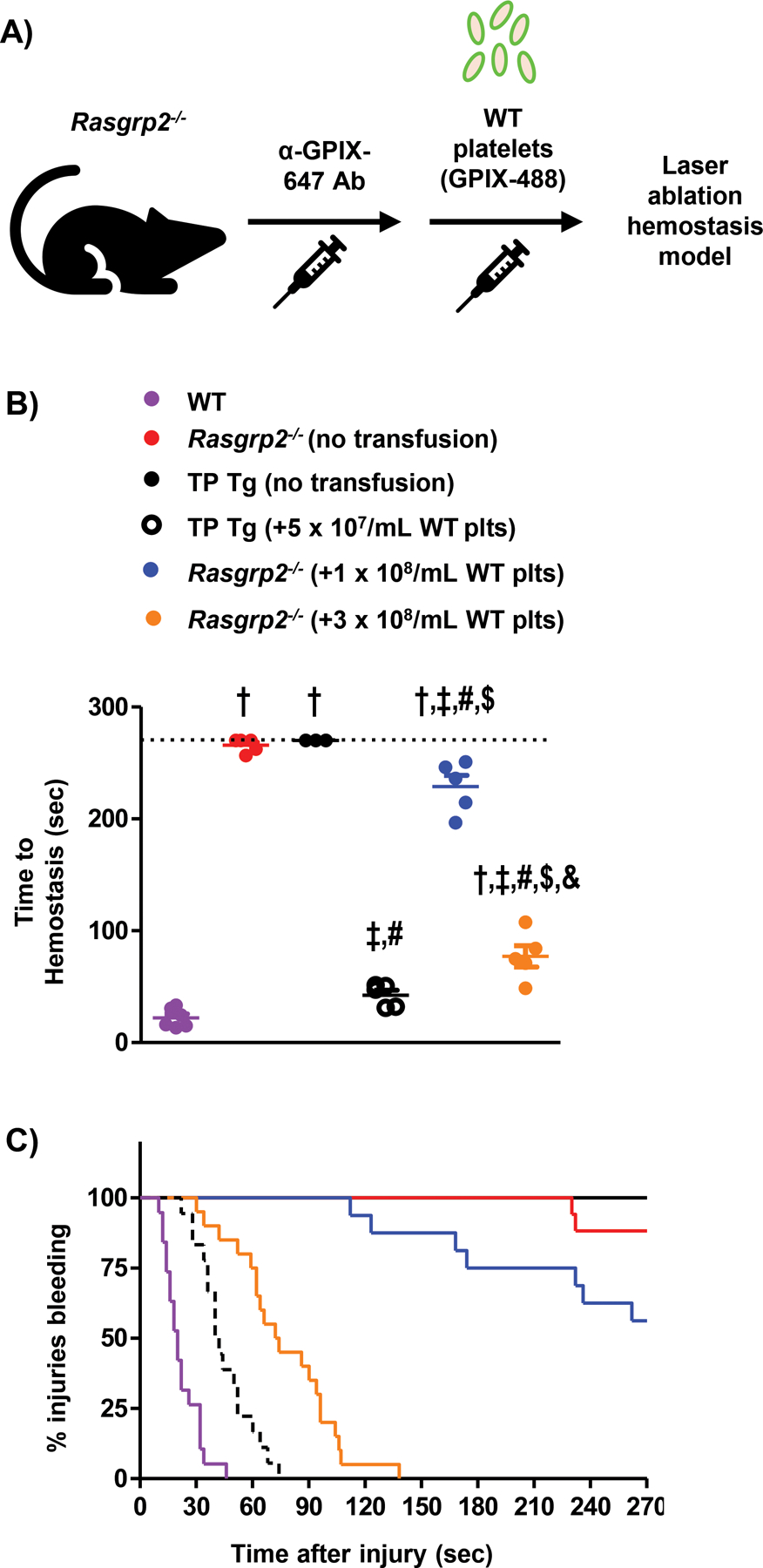
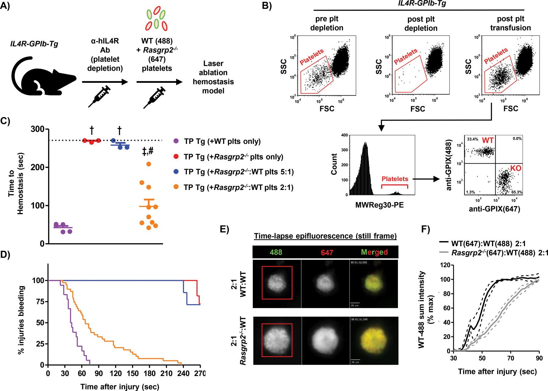
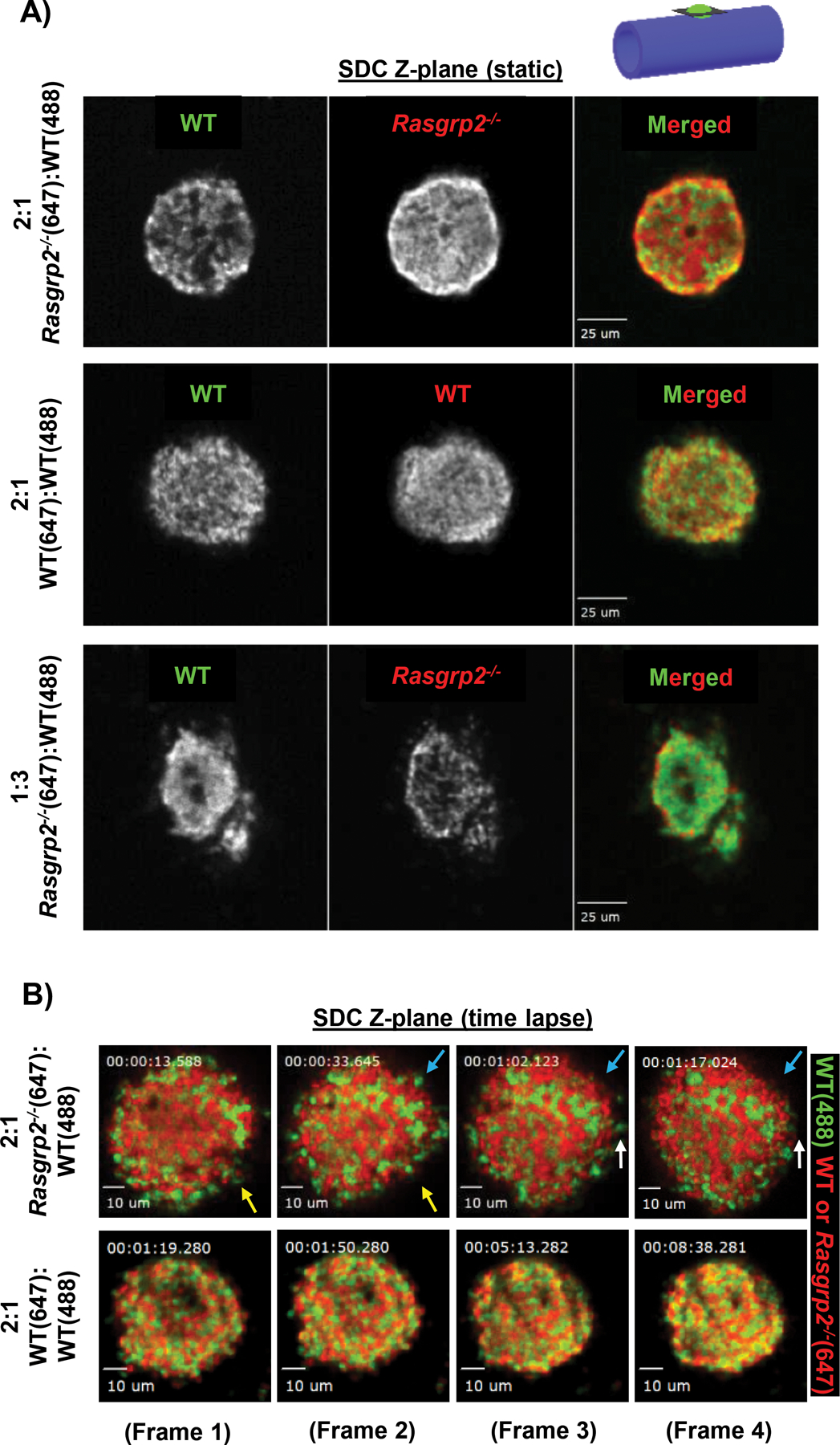
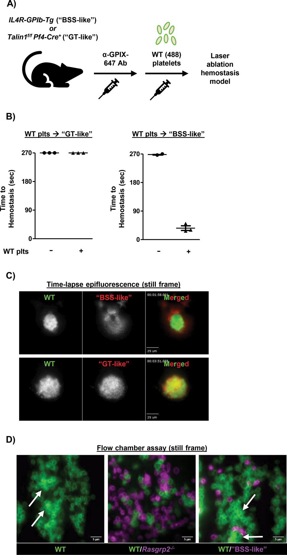
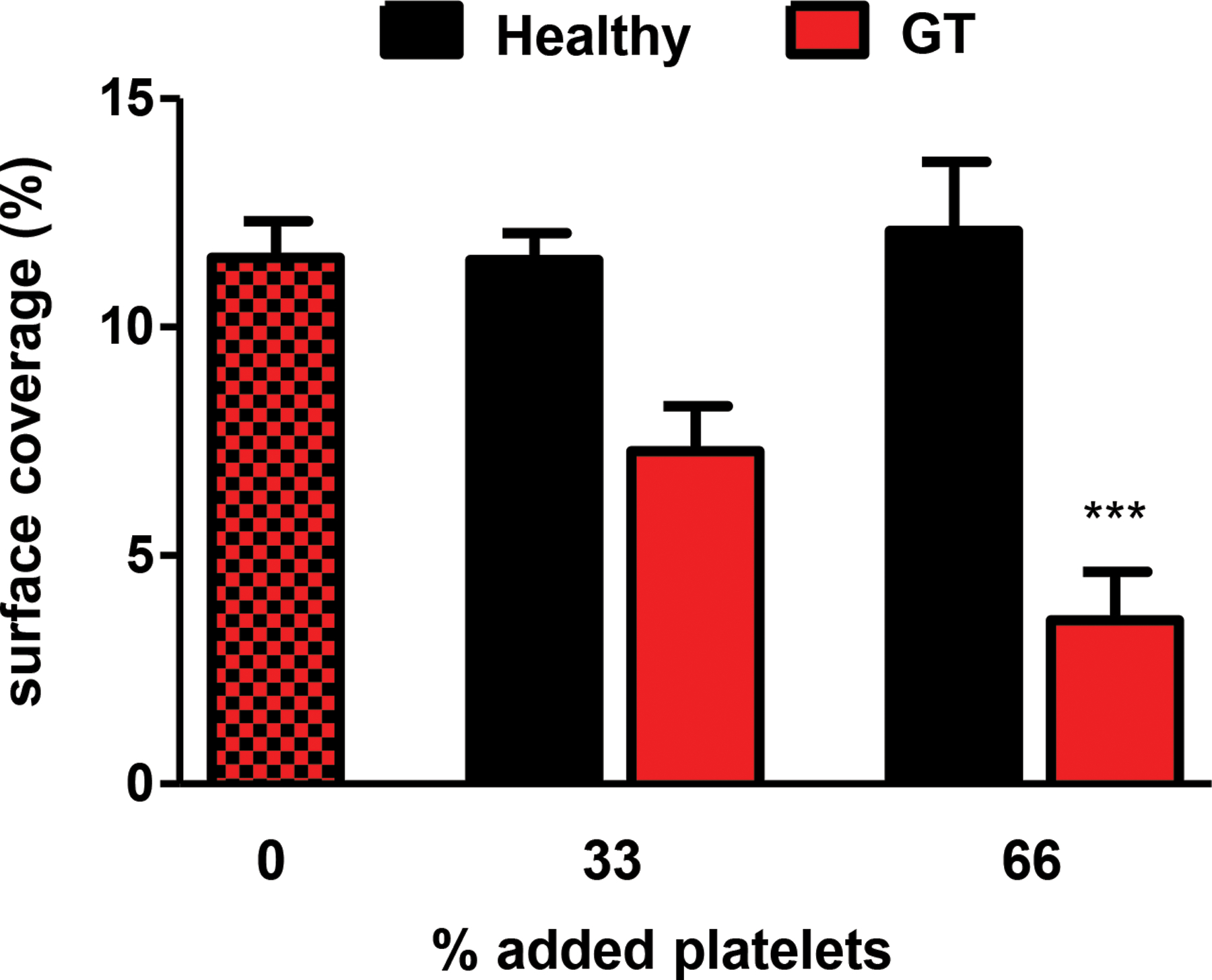
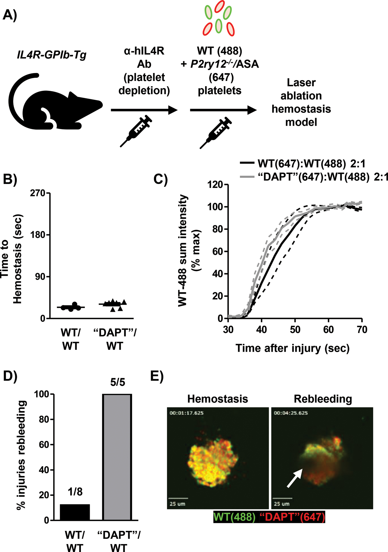
Platelet transfusions can fail to prevent bleeding in patients with inherited platelet function disorders (IPDs), such as Glanzmann's thrombasthenia (GT; integrin αIIbβ3 dysfunction), Bernard-Soulier syndrome [BSS; glycoprotein (GP) Ib/V/IX dysfunction], and the more recently identified nonsyndromic RASGRP2 variants. Here, we used IPD mouse models and real-time imaging of hemostatic plug formation to investigate whether dysfunctional platelets impair the hemostatic function of healthy donor [wild-type (WT)] platelets. In Rasgrp2-/- mice or mice with platelet-specific deficiency in the integrin adaptor protein TALIN1 ("GT-like"), WT platelet transfusion was ineffective unless the ratio between mutant and WT platelets was ~2:1. In contrast, thrombocytopenic mice or mice lacking the extracellular domain of GPIbα ("BSS-like") required very few transfused WT platelets to normalize hemostasis. Both Rasgrp2-/- and GT-like, but not BSS-like, platelets effectively localized to the injury site. Mechanistic studies identified at least two mechanisms of interference by dysfunctional platelets in IPDs: (i) delayed adhesion of WT donor platelets due to reduced access to GPIbα ligands exposed at sites of vascular injury and (ii) impaired consolidation of the hemostatic plug. We also investigated the hemostatic activity of transfused platelets in the setting of dual antiplatelet therapy (DAPT), an acquired platelet function disorder (APD). "DAPT" platelets did not prolong the time to initial hemostasis, but plugs were unstable and frequent rebleeding was observed. Thus, we propose that the endogenous platelet count and the ratio of transfused versus endogenous platelets should be considered when treating select IPD and APD patients with platelet transfusions.


Conflict of interest statement

Competing interests: All authors declare that they have no competing interests.

Figures

Fig. 1:
Fig. 1:. Rasgrp2−/− mice require a large number of transfused WT platelets for hemostasis.
(A) Model depicting platelet (plt) transfusion scheme in Rasgrp2−/− mice. Endogenous plts were labeled by injection of anti-GPIX-647 antibody prior to transfusion of GPIX-488 labeled WT plts. (B) Comparison of bleeding times in the saphenous vein laser ablation model. Rasgrp2−/− mice were transfused, or not, with a low (1 × 10⁸/mL) or high (3 × 10⁸/mL) number of WT plts, and compared with WT mice or plt-depleted IL4R-GPIb-Tg mice (thrombocytopenic (TP) Tg) transfused, or not, with a very low (5 × 10⁷/mL) number of WT plts. Each dot represents the average time-to-hemostasis for 4–6 individual injuries in one mouse; n=3–6 mice per group, data shown as mean ± SEM. P<0.05: vs. WT, vs. Rasgrp2−/− (no transfusion), # vs. TP Tg (no transfusion), $ vs. TP Tg (+5 × 10⁷/mL WT plts), & vs. Rasgrp2−/− (+1 × 10⁸/mL WT plts). (C) Kaplan-Meier curve representation of bleeding time data, including all individual injury sites. Note that >50% of injuries are still bleeding at the end of the experiment in Rasgrp2−/− mice receiving 1 × 10⁸/mL WT plts.
Fig. 2:
Fig. 2:. Co-transfusion of 2:1 Rasgrp2−/−:WT platelets is required for normal hemostasis.
(A) Model depicting platelet (plt) transfusion scheme. IL4R-GPIb-Tg mice were depleted of endogenous plts (thrombocytopenic (TP) Tg) and transfused with labeled WT and/or Rasgrp2−/− plts at the desired ratios, before undergoing the laser ablation hemostasis model. (B) Flow cytometry dot plots of whole blood from IL4R-GPIb-Tg mice before and after endogenous plt depletion, and after WT/Rasgrp2−/− plt transfusion. In blood collected post-transfusion, the anti-CD41 antibody MWReg30 was used to label all transfused platelets and distinct populations of differentially labeled WT(488) and Rasgrp2−/−(647) plts were observed. (C) TP Tg mice were transfused with WT or Rasgrp2−/− plts only, or ratios of Rasgrp2−/−:WT plts, and time-to-hemostasis was determined in the saphenous vein laser ablation model (movies S1–S4). n=3–10 per group, data shown as mean ± SEM. P<0.05: vs. TP Tg (+WT plts only), vs. TP Tg (+Rasgrp2−/− plts only), # vs. TP Tg (+Rasgrp2−/−:WT plts 5:1). (D) Kaplan-Meier curve representation of bleeding time data, including all individual injury sites. (E) Still frame images from epifluorescence videos at ~60 seconds post-laser injury in TP Tg mice transfused with WT(647):WT(488) (top panel) or Rasgrp2−/−(647):WT(488) plts (bottom panel) at a ratio of 2:1. Scale bar represents 25 μm. (F) Quantification of WT(488) plt adhesion (sum fluorescence intensity) in WT:WT or Rasgrp2−/−:WT plt transfused TP Tg mice following laser injury. Red boxes in (E) define area where 488 intensity was measured. Sum intensity was normalized to the maximum intensity at time-to-hemostasis for each injury. n=3, data graphed as mean ± SEM.
Fig. 3:
Fig. 3:. Rasgrp2−/− platelets disrupt hemostatic plug architecture.
(A) TP Tg recipient mice were transfused with differentially labeled WT and Rasgrp2−/− platelets, in the combinations and ratios noted to the left of the image panels. Laser injury was performed and hemostatic plugs allowed to form. Spinning disk confocal (SDC) Z-stacks were then acquired at the conclusion of time-lapse imaging. Shown here are representative single Z-planes from the center of the hemostatic plug (see cartoon in upper right), with individual channels shown in greyscale along with a colored merged image (see movies S5 and S6 for Z-stack series). Scale bars represent 25 μm. (B) Still frames from time-lapse SDC imaging in TP Tg mice transfused with either WT:WT or Rasgrp2−/−:WT plts at a 2:1 ratio. Colored arrows indicate several instances of loosely adhered platelets being shed from the edges of the Rasgrp2−/−:WT hemostatic plug (movie S7), whereas the WT:WT plug retains its structure with little to no platelet shedding (movie S8). Scale bars represent 10 μm.
Fig. 4:
Fig. 4:. Interference by dysfunctional platelets is dependent on GPIbα.
A) Model depicting platelet (plt) transfusion scheme. Endogenous plts in “GT-like” or “BSS-like” mice were labeled by injection of anti-GPIX-647 antibody prior to transfusion of GPIX-488 labeled WT plts. B) “GT-like” or “BSS-like” mice were transfused with WT plts to reach a circulating count of 1 × 10⁸/mL, for a ratio of ~ 7:1 endogenous:transfused plts in both mouse models. Time-to-hemostasis was then assessed in the laser injury model. n=3 per group. C) Mice were transfused with WT plts to reach a ratio ~ 2:1 endogenous:transfused plts, to allow for hemostatic plug formation in “GT-like” mice. Still frame images from epifluorescence videos (movies S9 and S11) demonstrate incorporation of “GT-like” (bottom panel) but not “BSS-like” (top panel) plts within the hemostatic plug. Some “BSS-like” plts can be seen adhered to the luminal side of the WT plug. Scale bars represent 25 μm. D) Flow chamber assay was performed on collagen-coated (200 μg/mL) coverslips at a shear rate of 1600 s-1. WT blood was flowed alone or after mixing at a 3:1 mutant:WT ratio with Rasgrp2−/− or “BSS-like” blood for 5 minutes over the collagen surface and visualized with a 100X oil objective. Plts were labeled with anti-GPIX antibody prior to mixing. Representative still frame images are shown. White arrows denote areas of WT thrombus formation. Scale bars represent 5 μm.
Fig. 5:
Fig. 5:. GT patient platelets interfere with the function of healthy donor platelets.
Blood collected from healthy donors was supplemented with platelet (plt) concentrates from other healthy donors, or from GT patients. Plt function was then tested using the Impact-R cone and plate analyzer. Plt surface coverage was determined by light microscopy following May-Grünwald staining of the well. n=6–7 per condition, data shown as mean ± SEM. ***P<0.001 compared to 0% added platelets. Red/black checkered bar represents control for both healthy and GT plts added, where healthy blood was run with no plt concentrations added.
Fig. 6:
Fig. 6:. “Dual anti-platelet therapy” platelets impair plug stability in the presence of WT platelets.
A) Model depicting platelet (plt) transfusion scheme. P2ry12−/− plts were treated with 2 mM acetylsalicylic acid (“DAPT”) ex vivo prior to transfusion. B) Time-to-hemostasis in TP Tg mice transfused with 2:1 “DAPT”:WT plts or 2:1 WT:WT plts was determined following laser injury. C) Adhesion of WT-488 plts was quantified in WT:WT or “DAPT”:WT plt transfused TP Tg mice. Data shown as mean ± SEM. D) Re-bleeding events (%) at individual injury sites during real-time SDC imaging. 5–8 injuries from 2–3 mice per group. E) Still frame images from real-time SDC video showing hemostasis and rebleeding at the same injury site (movie S13). White arrow indicates site of blood flow from hemostatic plug. Scale bars represent 25 μm.

References

    1. Savage B, Saldívar E, Ruggeri ZM, Initiation of platelet adhesion by arrest onto fibrinogen or translocation on von Willebrand factor, Cell 84, 289–297 (1996). - PubMed
    1. Stegner D, Nieswandt B, Platelet receptor signaling in thrombus formation, J. Mol. Med 89, 109–121 (2011). - PubMed
    1. Lee RH, Stefanini L, Bergmeier W, in Platelets, Michelson AD, Ed. (Elsevier, 2019), pp. 329–348.
    1. Stefanini L, Bergmeier W, RAP1-GTPase signaling and platelet function, J. Mol. Med 94, 13–19 (2016). - PMC - PubMed
    1. Zhu L, Yang J, Bromberger T, Holly A, Lu F, Liu H, Sun K, Klapproth S, Hirbawi J, Byzova TV, Plow EF, Moser M, Qin J, Structure of Rap1b bound to talin reveals a pathway for triggering integrin activation, Nat. Commun 8, 1744 (2017). - PMC - PubMed

Publication types