. 2020 Feb;122(4):578-589.
doi: 10.1038/s41416-019-0679-z. Epub 2019 Dec 12.

PLAGL2 promotes epithelial-mesenchymal transition and mediates colorectal cancer metastasis via β-catenin-dependent regulation of ZEB1

Affiliations

PLAGL2 promotes epithelial-mesenchymal transition and mediates colorectal cancer metastasis via β-catenin-dependent regulation of ZEB1

Liang Wu et al. Br J Cancer. 2020 Feb.

Abstract

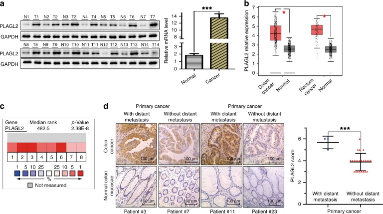
Background: We previously demonstrated that the pleomorphic adenoma gene like-2 (PLAGL2) is involved in the pathogenesis of Hirschsprung disease. Enhanced PLAGL2 expression was observed in several malignant tumours. However, the exact function of PLAGL2 and its underlying mechanism in colorectal cancer (CRC) remain largely unknown.

Methods: Immunohistochemical analysis of PLAGL2 was performed. A series of in vitro and in vivo experiments were conducted to reveal the role of PLAGL2 in the progression of CRC.

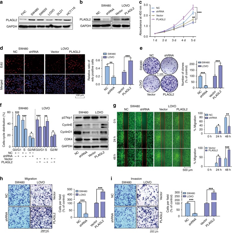
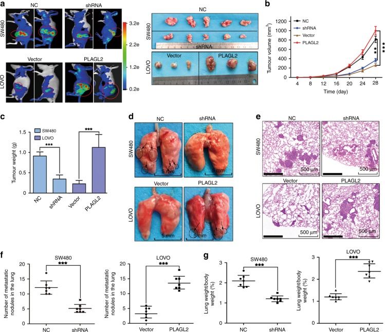
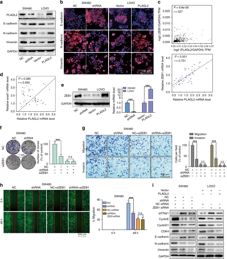
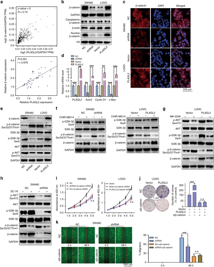
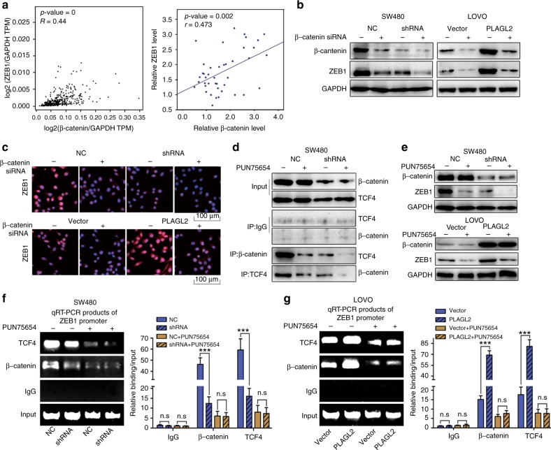
Results: Enhanced PLAGL2 expression was significantly associated with EMT-related proteins in CRC. The data revealed that PLAGL2 promotes CRC cell proliferation, migration, invasion and EMT both in vitro and in vivo. Mechanistically, PLAGL2 promoted the expression of ZEB1. PLAGL2 enhanced the expression and nuclear translocation of β-catenin by decreasing its phosphorylation. The depletion of β-catenin neutralised the regulation of ZEB1 that was caused by enhanced PLAGL2 expression. The small-molecule inhibitor PNU-74654, also impaired the enhancement of ZEB1 that resulted from the modified PLAGL2 expression. The depletion of ZEB1 could block the biological function of PLAGL2 in CRC cells.

Conclusions: Collectively, our findings suggest that PLAGL2 mediates EMT to promote colorectal cancer metastasis via β-catenin-dependent regulation of ZEB1.


Conflict of interest statement

The authors declare no competing interests.

Figures

Fig. 1
Fig. 1. PLAGL2 is overexpressed in CRC.
a The WB and qRT–PCR analysis showed that PLAGL2 overexpressed in CRC tissues compared to the expression in paired normal samples. T, CRC tissue; N, paired normal tissues. The data are presented as the mean ± SD from three independent experiments. *P < 0.05, **P < 0.01, ***P < 0.001, based on Student’s t-test. b The box plots were obtained from the GEPIA database to compare the expression of PLAGL2 in CRC specimens and matched normal specimens. c Meta-analysis of the PLAGL2 gene expression derived from the Oncomine database. d Representative immunohistochemistry images of the PLAGL2 expression levels in primary CRC tumours without metastasis vs CRC tumours with metastasis. Scale bars, 100 μm.
Fig. 2
Fig. 2. PLAGL2 promotes the proliferation, migration and invasion of CRC cells in vitro.
a PLAGL2 expression levels in five CRC cell lines and the normal colon epithelial cell line FHC. b The effects of PLAGL2 depletion and overexpression were determined by the WB analysis. ce The cell proliferation was regulated by modified PLAGL2 expression and was examined by CCK8 (c), EdU (d) and colony formation assays (e) in SW480 and LOVO cells. Scale bars, 100 μm (d). Scale bars, 1 cm (e). f The cell cycle results showed that PLAGL2 regulates cell cycle progression. PLAGL2 depletion increased the G0G1 fraction and decreased the S and G2M fraction. The expression of key cell cycle regulatory proteins was regulated by modified PLAGL2 expression. g The migration capacity of PLAGL2 in SW480 and LOVO cells was detected with a wound-healing assay. The cells migrating into the wounded areas were photographed at 0, 24, and 48 h. Scale bars, 500 μm. h, i The migration and invasion capacity of PLAGL2 in SW480 and LOVO cells were also evaluated with transwell assays. Scale bars, 200 μm. *P < 0.05, **P < 0.01, ***P < 0.001, based on Student’s t-test. The data are presented as the mean ± SD from three independent experiments.
Fig. 3
Fig. 3. PLAGL2 promotes CRC cell proliferation and metastasis in vivo.
a Representative bioluminescence pictures of the nude mice 28 days after injection. In all, 5 × 106 stable SW480 and LOVO cells were injected subcutaneously into the groin of nude mice (n = 7 per group). Representative images of the corresponding xenograft 28 days after inoculation. b Tumour volumes in the different groups. The growth curve of the tumours that formed after subcutaneous injection. c Enhanced PLAGL2 expression increased the tumour weights. d Lung metastasis models. Representative images of visible lung metastases. The metastatic nodules are indicated with arrows. Scale bars, 5 mm. e Representative images of the corresponding HE staining. Scale bars, 500 μm. f The numbers of metastatic nodules. g The depletion of PLAGL2 significantly reduced the lung weight. *P < 0.05, **P < 0.01, ***P < 0.001, based on Student’s t-test.
Fig. 4
Fig. 4. PLAGL2 induces ZEB1-mediated EMT.
a The levels of three EMT-related proteins in SW480 and LOVO cells. b Immunofluorescence assay was performed to detect EMT markers in SW480 and LOVO cells. Targeted proteins were stained red, and the nuclei were stained blue with 4′,6-diamidino-2-phenylindole (DAPI). Scale bars, 100 μm. c Data derived from the GEPIA database showed that there was a significant positive correlation between PLAGL2 and ZEB1 in CRC tissues. The data from this study also revealed a significant positive correlation between PLAGL2 and ZEB1. d The correlation between PLAGL2 and snail1 was also detected. There was no significant correlation between PLAGL2 and snail1 in CRC tissues. e The WB and qRT–PCR analysis showed that enhanced PLAGL2 expression increased ZEB1 expression. fh The depletion of PLAGL2 did not further decrease proliferation (f), migration (g, h) and invasion (g) in ZEB1-knockdown SW480 cells. i The downregulation of ZEB1 could rescue the levels of the EMT-related and cell cycle regulatory proteins in SW480 and LOVO cells, which was examined by the WB analysis. The data are presented as the mean ± SD from three independent experiments. n.s: no significance. *P < 0.05, **P < 0.01, ***P < 0.001, based on Student’s t-test.
Fig. 5
Fig. 5. PLAGL2 regulates β-catenin expression by modulating AKT/GSK-3β signalling.
a The GEPIA database showed that a significantly positive correlation between PLAGL2 and β-catenin could be observed in CRC tissues. The data from this study also revealed a significant positive correlation between PLAGL2 and β-catenin. b The nuclear β-catenin and total β-catenin protein levels in SW480 and LOVO cells. GAPDH and H3 were used as cytoplasmic or nuclear protein controls, respectively. c The immunofluorescence assays demonstrated that PLAGL2 increased the expression of β-catenin and the nuclear translocation of β-catenin. Scale bars, 100 μm. d The depletion of PLAGL2 markedly diminished the expression of the β-catenin target genes Axin2, c-Myc and Cyclin-D1, which were examined by the qRT–PCR analysis. e The WB analysis revealed that enhanced PLAGL2 expression promoted AKT and GSK-3β phosphorylation, impeding β-catenin phosphorylation. No significant difference was observed in the total GSK-3β and AKT levels. f CHIR98014, a small-molecule GSK-3β inhibitor, partly blocked the effect of modified PLAGL2 expression on β-catenin. g Stable LOVO cells were treated with the AKT specific inhibitor MK-2206. The corresponding protein levels were detected by the WB analysis. h Stable SW480 cells were treated with the AKT specific activator SC79. The corresponding protein levels were detected by the WB analysis. ik The downregulation of β-catenin could rescue the regulatory effect of PLAGL2 on cell proliferation and migration. The cell proliferation was examined by CCK8 (i) and colony formation assays (j). Scale bars, 1 cm. The migration capacity was detected with a wound-healing assay (k). Scale bars, 500 μm. The data are presented as the mean ± SD from three independent experiments. n.s: no significance. *P < 0.05, **P < 0.01, ***P < 0.001, based on Student’s t-test.
Fig. 6
Fig. 6. PLAGL2 modulates ZEB1 expression through a β-catenin-dependent pathway.
a The GEPIA database showed that a significant positive correlation between ZEB1 and β-catenin could be observed in CRC tissues. The data from this study also revealed a significant positive correlation between ZEB1 and β-catenin. b The WB analysis showed that the depletion of β-catenin neutralised the promotion of ZEB1 caused by enhanced PLAGL2 expression. (c) Depletion of β-catenin neutralised the promotion of ZEB1 caused by enhanced PLAGL2 expression, which was confirmed by immunofluorescence assays. Scale bars, 100 μm. d The validity of the small-molecule inhibitor PNU-74654 was verified by the WB analysis. e The WB analysis revealed that the PNU-74654, which blocked the interaction between β-catenin and TCF4, thereby neutralising the promotion of ZEB1 caused by enhanced PLAGL2 expression. f, g The ChIP assays were performed to verify the binding between β-catenin/TCF4 complexes and ZEB1 promoter in SW480 and LOVO cells. The bound DNA fragments were amplified by qRT-PCR. Then the products of qRT-PCR were examined by gel electrophoresis on 2% agarose gels. PNU-74654 blocked β-catenin/TCF4 complexes from directly binding to the ZEB1 promoter and neutralised the promotion of ZEB1 caused by enhanced PLAGL2 expression. The data are presented as the mean ± SD from three independent experiments. n.s: no significance. *P < 0.05, **P < 0.01, ***P < 0.001, based on Student’s t-test.

References

    1. Siegel RL, Miller KD, Fedewa SA, Ahnen DJ, Meester R, Barzi A, et al. Colorectal cancer statistics, 2017. CA Cancer J. Clin. 2017;67:177–93.. - PubMed
    1. Siegel RL, Miller KD, Jemal A. Cancer statistics, 2019. CA Cancer J. Clin. 2019;69:7–34. - PubMed
    1. Chaffer CL, Weinberg RA. A perspective on cancer cell metastasis. Science. 2011;331:1559–1564. - PubMed
    1. Thiery JP, Acloque H, Huang RY, Nieto MA. Epithelial-mesenchymal transitions in development and disease. Cell. 2009;139:871–90.. - PubMed
    1. Dongre A, Weinberg RA. New insights into the mechanisms of epithelial-mesenchymal transition and implications for cancer. Nat. Rev. Mol. Cell Biol. 2019;20:69–84. - PubMed

Publication types

MeSH terms